. 2025 Apr 24;15(5):441.
doi: 10.3390/brainsci15050441.

The Size and Localization of Ribeye and GluR2 in the Auditory Inner Hair Cell Synapse of C57BL/6 Mice Are Affected by Short-Pulse Corticosterone in a Sex-Dependent Manner

Affiliations

The Size and Localization of Ribeye and GluR2 in the Auditory Inner Hair Cell Synapse of C57BL/6 Mice Are Affected by Short-Pulse Corticosterone in a Sex-Dependent Manner

Ewa Domarecka et al. Brain Sci. .

Abstract

Background: Inner hair cell (IHC) ribbon synapses are the initial synapses in the auditory pathway, comprising presynaptic ribbons and postsynaptic glutamate receptors on the peripheral afferent fibers. The excitatory neurotransmitter glutamate primarily signals through AMPA-type heterotetrameric receptors (AMPARs), composed of GluR1, GluR2, GluR3, and GluR4 subunits. Research shows that corticosterone affects AMPA receptor subunits in the central nervous system. The present study investigates the effects of corticosterone on AMPA receptor subunits in the murine cochlea. Methods: Cochlear explants were isolated from male and female C57BL/6 pups (postnatal days 4-5), treated for 20 min with 100 nM corticosterone, and cultured for an additional 24 h. The concentration of AMPAR protein subunits was quantified using an ELISA assay, while gene expression was analyzed using RT-PCR. The synaptic localization patterns of GluR2 and Ribeye were examined using immunofluorescence and confocal microscopy. Results: Male C57BL/6 mice have a significantly greater basal concentration of the GluR2 subunit than females and more GluR2 puncta per IHC than females. Corticosterone increases the size of Ribeye in males and increases twofold GluR2/Ribeye colocalization in the apical region of females. Conclusions: Exposure of membranous cochleae to corticosterone induces changes consistent with neuroplasticity in the auditory periphery. The observed effect is sex-dependent.

Keywords: AMPA; cochlea; corticosterone; glutamate receptor subunits; inner hair cell synapse; stress hormones.


Conflict of interest statement

The authors declare no conflicts of interest.

Figures

Figure 1
Figure 1
Cochlear tissues were dissected from postnatal day (P4–P5) male and female pups and placed into culture for 24 h. Subsequently, the tissues were subjected to a 20 min pulse exposure to 100 nM CORT, followed by three washes and a 24 h incubation period. Next, protein concentration was analyzed using ELISA, and relative gene expression was determined by semiquantitative real-time RT-PCR. Protein localization was visualized using confocal microscopy. The manuscript uses pink to represent female data and blue for male data. Created with Biorender.com.
Figure 2
Figure 2
(a) Concentration of AMPA receptor subunits in the membranous cochlea of P4–P5 C57BL/6 mice; (b) sex influences the baseline concentration of AMPA receptor subunits [ng/µg] in cochlear tissues. The differences in the concentration of GluR1 protein between male and female mice, calculated using t-test, were not statistically significant (M: 33.23 ± 13.82, F: 22.62 ± 9.63; t(15) = −1.813, p = 0.090, n = 8–9 for each experimental group). The concentration of GluR2 was significantly higher in male mice compared to females (F: 2.82 ± 0.57, M: 6.16 ± 1.89, t(9.296) = −5.112, *** p < 0.001, Cohen’s d = 2.46, strong effect, n = 9–10 cochlear tissues per group). The differences in concentration of GluR3 between males and females were insignificant (M: 2.99 ± 0.94, F: 2.48 ± 0.74, n = 4–6 cochlear tissues per group). Mean ± SD is reported. The concentration of GluR4 was below the assay threshold sensitivity in 90% of the samples.
Figure 3
Figure 3
The total number of GluR2 puncta in inner hair cells (IHCs) varies between sexes. (a) Overview of the mouse cochlea highlighting the three parts used in further analyses: apical, middle, and basal regions; (b) representative confocal images of IHC synapses in the apex, mid, and base areas. The cochleae were labeled with antibodies against Ribeye (red), GluR2 (green), and with DAPI (blue). Scale bar: 10 µm; (c) no difference was observed in the numbers of Ribeye puncta per IHC based on sex; (d) the number of GluR2 puncta per IHC in the cochlea was significantly higher in male mice compared to female mice (Mann–Whitney U test U = 582, p < 0.001, Cohen’s d = 0.35, modest effect). The median ± 95% CI of the median confidence interval is reported; (e) a significant difference in GluR2 puncta numbers between male and female animals was observed in the apical part (M: 6.25 ± 1.92, F: 4.11 ± 1.74; t(18) = 2.619, p = 0.017, Cohen’s d = 1.17, strong effect), the middle part (M: 7.22 ± 2.10, F: 5.99 ± 1.89; t(43) = 2.077, p = 0.044, Cohen’s d = 0.62, moderate effect), and the basal part of the cochleae (M: 5.59 ± 2.74, F: 3.56 ± 0.93; t(13.504) = 2.433, p = 0.030, Cohen’s d = 0.99, moderate effect). Mean ± SD is reported, with n = 6–8 cochleae for each experimental group, number of analyzed IHCs in total = 1000, average number of analyzed Ribeye puncta per group = 5824, and average number of analyzed GluR2 puncta per group = 2983. Statistical significance is shown in the figure: * p < 0.05; *** p < 0.001.
Figure 4
Figure 4
CORT exposure affects the numbers and colocalization of Ribeye and GluR2 puncta in IHCs of female but not male cochlear explants. (a) The image depicts the middle part of the cochlea. The explants were cultured in either tissue culture medium alone or exposed to 100 nM CORT for 20 min and cultured for 24 h. The tissues were labeled with anti-Ribeye (red), anti-GluR2 (green), and DAPI (blue), with yellow color indicating the colocalization of the fluorescence signals (Ribeye/GluR2). The scale bar represents 10 µm; (b1) reduction of Ribeye puncta number in females after CORT in the middle region of the cochlea (control: 13.26 ± 2.74, CORT: 11.33 ± 2.50; t(45) = −2.532, p = 0.015, Cohen’s d= 0.74, moderate effect); (b2) no changes in the numbers of GluR2 puncta after CORT; (b3) increased colocalization of Ribeye and GluR2 after CORT exposure in the apical region of the cochlea, rising from 22% to 37% (t(14.627) = 2.745, p = 0.015, Cohen’s d = 1.10, strong effect); (c1c3) CORT does not induced changes in the number of Ribeye and GluR2 puncta or in the colocalization pattern. Mean values are presented ± SD, with n = 6–8 cochleae in each experimental group. Total number of analyzed IHCs: 2080; average number of analyzed Ribeye/GluR2 puncta in female group: 6266/2818; average number of analyzed Ribeye/GluR2 puncta in male group: 5880/3297. Statistical significance is shown in the figure: * p < 0.05.
Figure 5
Figure 5
CORT exposure increases the colocalization of Ribeye and GluR2 in cochlear synapses. The changes in the overlapping area of Ribeye and GluR2 were analyzed in control versus CORT-exposed cochleae. (a) Female cochlear explants. In CORT-exposed female explants, an increase in the overlapping area was observed at the apex and middle regions. At the same time, no change was seen at the base (apical: 15% to 20%, medial: 15% to 18%, basal: 14% to 15%); (b) male cochlear explants. An increase in the overlap between Ribeye and GluR2 was noted in the medial region of the cochlea, rising from 13% to 15%. For all significant changes, Cohen’s effect size was weak (0.1–0.2). The data are presented as median values ± 95% confidence interval (CI) of the median, with sample sizes ranging from n = 6 to 8 cochleae for each experimental group. The total number of analyzed IHCs is 2080, with the average number of analyzed Ribeye/GluR2 puncta being 6266/2818 in females and 5880/3297 in males. Statistical significance is shown in the figure: ** p < 0.01; *** p < 0.001.
Figure 6
Figure 6
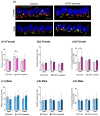
Exposure to CORT influences the size of Ribeye and GluR2 in a sex-dependent manner. The enlargement of GluR2 was observed in females and males, while changes in Ribeye size were seen exclusively in male cochlear explants. (a) Significant changes in GluR2 size were observed in the middle part of the cochlea from female mice (control: 0.13 µm2, CORT: 0.17 µm2 (median values); Mann–Whitney U test, p < 0.001) and in the basal region of the cochlea from male mice (control: 0.13 µm2; CORT: 0.15 µm2 (median values); Mann–Whitney U test, p = 0.011); (b) changes in Ribeye size in male cochlear explants (an increase of 0.04 µm2) occurred throughout the entire cochlear explant (Mann–Whitney U test). For all significant changes, Cohen’s effect size was weak (0.1–0.2). The data are expressed as median values ± 95% CI of median (n = 6–8 cochleae for each experimental group, number of analyzed IHCs in total = 2080, average number of analyzed Ribeye/GluR2 puncta in female groups = 6266/2818, average number of analyzed Ribeye/GluR2 puncta in male groups = 5880/3297). Statistical significance is shown in the figure: * p < 0.05; *** p < 0.001.
Figure 6
Figure 6
Exposure to CORT influences the size of Ribeye and GluR2 in a sex-dependent manner. The enlargement of GluR2 was observed in females and males, while changes in Ribeye size were seen exclusively in male cochlear explants. (a) Significant changes in GluR2 size were observed in the middle part of the cochlea from female mice (control: 0.13 µm2, CORT: 0.17 µm2 (median values); Mann–Whitney U test, p < 0.001) and in the basal region of the cochlea from male mice (control: 0.13 µm2; CORT: 0.15 µm2 (median values); Mann–Whitney U test, p = 0.011); (b) changes in Ribeye size in male cochlear explants (an increase of 0.04 µm2) occurred throughout the entire cochlear explant (Mann–Whitney U test). For all significant changes, Cohen’s effect size was weak (0.1–0.2). The data are expressed as median values ± 95% CI of median (n = 6–8 cochleae for each experimental group, number of analyzed IHCs in total = 2080, average number of analyzed Ribeye/GluR2 puncta in female groups = 6266/2818, average number of analyzed Ribeye/GluR2 puncta in male groups = 5880/3297). Statistical significance is shown in the figure: * p < 0.05; *** p < 0.001.
Figure 7
Figure 7
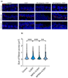
(a) Representative confocal images display male cochlear explants with inner hair cell (IHC) synapses in the apex, mid, and base regions. The cochleae were stained with anti-Ribeye (red), anti-GluR2 (green), and DAPI (blue). Scale bar: 10 µm; (b) in the middle region of the male cochlea, the Kruskal–Wallis test detected significant differences between CORT and MIFE + CORT groups (CORT: 0.24, MIFE + CORT: 15, p < 0.001), but not between CORT and SPIRO + CORT (CORT: 0.24, 0.22, p = ns). Median values are presented, with n ranging from 2 to 3 cochleae in each experimental group, a total number of analyzed IHCs of 100, and a total number of analyzed Ribeye puncta of 1554. Statistical significance is shown in the figure: ** p < 0.01; *** p < 0.001.

Similar articles

References

    1. Glowatzki E., Fuchs P.A. Transmitter release at the hair cell ribbon synapse. Nat. Neurosci. 2002;5:147–154. doi: 10.1038/nn796. - DOI - PubMed
    1. Meyer A.C., Frank T., Khimich D., Hoch G., Riedel D., Chapochnikov N.M., Yarin Y.M., Harke B., Hell S.W., Egner A., et al. Tuning of synapse number, structure and function in the cochlea. Nat. Neurosci. 2009;12:444–453. doi: 10.1038/nn.2293. - DOI - PubMed
    1. Rutherford M.A. Resolving the structure of inner ear ribbon synapses with STED microscopy. Synapse. 2015;69:242–255. doi: 10.1002/syn.21812. - DOI - PubMed
    1. Floriou-Servou A., von Ziegler L., Waag R., Schläppi C., Germain P.-L., Bohacek J. The Acute Stress Response in the Multiomic Era. Biol. Psychiatry. 2021;89:1116–1126. doi: 10.1016/j.biopsych.2020.12.031. - DOI - PubMed
    1. de Kloet E.R., Joëls M., Holsboer F. Stress and the brain: From adaptation to disease. Nat. Rev. Neurosci. 2005;6:463–475. doi: 10.1038/nrn1683. - DOI - PubMed

LinkOut - more resources