. 2025 May 8;13(5):1142.
doi: 10.3390/biomedicines13051142.

Reduced Expression of UPRmt Proteins HSP10, HSP60, HTRA2, OMA1, SPG7, and YME1L Is Associated with Accelerated Heart Failure in Humans

Affiliations

Reduced Expression of UPRmt Proteins HSP10, HSP60, HTRA2, OMA1, SPG7, and YME1L Is Associated with Accelerated Heart Failure in Humans

Petra Bakovic et al. Biomedicines. .

Abstract

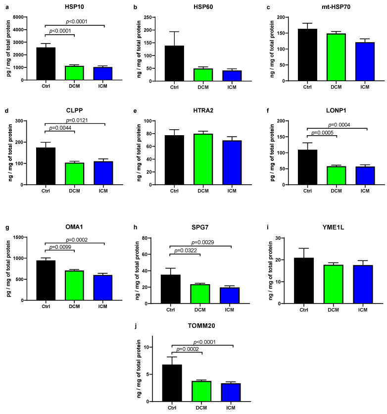
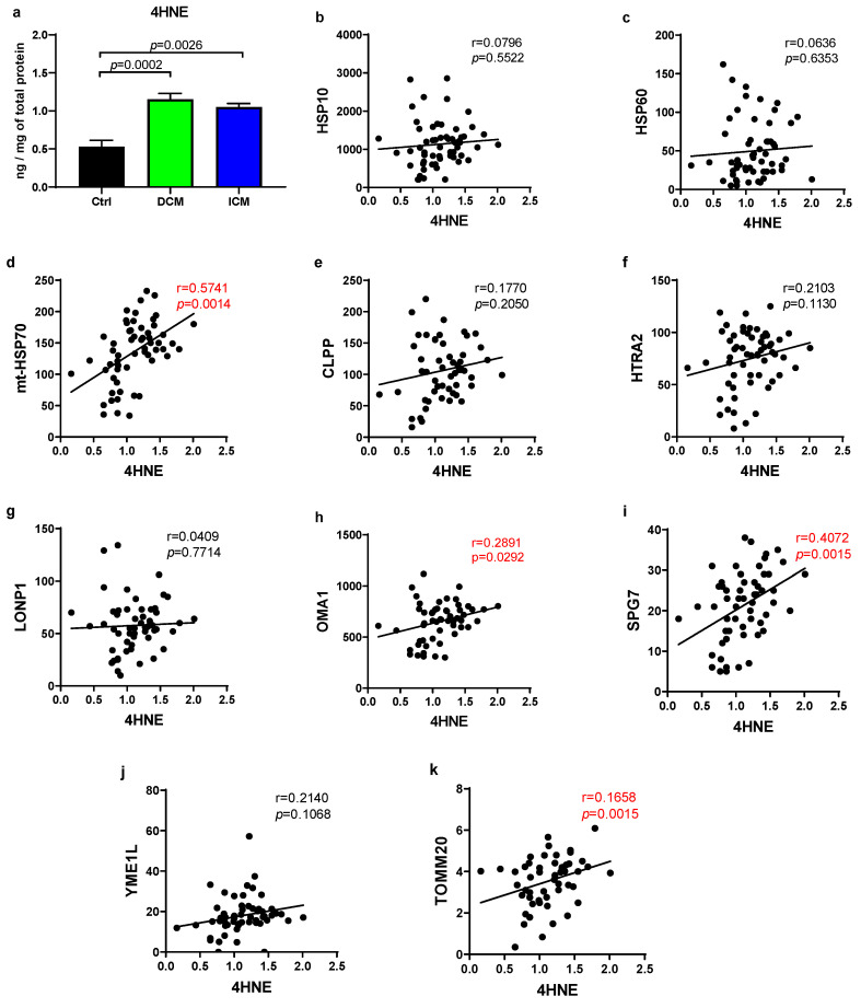
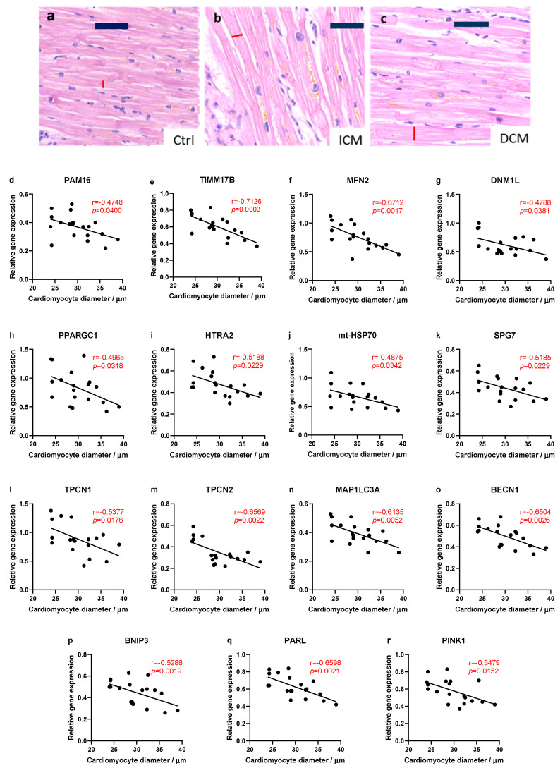
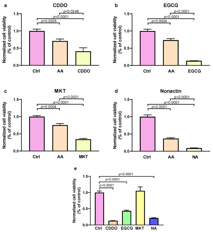
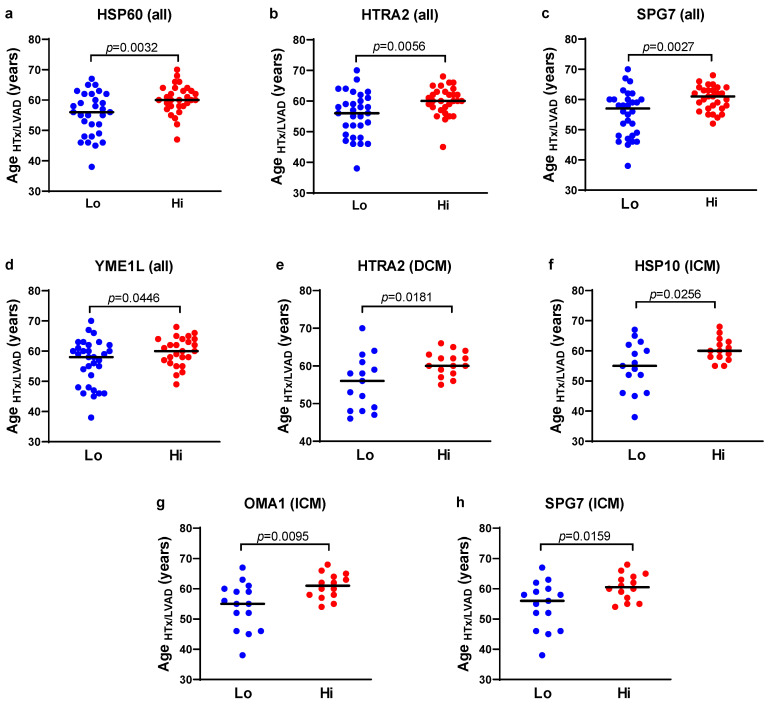
Background/Objectives: The mitochondrial unfolded protein response (UPRmt) is one of the mitochondrial quality control mechanisms that is responsible for reparation and removal of damaged proteins in mitochondria. Methods: Here we investigated the role of the UPRmt in the myocardium of humans with and without heart failure and in the cell culture model. Results: The analysis of myocardial samples by ELISA from patients with ischemic cardiomyopathy (ICM) and dilated cardiomyopathy (DCM), as well as healthy donors, revealed a significantly reduced expression of the UPRmt proteins HSP10, CLPP, LONP1, OMA1, and SPG7 in patients with DCM and ICM. Furthermore, patients with DCM and ICM exhibited elevated levels of myocardial reactive oxygen species (ROS, tested by 4-hydroxynonenal) compared to controls, and a positive correlation between ROS production and mt-HSP70, OMA1, and SPG7 protein expression. The correlation analysis indicated a negative correlation between cardiomyocyte hypertrophy and the expression of several UPRmt genes. The inhibition of four tested UPRmt effector proteins exacerbated the injury of cultured cells under oxidative stress. The patients with ICM, DCM, or both, who showed lower myocardial expression of HSP10, HSP60, HTRA2, OMA1, SPG7, and YME1L, underwent heart transplantation or implantation of a left ventricular assist device earlier in life compared to those with the higher protein expression. Conclusions: In conclusion, our findings indicate that the reduced expression of several UPRmt effector proteins is associated with accelerated heart failure in patients, which, together with other results, indicates that impaired UPRmt may contribute to the pathogenesis of heart failure in humans.

Keywords: ROS; UPRmt; cardiomyopathy; heart; mitochondria.


Conflict of interest statement

Authors declare no conflicts of interest.

Figures

Figure 1
Figure 1
Protein expression of UPRmt elements and TOMM20 in failing human hearts. (aj) Protein levels in human myocardial samples were quantified using ELISA kits. Analyses included UPRmt chaperones (HSP10, HSP60, and mt-HSP70), proteases (CLPP, HTRA2, LONP1, OMA1, SPG7, and YME1L), and translocase of the outer mitochondrial membrane TOMM20, which was used to assess mitochondrial content in the myocardium. Summary data are presented as mean value ± SEM. Abbreviations: Ctrl (control; n = 6); DCM (heart failure caused by dilated cardiomyopathy; n = 30); and ICM (heart failure caused by ischemic cardiomyopathy; n = 29). Significant p-values are indicated in graphs.
Figure 2
Figure 2
Production of ROS and UPRmt in human hearts. (a) Levels of 4-hydroxynonenal (4HNE) were quantified using an ELISA kit in samples of failing human hearts. (bk) Scatter plots illustrating a correlation between 4HNE levels and UPRmt protein levels in failing human hearts. Pearson or Spearman coefficient correlation and p-value are given on each graph, where red color indicates a statistically significant correlation (p < 0.05). Abbreviations: Ctrl (control; n = 6); DCM (heart failure caused by dilated cardiomyopathy; n = 30); and ICM (heart failure caused by ischemic cardiomyopathy; n = 29).
Figure 3
Figure 3
Relationship between myocardial hypertrophy and the UPRmt and MQC gene expression. (ac) Representative images of hematoxylin and eosin (H&E)-stained sections of myocardium that were used to measure cardiomyocyte diameters in formalin-fixed paraffin-embedded slices of hearts of patients with DCM or ICM. Scale bars = 50 µm (dr) Scatter plots illustrate the correlation between cardiomyocyte’s diameter and a relative gene expression in failing human hearts (ICM and DCM samples combined). Pearson or Spearman coefficient correlation and p-values are given on each graph, where red color indicates statistically significant difference (p < 0.05). Abbreviations: Ctrl (control; n = 6); DCM (heart failure caused by dilated cardiomyopathy; n = 30); and ICM (heart failure caused by ischemic cardiomyopathy; n = 29).
Figure 4
Figure 4
Effect of UPRmt inhibitors on human cultured cells exposed to oxidative stress. An MTT assay was used to assess viability of cells without any treatment (control, Ctrl), cells treated with antimycin A to induce oxidative stress (AA), and cells treated with combination of AA and one of four different UPRmt inhibitors: (a) EGCG (inhibitor of OMA1); (b) MKT-077 (MKT; inhibitor of mt-HSP70); (c) CDDO (inhibitor of LONP); and (d) nonactin (NA; inhibitor of HSP60). Effects of inhibitors alone, without AA are shown in (e). Data are expressed as means ± SEM with n = 8 for each group. Data are normalized to control (Ctrl). Significant p-values are indicated in graphs.
Figure 5
Figure 5
The age of patients undergoing heart transplantation or LVAD implantation according to the myocardial expression of UPRmt proteins. (ah) Individual data points represent the age when patients underwent heart transplantation or LVAD implantation (ageHTx/LVAD), and the horizontal line indicates mean values. Patients were divided into two groups according to the myocardial expression of respective UPRmt proteins (CLPP, HSP10, HSP60, mt-HSP70, HTRA2, LONP1, OMA1, SPG7, TOMM20, and YME1L), where half of the patients with a lower expression were assigned to the low group (Lo) group, and half of the patients with a higher expression were assigned to the higher group (Hi). The patients with dilated cardiomyopathy (DCM) and ischemic cardiomyopathy (ICM) were analyzed separately or together (all). Only significant differences (p < 0.05) are shown.

Similar articles

References

    1. Briceno N., Schuster A., Lumley M., Perera D. Ischaemic Cardiomyopathy: Pathophysiology, Assessment and the Role of Revascularisation. Heart. 2016;102:397–406. doi: 10.1136/heartjnl-2015-308037. - DOI - PubMed
    1. Heymans S., Lakdawala N.K., Tschöpe C., Klingel K. Dilated Cardiomyopathy: Causes, Mechanisms, and Current and Future Treatment Approaches. Lancet. 2023;402:998–1011. doi: 10.1016/S0140-6736(23)01241-2. - DOI - PubMed
    1. Arbelo E., Protonotarios A., Gimeno J.R., Arbustini E., Barriales-Villa R., Basso C., Bezzina C.R., Biagini E., Blom N.A., de Boer R.A., et al. 2023 ESC Guidelines for the Management of Cardiomyopathies: Developed by the Task Force on the Management of Cardiomyopathies of the European Society of Cardiology (ESC) Eur. Heart J. 2023;44:3503–3626. doi: 10.1093/eurheartj/ehad194. - DOI - PubMed
    1. Gallo G., Rubattu S., Volpe M. Mitochondrial Dysfunction in Heart Failure: From Pathophysiological Mechanisms to Therapeutic Opportunities. Int. J. Mol. Sci. 2024;25:2667. doi: 10.3390/ijms25052667. - DOI - PMC - PubMed
    1. Liu J., He X., Zheng S., Zhu A., Wang J. The Mitochondrial Unfolded Protein Response: A Novel Protective Pathway Targeting Cardiomyocytes. Oxid. Med. Cell. Longev. 2022;2022:6430342. doi: 10.1155/2022/6430342. - DOI - PMC - PubMed

LinkOut - more resources