Review
. 2025 May 10;13(5):1160.
doi: 10.3390/biomedicines13051160.

Biomarker-Driven Approaches to Bone Metastases: From Molecular Mechanisms to Clinical Applications

Affiliations
Review

Biomarker-Driven Approaches to Bone Metastases: From Molecular Mechanisms to Clinical Applications

Youssef Elshimy et al. Biomedicines. .

Abstract

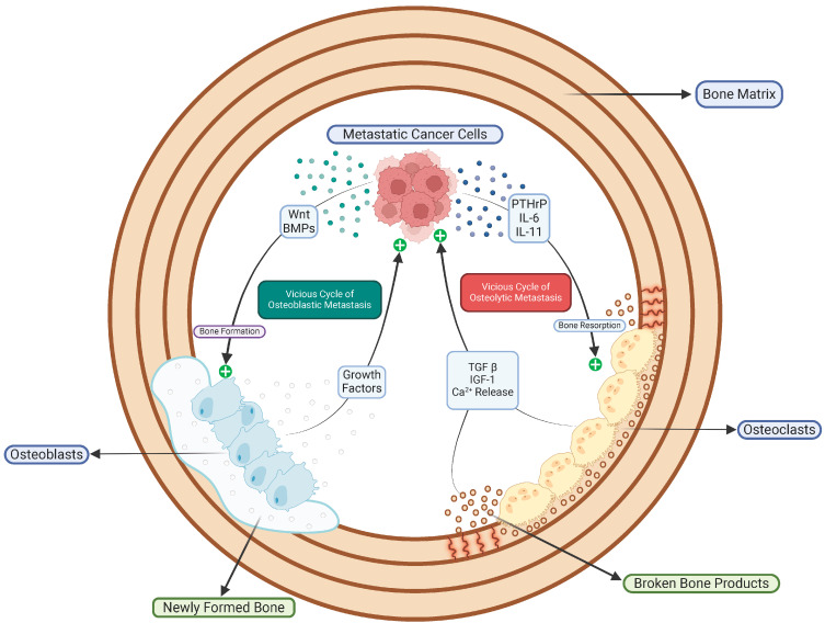
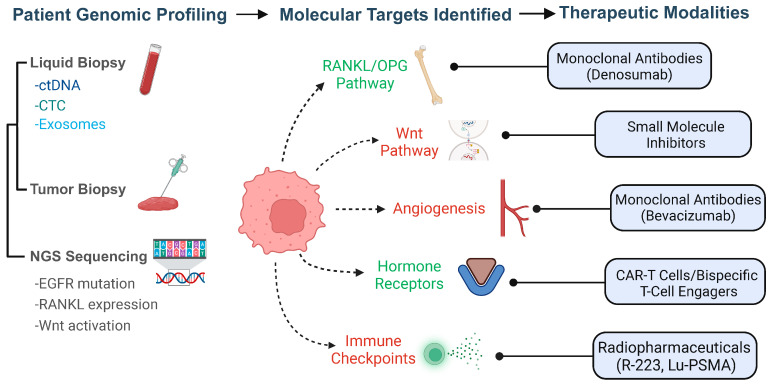
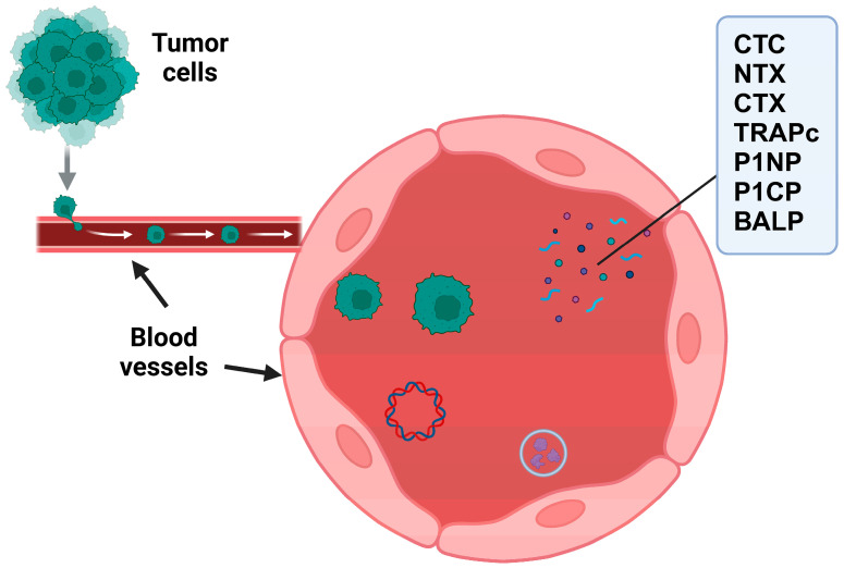
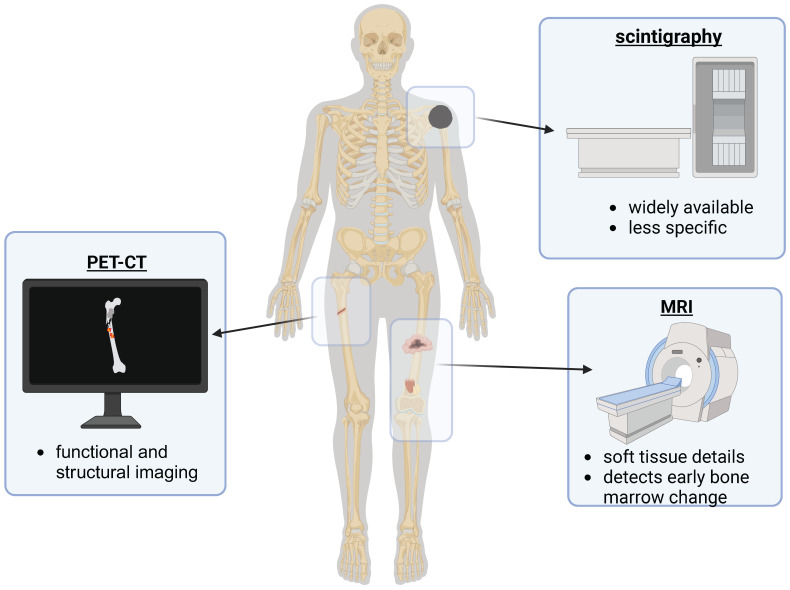
Bone metastases represent a critical complication in oncology, frequently indicating advanced malignancy and substantially reducing patient quality of life. This review provides a comprehensive analysis of the complex interactions between tumor cells and the bone microenvironment, emphasizing the relevance of the "seed and soil" hypothesis, the RANK/RANKL/OPG signaling axis, and Wnt signaling pathways that collectively drive metastatic progression. The molecular and cellular mechanisms underlying the formation of osteolytic and osteoblastic lesions are examined in detail, with a particular focus on their implications for bone metastases associated with breast, prostate, lung, and other cancers. A central component of this review is the categorization of pathological biomarkers into four types: diagnostic, prognostic, predictive, and monitoring. We provide a comprehensive evaluation of circulating tumor cells (CTCs), bone turnover markers (such as TRACP-5b and CTX), advanced imaging biomarkers (including PET/CT and MRI), and novel genomic signatures. These biomarkers offer valuable insights for early detection, enhanced risk stratification, and optimized therapeutic decision-making. Furthermore, emerging strategies in immunotherapy and bone-targeted treatments are discussed, highlighting the potential of biomarker-guided precision medicine to enhance personalized patient care. The distinctiveness of this review lies in its integrative approach, combining fundamental pathophysiological insights with the latest developments in biomarker discovery and therapeutic innovation. By synthesizing evidence across various cancer types and biomarker categories, we provide a cohesive framework aimed at advancing both the scientific understanding and clinical management of bone metastases.

Keywords: BTMs; CTCs; biomarkers in precision oncology; bone metastasis; circulating biomarkers; ctDNA; precision medicine; therapeutic targets.


Conflict of interest statement

The authors declare no conflict of interest.

Figures

Figure 1
Figure 1
Schematic representation of the vicious cycle of bone metastasis. This figure illustrates the dynamic interaction between metastatic cancer cells and the bone microenvironment, emphasizing the dual nature of bone remodeling in metastasis. Metastatic cancer cells secrete various factors, including parathyroid hormone-related protein (PTHrP), interleukin-6 (IL-6), and interleukin-11 (IL-11), which stimulate osteoclasts to enhance bone resorption. This resorption releases growth factors such as transforming growth factor-beta (TGF-β), insulin-like growth factor 1 (IGF-1), and calcium, further promoting tumor growth and metastatic activity. Simultaneously, tumor-secreted factors such as Wnt proteins and bone morphogenetic proteins (BMPs) stimulate osteoblasts, leading to new bone formation, but structurally flawed and disorganized, characteristic of osteoblastic metastasis. The figure encapsulates the interconnected cycles of osteolytic and osteoblastic metastasis that contribute to the progression of bone metastases and highlights potential therapeutic targets within these pathways.
Figure 2
Figure 2
Interactions between tumor cells, bone microenvironment, and angiogenesis in bone metastases: This figure illustrates the complex interplay of cellular and molecular mechanisms involved in bone metastasis. Tumor cells in the bone microenvironment secrete interleukins (IL-8 and IL-11), which stimulate pre-osteoclasts’ maturation into osteoclasts. The receptor activator of nuclear factor kappa-B ligand (RANKL) expressed by osteoblasts binds to RANK on osteoclasts, promoting osteoclast activity and bone resorption. This process is crucial for the remodeling of the bone matrix and the establishment of metastases. Additionally, the Wnt signaling pathway, depicted in the bottom left, influences the regulation of osteoblast and osteoclast functions, critical for maintaining bone homeostasis and metastatic progression. The top right section highlights the role of angiogenesis in supporting tumor growth and metastasis, mediated by vascular endothelial growth factor (VEGF) and its receptors (VEGFRs), regulated through hypoxia-inducible factors (HIF-1α and HIF-1β). These interactions underscore potential biomarkers and therapeutic targets in managing bone metastases.
Figure 3
Figure 3
Integration of patient genomic profiling and therapeutic targeting in bone metastasis: This figure outlines the process of patient genomic profiling leading to personalized therapeutic interventions for bone metastasis. It begins with the collection of genetic material through liquid biopsy (ctDNA, CTCs, exosomes) and tumor biopsy, followed by next-generation sequencing (NGS), which identifies specific mutations and pathway activations such as EGFR mutations, RANKL expression, and Wnt pathway activation. Identified molecular targets include the RANKL/OPG pathway, Wnt signaling, angiogenesis pathways, hormone receptors, and immune checkpoints. These targets correspond to various therapeutic modalities, including monoclonal antibodies (e.g., Denosumab for RANKL, Bevacizumab for angiogenesis), small-molecule inhibitors, CAR-T cells and bispecific T-cell engagers for immune modulation, and radiopharmaceuticals (e.g., R-223, Lu-PSMA). This strategic approach underscores the potential for precision medicine in treating bone metastases by tailoring therapies based on specific genetic profiles and molecular characteristics.
Figure 4
Figure 4
Circulating tumor cells (CTCs) and biomarkers of bone remodeling in the context of metastasis: This diagram depicts the dissemination of tumor cells from the primary site into the bloodstream, where they become circulating tumor cells (CTCs). It highlights the interaction between CTCs and the bone environment, where they can contribute to metastatic bone disease. The right-hand panel lists key biomarkers associated with bone remodeling and metastasis: C-terminal telopeptide (CTX) and N-terminal telopeptide (NTX) for bone resorption, tartrate-resistant acid phosphatase (TRAPc), procollagen type I N propeptide (P1NP), procollagen type I C propeptide (P1CP), and bone-specific alkaline phosphatase (BALP). These biomarkers serve as crucial indicators for the diagnosis, monitoring, and understanding of the metastatic process, as well as potential therapeutic targets.
Figure 5
Figure 5
Diagnostic imaging modalities for bone metastasis detection. This figure provides an overview of the primary imaging techniques used in the diagnosis and monitoring of bone metastases. Each modality is linked to specific features and uses within the clinical context: PET-CT combines positron emission tomography (PET) and computed tomography (CT) to offer both functional and structural imaging. This modality is crucial for identifying active metastatic sites and assessing metabolic activity. Scintigraphy: A widely available nuclear medicine test that uses radioactive substances to detect abnormalities in bone metabolism, though it is less specific compared to other modalities. MRI: Magnetic resonance imaging provides detailed images of soft tissues and is highly sensitive for detecting early changes in the bone marrow associated with metastasis. These imaging techniques are integral to the comprehensive assessment of bone metastases, aiding in the precise localization of lesions and guiding subsequent therapeutic strategies.

Similar articles

References

    1. Zhao C., Lou Y., Wang Y., Wang D., Tang L., Gao X., Zhang K., Xu W., Liu T., Xiao J. A Gene Expression Signature-Based Nomogram Model in Prediction of Breast Cancer Bone Metastases. Cancer Med. 2019;8:200–208. doi: 10.1002/cam4.1932. - DOI - PMC - PubMed
    1. Hernandez R.K., Wade S.W., Reich A., Pirolli M., Liede A., Lyman G.H. Incidence of Bone Metastases in Patients with Solid Tumors: Analysis of Oncology Electronic Medical Records in the United States. BMC Cancer. 2018;18:44. doi: 10.1186/s12885-017-3922-0. - DOI - PMC - PubMed
    1. Jiang W., Rixiati Y., Zhao B., Li Y., Tang C., Liu J. Incidence, Prevalence, and Outcomes of Systemic Malignancy with Bone Metastases. J. Orthop. Surg. Hong Kong. 2020;28:2309499020915989. doi: 10.1177/2309499020915989. - DOI - PubMed
    1. Rieunier G., Wu X., Macaulay V.M., Lee A.V., Weyer-Czernilofsky U., Bogenrieder T. Bad to the Bone: The Role of the Insulin-Like Growth Factor Axis in Osseous Metastasis. Clin. Cancer Res. 2019;25:3479–3485. doi: 10.1158/1078-0432.CCR-18-2697. - DOI - PubMed
    1. Brown J.E., Cook R.J., Major P., Lipton A., Saad F., Smith M., Lee K.-A., Zheng M., Hei Y.-J., Coleman R.E. Bone Turnover Markers as Predictors of Skeletal Complications in Prostate Cancer, Lung Cancer, and Other Solid Tumors. J. Natl. Cancer Inst. 2005;97:59–69. doi: 10.1093/jnci/dji002. - DOI - PubMed

LinkOut - more resources