. 2025 May 14;13(5):1195.
doi: 10.3390/biomedicines13051195.

Laser-Induced Cortical Lesions in Mice as a Model for Progressive Multiple Sclerosis Pathology

Affiliations

Laser-Induced Cortical Lesions in Mice as a Model for Progressive Multiple Sclerosis Pathology

Bhavya Ojha et al. Biomedicines. .

Abstract

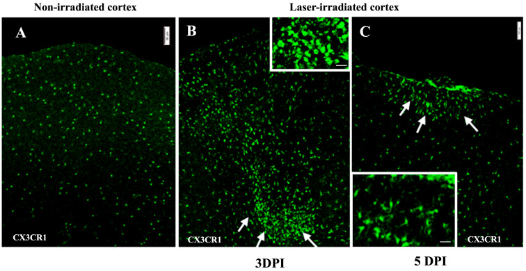
Background: The current animal models of multiple sclerosis (MS) predominantly emphasize white matter inflammation, reflecting early-stage disease. However, progressive MS (PMS) is characterized by cortical pathology, including subpial demyelination, chronic meningeal inflammation, and microglial activation, which are underrepresented in the existing models. While alternative mouse models replicate the relapsing-remitting phenotype and gray matter pathology, pathology is frequently dispersed throughout the brain, complicating the analysis of the specific lesion sites. Methods: To address this gap, we developed a novel model that integrates laser-induced focal demyelination with cytokine-driven meningeal inflammation to replicate the key aspects of PMS cortical pathology. Results: Using two-photon laser irradiation, we induced controlled subpial cortical lesions in CX3CR1-GFP mice, leading to microglial activation, astrocytosis, and focal demyelination. The addition of IFNγ-expressing adenovirus to promote meningeal inflammation which resulted in prolonged glial responses, increased immune cell infiltration, and exacerbated demyelination, mimicking the PMS-associated pathology. Conclusions: This model provides a powerful tool to investigate the mechanisms underlying the cortical lesion development and immune-mediated neurodegeneration in PMS. By capturing the critical aspects of cortical pathology, it enables the evaluation of therapeutic strategies targeting neuroinflammation and demyelination, ultimately aiding in the development of new treatments of progression in PMS patients.

Keywords: cortical pathology; demyelination; meningeal inflammation; progressive multiple sclerosis.


Conflict of interest statement

The authors declare no conflicts of interest.

Figures

Figure 1
Figure 1
Histopathological analysis of brain tissue. (A) Laser-induced bleeding in the irradiated cortex is visible in isolated brains as well as in cryostat sections at 3 DPI. (B) Hematoxylin and eosin (H&E) staining of non-irradiated cortex of mouse brain showing no histological changes. (D) Micrograph shows higher magnification of inset box in (B). (C) Histological examination using H&E staining confirms loss of tissue integrity and increased cellularity in the irradiated cortex (white outline indicates lesion area). Box inset indicates lesion rim, visible in higher magnification in (E), showing cellular aggregates in the lesion rim. Inset in (E) shows cellular aggregates in laser-irradiated cortex (arrows indicate possible PMNs). (n = 9). Scale bar: 200 μm (B,C), 100 µm (D,E), 10 µm (inset).
Figure 2
Figure 2
Microglia in laser-irradiated mice: the micrographs show green microglia in CX3CR1GFP/+ mice. (A) Microglia in the unirradiated cortex appear unaffected by skull thinning. (B) Microglia cluster around lesions in the irradiated cortex at 3 DPI. The microglia form aggregates, indicated by white arrows. Inset shows higher magnification. (C) Microglia in irradiated cortex at 5 DPI. The cluster of microglia is more compact and localized to a smaller area indicated by white arrows. Inset: microglia within clusters show amoeboid morphology. The experiment was independently performed twice (n = 12). Scale bar, 50 µm, 10 µm (inset).
Figure 3
Figure 3
Astrogliosis after irradiation. (A) Micrograph of non-irradiated cortex showing normal low-level GFAP staining. (B,C) Astrocytic activity in irradiated cortex indicated by GFAP-stained astrocytes in red at 3 DPI (B) and 5 DPI (C). (B) GFAP reactive astrocytes align around the lesion region and at the glia limitans, localizing at the periphery of the lesion, indicated by white arrows. (C) At 5 DPI, the region of astrocytic activity has condensed, with a reduced area of gliosis proximal to the subpial region, indicated by white arrows. Activated astrocytes were not observed deeper in the cortex. The experiment was independently performed twice (n = 12). Scale bar, 50 µm.
Figure 4
Figure 4
Demyelination: micrographs show anti-MBP staining (red). (A) Undisrupted myelin in the non-irradiated cortex. (B) Demyelination at 3 DPI in the irradiated cortex. MBP staining shows disrupted myelin, with myelin debris in the lesion, indicated by the dashed line circle. (C) MBP staining at 5 DPI indicates no loss of myelin. The experiment was independently performed twice (n = 12). Scale bar, 50 µm.
Figure 5
Figure 5
Leukocyte infiltration (red) in cortex after irradiation: (A) CD45 staining in non-irradiated cortex showing no infiltration. (B,C) Intense infiltration at 3 DPI, lessening at 5 DPI (C). Inset in (B) shows GR1 + (green) and CD45+ (red) colocalization (arrow), DAPI (nuclei, blue). Scale bar, 100 µm. (D) BBB disruption in irradiated cortex at 3 DPI: micrograph shows HRP that leaked from peripheral blood following IV injection 15 min earlier. The experiment was independently performed twice (n = 12). Scale bar, 200 µm. 10 µm (inset).
Figure 6
Figure 6
Gliosis and infiltration in irradiated mice with meningeal inflammation (intrathecal Ad-IFN-γ 1 week before irradiation). (AC) Microglia in CX3CR1gfp/+ mice. (A) Non-irradiated cortex. (B) At 3 DPI, microglia aggregated at the periphery of the lesion. (C) The size of the cluster and the density remained extensive at 5 DPI. (DF) Astrocytes stained with anti-GFAP. (D) Non-irradiated cortex. (E,F) Activated astrocytes at the periphery of the lesion (white arrows) at 3 DPI (E) and 5 DPI (F), aligning with microglia in (B,C). (G) MBP staining in non-irradiated cortex. (H,I) Myelin disruption at 3 DPI (H) and 5DPI (I), indicated by the dashed line circles. (JL) Leukocyte infiltration (CD45 staining). (J) Non-irradiated cortex. (K,L) Intense infiltration at 3 DPI (K) and 5DPI (L). The experiment was independently performed twice (n = 16), scale bar, 50 µm.
Figure 7
Figure 7
(A) Representative flow cytometry profiles from individual ipsilateral hemisphere suspension from irradiated mice. Flow cytometry gating strategy to distinguish CD45high leukocytes from Cd45dim microglia. Monocytes (CD45highCD11bhighLy6Chigh) and granulocytes (CD45highCD11bhighLy6Ghigh) were gated from CD45high. (B) Infiltration of leukocytes is enhanced by meningeal IFNγ in laser irradiated group. The total number of infiltrating CD45high leukocytes is enhanced at both 3 DPI ((i), p = 0.0242) and 5 DPI ((ii), p = 0.0121). (iii) The total number of Ly6chighcells, representing monocytes, in the irradiated cortex was lower at 5 DPI than at 3 DPI (p = 0.0207). (iv) Significantly more Ly6chigh monocytes infiltrated the irradiated than the non-irradiated cortex (p = 0.0295). The experiment was independently performed twice (n = 4).

References

    1. Lassmann H., Bruck W., Lucchinetti C.F. The immunopathology of multiple sclerosis: An overview. Brain Pathol. 2007;17:210–218. doi: 10.1111/j.1750-3639.2007.00064.x. - DOI - PMC - PubMed
    1. Reich D.S., Lucchinetti C.F., Calabresi P.A. Multiple Sclerosis. N. Engl. J. Med. 2018;378:169–180. doi: 10.1056/NEJMra1401483. - DOI - PMC - PubMed
    1. Kuhlmann T., Ludwin S., Prat A., Antel J., Bruck W., Lassmann H. An updated histological classification system for multiple sclerosis lesions. Acta Neuropathol. 2017;133:13–24. doi: 10.1007/s00401-016-1653-y. - DOI - PubMed
    1. Choi M., Ku T., Chong K., Yoon J., Choi C. Minimally invasive molecular delivery into the brain using optical modulation of vascular permeability. Proc. Natl. Acad. Sci. USA. 2011;108:9256–9261. doi: 10.1073/pnas.1018790108. - DOI - PMC - PubMed
    1. Magliozzi R., Howell O.W., Nicholas R., Cruciani C., Castellaro M., Romualdi C., Rossi S., Pitteri M., Benedetti M.D., Gajofatto A., et al. Inflammatory intrathecal profiles and cortical damage in multiple sclerosis. Ann. Neurol. 2018;83:739–755. doi: 10.1002/ana.25197. - DOI - PubMed

LinkOut - more resources