Review
. 2025 Apr 28;14(5):527.
doi: 10.3390/antiox14050527.

Ferroptosis and Iron Homeostasis: Molecular Mechanisms and Neurodegenerative Disease Implications

Affiliations
Review

Ferroptosis and Iron Homeostasis: Molecular Mechanisms and Neurodegenerative Disease Implications

Nurzhan Abdukarimov et al. Antioxidants (Basel). .

Abstract

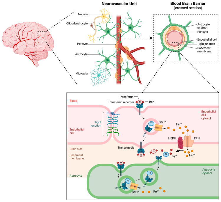
Iron dysregulation has emerged as a pivotal factor in neurodegenerative pathologies, especially through its capacity to promote ferroptosis, a unique form of regulated cell death driven by iron-catalyzed lipid peroxidation. This review synthesizes current evidence on the molecular underpinnings of ferroptosis, focusing on how disruptions in iron homeostasis interact with key antioxidant defenses, such as the system Xc--glutathione-GPX4 axis, to tip neurons toward lethal oxidative damage. Building on these mechanistic foundations, we explore how ferroptosis intersects with hallmark pathologies in Alzheimer's disease (AD) and Parkinson's disease (PD) and examine how iron accumulation in vulnerable brain regions may fuel disease-specific protein aggregation and neurodegeneration. We further surveyed the distinct components of ferroptosis, highlighting the role of lipid peroxidation enzymes, mitochondrial dysfunction, and recently discovered parallel pathways that either exacerbate or mitigate neuronal death. Finally, we discuss how these insights open new avenues for neuroprotective strategies, including iron chelation and lipid peroxidation inhibitors. By highlighting open questions, this review seeks to clarify the current state of knowledge and proposes directions to harness ferroptosis modulation for disease intervention.

Keywords: Alzheimer’s disease; Parkinson’s disease; cell death; ferroptosis; iron homeostasis.


Conflict of interest statement

The authors declare no conflict of interest.

Figures

Figure 1
Figure 1
The system Xc-GSH-GPX4 axis negatively regulates ferroptosis by suppressing excessive peroxidation of phospholipids. Lipid peroxidation is maintained by ferroptosis defense mechanisms mediated by GSH and GPX4, with GPX4 utilizing GSH to neutralize lipid peroxides, and cystine uptake mediated by system Xc provides the key precursor cysteine for GSH synthesis. The upregulation of either MRP1 or CDO1 counteracts this activity and decreases GSH synthesis. Suppression of system Xc-GSH-GPX4 activity by the ferroptosis inducers erastin and RSL3 results in excessive lipid peroxidation, leading to membrane rupture and cell death. Abbreviations: CDO1, cysteine dioxygenase 1; GSH, reduced glutathione; GPX4, glutathione peroxidase 4; LOX, lipoxygenase; MRP1, multidrug-resistance-associated protein 1; RSL3, RAS-selective lethal. Created at https://BioRender.com (accessed on 10 March 2025).
Figure 2
Figure 2
Lipid metabolism generates substrates for lipid peroxidation during ferroptosis. PUFA remodeling and incorporation into phospholipids through the actions of ACSL4 and LPCAT3 generate PUFA-PLs, the primary substrates of peroxidation in ferroptosis. The accumulation of lipid peroxides leads to profound cellular consequences, beginning with alterations in membrane properties. These changes include decreased membrane fluidity, disrupted membrane protein function, altered membrane organization, and, ultimately, membrane rupture and ferroptosis. Abbreviations: ACSL4, acyl-coenzyme A synthetase long-chain family member 4; GPX4, glutathione peroxidase 4; iPLA2β, calcium-independent phospholipase A2β; LPCAT3, lysophosphatidylcholine acyltransferase 3; LOX, lipoxygenase; PUFA, polyunsaturated fatty acid; PL, phospholipid. Created at https://BioRender.com (accessed on 10 March 2025).
Figure 3
Figure 3
Iron metabolism facilitates lipid peroxidation in ferroptosis. Extracellular Fe3+ attached to transferrin (Tf) binds to the transferrin receptor, leading to the internalization of the transferrin–receptor complex (TFRC). Once inside the endosome, STEAP3, a metalloreductase, converts Fe3+ into Fe2+. DMT1 then shuttles Fe2+ from the endosome to a cytoplasmic labile iron pool, where Fe2+ generates ROS through the Fenton reaction. Fe2+ can also be exported via the FPN. Abbreviations: DMT1, divalent metal transporter 1; FPN, ferroportin; LOX, lipoxygenase; ROS, reactive oxygen species; STEAP3, six-transmembrane epithelial antigen of the prostate 3; Tf, transferrin; TFRC, transferrin–receptor complex. Created at https://BioRender.com (accessed on 10 March 2025).
Figure 4
Figure 4
Summary of the key mechanism underlying ferroptotic cell death. Ferroptosis is primarily governed by three interrelated pathways: the GSH/GPX4 antioxidant system, iron metabolism, and lipid peroxidation. The initiation of ferroptosis is driven by two critical events: suppression of the SLC7A11/GSH/GPX4 axis and the accumulation of intracellular free iron. Iron is imported into cells via transferrin-mediated endocytosis after binding to the transferrin receptor (TFR), forming the TFR complex (TFRC). Once inside the cell, ferric iron (Fe3+) is converted to ferrous iron (Fe2+) and transported into the cytoplasm via the action of STEAP3 and DMT1, respectively. Fe2+, once imported into the cytoplasm, either enters the labile iron pool, where it participates in redox reactions such as the Fenton reaction, or is sequestered and stored in a redox-inactive form by ferritin to prevent oxidative damage. The accumulation of redox-active iron promotes the formation of lipid peroxides and thus contributes to ferroptotic cell death. The system Xc antiporter imports extracellular cystine in exchange for intracellular glutamate at a 1:1 ratio. Intracellularly, cystine is reduced to cysteine, which serves as a precursor for glutathione (GSH) synthesis. This process is catalyzed by glutathione synthase (GSS). Glutathione peroxidase 4 (GPX4) plays a central role by reducing lipid hydroperoxides (PL-OOH) to nontoxic lipid alcohols (PL-OH) using GSH as a reducing agent. The lipid composition of cellular membranes also determines susceptibility to ferroptosis. Long-chain fatty acyl-CoA synthetase 4 (ACSL4) and lysophosphatidylcholine acyltransferase 3 (LPCAT3) promote the incorporation of polyunsaturated fatty acids (PUFAs) into membrane phospholipids, generating PUFA-containing phospholipids (PUFA-PLs), which are highly susceptible to oxidation by reactive oxygen species (ROS), thereby promoting lipid peroxidation and ferroptosis. Several chemical and genetic modulators influence ferroptotic sensitivity. Iron chelators such as deferoxamine inhibit ferroptosis by reducing the labile iron pool and preventing iron-catalyzed oxidative damage. Erastin promotes ferroptosis by inhibiting system Xc, leading to cystine and glutathione depletion and impaired GPX4 function. Similarly, RSL3 directly inhibits GPX4, resulting in lipid peroxide accumulation. Multidrug-resistance-associated protein 1 (MRP1) exacerbates ferroptosis by exporting intracellular glutathione, diminishing antioxidant capacity. Cysteine dioxygenase 1 (CDO1) shifts cysteine metabolism away from glutathione synthesis toward taurine production, thereby reducing cellular defense against oxidative stress. In contrast, calcium-independent phospholipase A2 beta (iPLA2β) acts as a suppressor of ferroptosis by hydrolyzing oxidized phospholipids and attenuating lipid peroxidation. Created via BioRender.com (accessed 1 December 2024).
Figure 5
Figure 5
Model depicting the neovascular unit of the brain with a cross-section delineating the arrangement of cell types forming the blood-brain barrier (BBB). A magnified image of a section of the BBB is shown, depicting two capillary endothelial cells connected by tight junctions along with a section of an astrocyte end-foot. The main routes of iron uptake by capillary endothelial cells involve three main processes: uptake by endocytosis, transcytosis, and FPN-mediated export. Initially, transferrin (Tf, red), which is iron-bound, binds to the transferrin receptor (TfR) on the luminal membrane of endothelial cells, and the resulting transferrin–receptor complex (TFRC) is subsequently endocytosed. Following internalization, the complex can proceed via two distinct routes. In the first route, termed transcytosis (1), the TFRC is directly shuttled to the abluminal membrane, where Tf is then released into the extracellular fluid of the brain. Alternatively, the TFRC is dissociated within the acidic environment of the endosome. In this case, ferric (Fe3+) iron is released from Tf and reduced to ferrous (Fe2+) iron via the action of STEAP proteins, yielding apo-transferrin (apo-Tf, blue). The apo-Tf is then recycled back to the luminal membrane along with the TfR. Ferrous (Fe2+) iron is subsequently released into the cytoplasm via DMT1, where it can be exported via the ferroportin (FPN) export pathway (2). Hephaestin (HEPH), a membrane-bound homologue of ceruloplasmin, acts as a ferroxidase, converting Fe2+ to ferric (Fe3+) iron to facilitate iron export via ferroportin. The action of HEPH enables apo-Tf to bind iron for utilization by other cells. Created at https://BioRender.com (accessed on 12 March 2025).

References

    1. Galluzzi L., Vitale I., Aaronson S.A., Abrams J.M., Adam D., Agostinis P., Alnemri E.S., Altucci L., Amelio I., Andrews D.W., et al. Molecular mechanisms of cell death: Recommendations of the Nomenclature Committee on Cell Death 2018. Cell Death Differ. 2018;25:486–541. doi: 10.1038/s41418-017-0012-4. - DOI - PMC - PubMed
    1. Kerr J.F., Wyllie A.H., Currie A.R. Apoptosis: A basic biological phenomenon with wide-ranging implications in tissue kinetics. Br. J. Cancer. 1972;26:239–257. doi: 10.1038/bjc.1972.33. - DOI - PMC - PubMed
    1. Dixon S.J., Lemberg K.M., Lamprecht M.R., Skouta R., Zaitsev E.M., Gleason C.E., Patel D.N., Bauer A.J., Cantley A.M., Yang W.S., et al. Ferroptosis: An iron-dependent form of nonapoptotic cell death. Cell. 2012;149:1060–1072. doi: 10.1016/j.cell.2012.03.042. - DOI - PMC - PubMed
    1. Yu P., Zhang X., Liu N., Tang L., Peng C., Chen X. Pyroptosis: Mechanisms and diseases. Signal Transduct. Target. Ther. 2021;6:128. doi: 10.1038/s41392-021-00507-5. - DOI - PMC - PubMed
    1. Levine B., Kroemer G. Autophagy in the pathogenesis of disease. Cell. 2008;132:27–42. doi: 10.1016/j.cell.2007.12.018. - DOI - PMC - PubMed

LinkOut - more resources