. 2025 May 11;14(5):580.
doi: 10.3390/antiox14050580.

Tea Polyphenols Mitigate Radiation-Induced Ferroptosis and Intestinal Injury by Targeting the Nrf2/HO-1/GPX4 Signaling Pathway

Affiliations

Tea Polyphenols Mitigate Radiation-Induced Ferroptosis and Intestinal Injury by Targeting the Nrf2/HO-1/GPX4 Signaling Pathway

Runtian Li et al. Antioxidants (Basel). .

Abstract

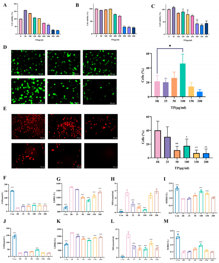
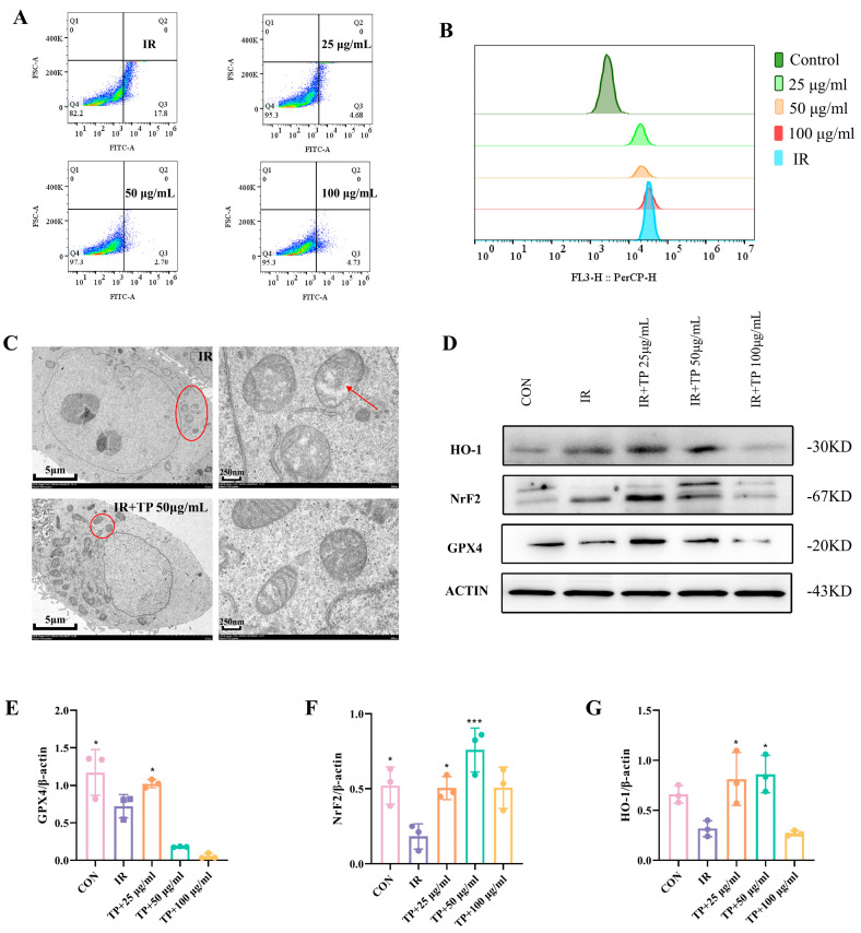
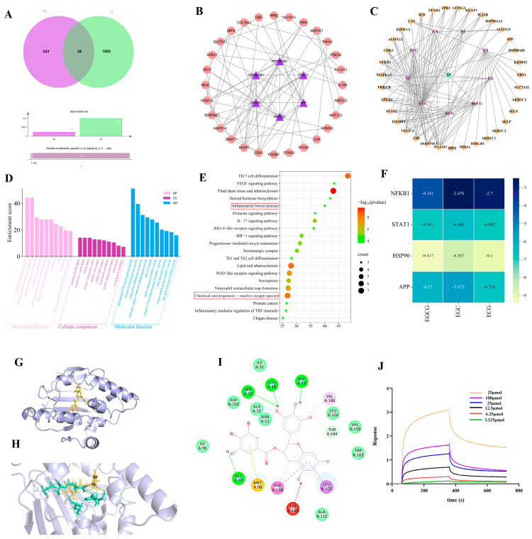
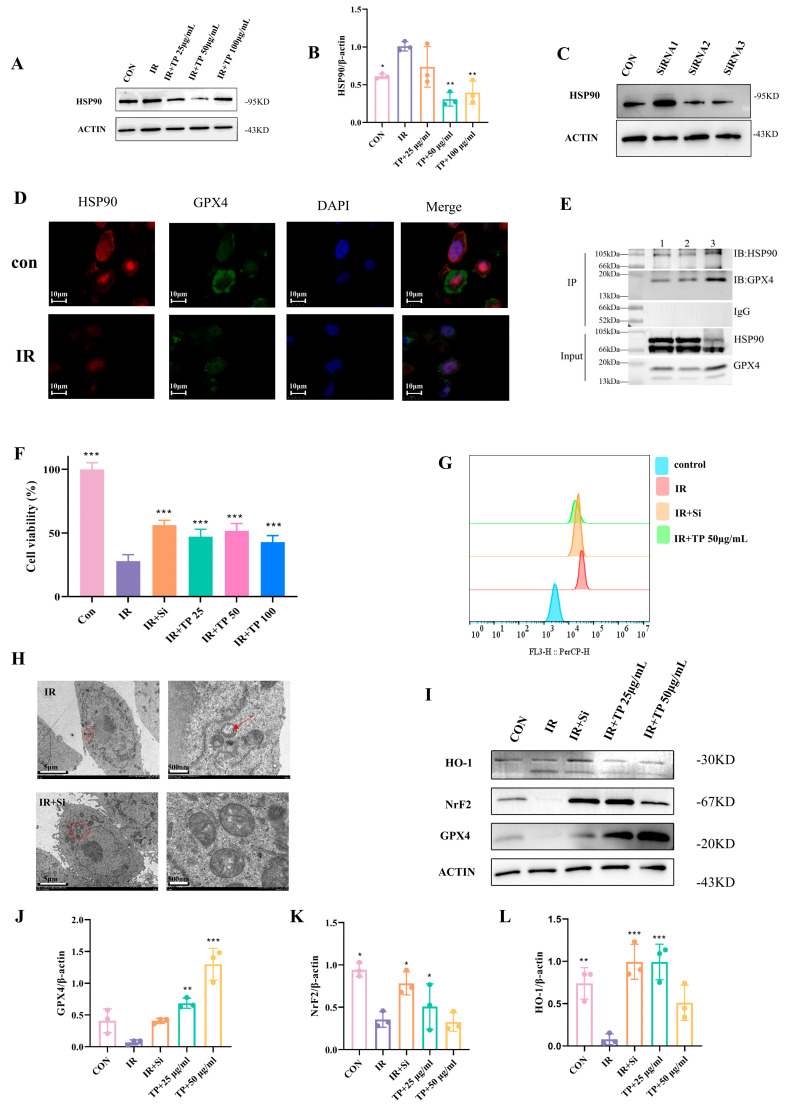
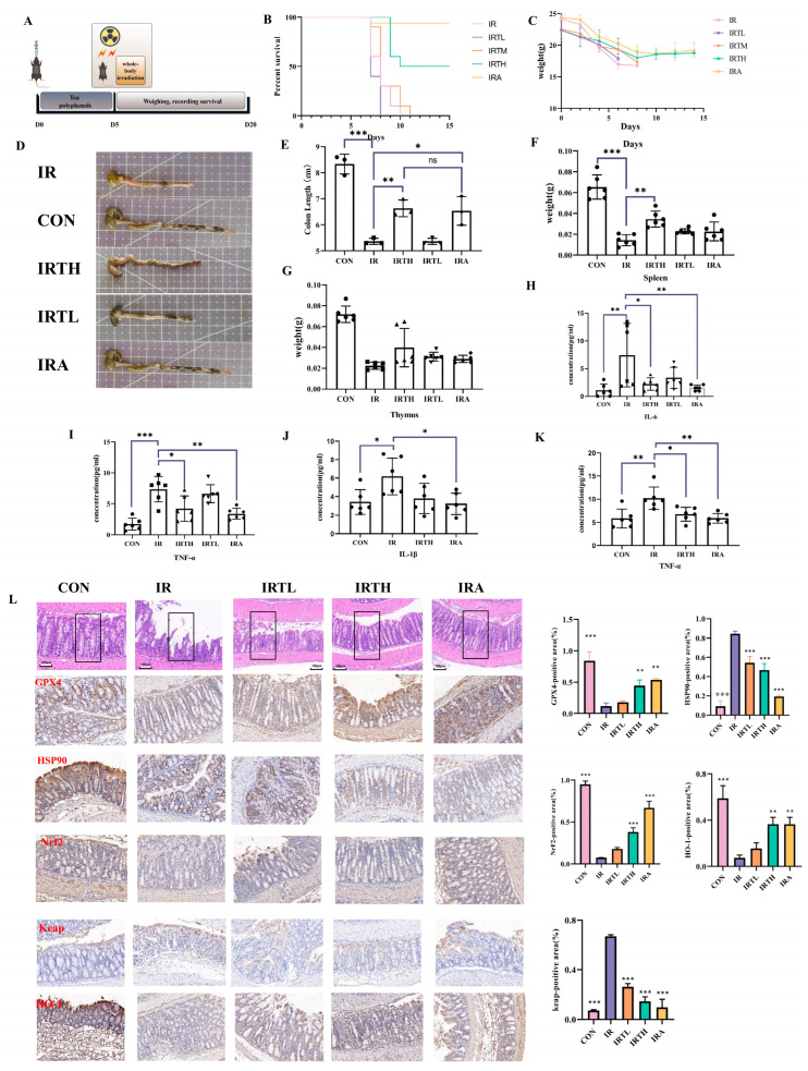
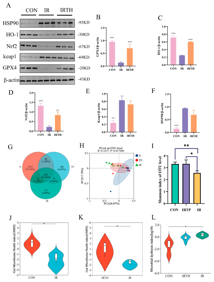
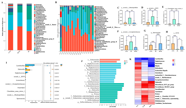
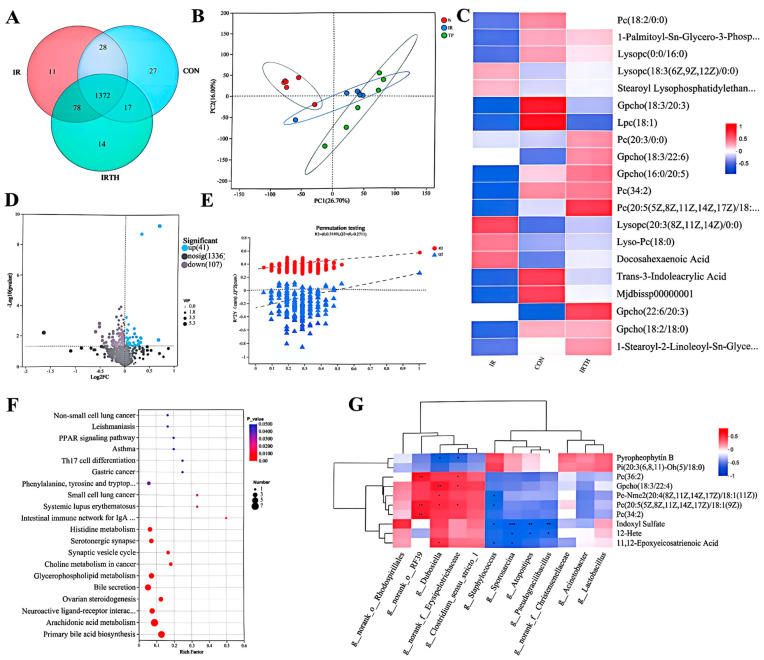
Radiation-induced intestinal injury (RIII) is a significant concern for cancer patients receiving radiation therapy, as it can lead to complications such as radiation enteropathy. Presently, there are limited options for preventing or treating RIII. Tea polyphenols (TP), found in tea, provide various health benefits, but their antiradiation mechanisms are not fully understood. C57BL/6 mice pre-treated with TP for five days showed a significant improvement in survival rates after being exposed to 10 Gy of 60Co radiation. In the same way, abdominal exposure to 15 Gy of 60Co radiation effectively mitigated radiation-induced colon shortening, damage to intestinal tissues, oxidative stress, the release of inflammatory factors, and disruptions in intestinal microbial balance. In addition, TP treatment lowered the elevation of reactive oxygen species (ROS), iron imbalance, mitochondrial damage, and ferroptosis in IEC-6 cells post-irradiation. Utilizing network pharmacology, molecular docking, and affinity testing, we identified that TP has the capability to target the Nrf2/HO-1/GPX4 signaling pathway, while EGCG, a principal constituent of TP, interacts with HSP90 and mitigates radiation-induced ferroptosis. These findings suggest that TP may serve as a promising therapeutic agent to alleviate radiation-induced intestinal injury (RII).

Keywords: HSP90; ferroptosis; gut microbiota; metabolites; radiation-induced intestinal injury; tea polyphenol.


Conflict of interest statement

The authors declare no conflicts of interest.

Figures

Figure 1
Figure 1
TP promoted IEC-6 cell proliferation after IR. (AC) CCK-8 assays showed TP toxicity on IEC-6 cells at 24, 48, and 72 h (n = 6). (D) Calcein-AM stained with green representing live cell. Representative. Scale bar = 50 μm. The AM ratio was calculated. (E) The red representative fluorescent images of DCFH-DA in IEC-6 cells (n = 3). A quantitative analysis was performed. Cellular ROS was stained by red fluorescence. Scale bar = 100 μm. * p < 0.05, ** p < 0.01 vs. IR group. (FI) GSH, LDH, MDA, and SOD levels in IEC-6 cells were measured 24 h after TP treatment post-radiation (n = 3, 8 Gy), * p < 0.05, *** p < 0.001 vs. IR group. (JM) GSH, LDH, MDA, and SOD levels in IEC-6 cells were measured 24 h after TP treatment post-radiation (n = 3, 12 Gy), *** p < 0.001 vs. IR group.
Figure 2
Figure 2
TP alleviated ferroptosis in IEC−6 cells post-irradiation. (A) The lipid ROS levels in IEC−6 cells were measured 24 h after TP treatment post-radiation and analyzed using a flow cytometer (n = 3). (B) The lipid Fe2+ levels in IEC−6 cells were measured 24 h after TP treatment post-radiation and analyzed using a flow cytometer (n = 3). (C) TEM imaging of irradiated cells was performed and measured 24 h after TP treatment post-radiation. Red circles indicate mitochondria, while red arrows highlight their enlarged structure. (DG) Protein levels of HO−1, Nrf2, and GPX4 in IEC−6 cells were assayed by Western blotting, * p < 0.05, *** p < 0.001 vs. IR group.
Figure 3
Figure 3
TP−IR network pharmacology analysis. (A) Venn diagram. (B) PPI network; purple represents the core target. (C) Drug–disease–target interaction. (D) GO enrichment analysis of TP for the treatment of RII. (E) Enrichment analysis of KEGG pathway for TP treatment of RII. (F) The heatmap illustrating the magnitude of binding energy of components in TP with respect to core target proteins. (G) Possible binding modes of EGCG to HSP90. (H) Structures of the binding pockets are shown by PyMOL 3.1 software. (I) Two-dimensional interactions of compounds and their targets. (J) BLI assay for EGCG and HSP90 binding affinity detection (Kd = 25 μmmol/L).
Figure 4
Figure 4
Mechanism of TP’s protection against radiation-induced damage. (A,B) WB analysis of HSP90 in IEC-6 cells after TP treatment, * p < 0.05, ** p < 0.01, vs. IR group. (C) WB of HSP90 in IEC−6 cells. (D) Cell viability was measured by CCK−8 in HSP90-siRNA transfected cells with TP treatment (n = 6), *** p < 0.001 vs. IR group. (E) Fe2+ levels in irradiated cells transfected with SiHSP90 and treated with TP were measured by flow cytometry. (F) Co−localization of HSP90 (red), GPX4 (green), and DAPI (blue). (G) Interaction between GPX4 and HSP90 was detected by immunoprecipitation. (H) TEM of irradiated IEC−6 cells: red circles indicate mitochondria, while red arrows highlight their enlarged structure. (IL) HO−1, GPX4, and Nrf2 expression in irradiated cells (IR), SiHSP90-transfected cells (IR+Si), and cells treated with TP was analyzed by WB, * p < 0.05, ** p < 0.01, *** p < 0.001 vs. IR group.
Figure 5
Figure 5
TP treatment alleviates radiation-induced damage in mice. (A) Experimental setup and treatment protocol. (B) Survival rates of mice post-radiation. (C) Average body weight post-radiation. (D,E) Colon length after radiation exposure (n = 6). (F) Average spleen weight after radiation (n = 6). (G) Average thymus weight post-radiation (n = 6). (H) Serum IL-6 levels (n = 6). (I) Serum TNF-α levels (n = 6). (J) Spleen IL-1β levels (n = 6). (K) Spleen TNF-α levels (n = 6). (L) HE staining of intestinal tissues post-radiation, GPX4, HSP90, keap, HO-1, NrF2 immunofluorescence in intestinal tissues post-radiation. * p < 0.05, ** p < 0.01, *** p < 0.001 vs. IR group.
Figure 6
Figure 6
Gut microbiota diversity in IR mice. (AF) Protein levels of HSP90, HO−1, Nrf2, keap1, and GPX4 in intestine were assayed by Western blotting. (G) Venn diagram. (H) PCoA plot. (I) Shannon index (n = 6). (JL) GMHI and MDI analysis (n = 6). * p < 0.05, ** p < 0.01, *** p < 0.001 vs. IR group.
Figure 7
Figure 7
Gut microbiota analysis. (A,B) Phylum-level comparisons. (CH) Kruskal−Wallis test for identifying changes in abundance of Erysipelotrichaceae, Clostridium_sensu_stricto_1, Rhodospirillales, RF39, Sporosarcina, and ASF356. n = 6, * p < 0.05, ** p < 0.01, *** p < 0.001 vs. IR group. (I) Abundance differences between IR and TP groups. (J) LEfSe analysis of group differences. (K) Correlation between microbiota and inflammatory factors (top 20 genera).
Figure 8
Figure 8
HPLC−MS untargeted metabolomics. (A) Venn diagram. (B) PCA analysis for all groups in both ion modes. (C) Heatmap of top 20 differential metabolites. (D) PLS−DA models with displacement test results. (E) Volcano plot: IR vs. IRTH groups. (F) Pathway analysis of differential metabolites. (G) Correlation of metabolites and microbiota at phylum/genus levels (top 20). Red: upregulated, blue: downregulated. Significant positive and negative correlations are indicated by red and blue squares, respectively. * p < 0.05, ** p < 0.01, *** p < 0.001.

Similar articles

References

    1. Jian Y., Zhang D., Liu M., Wang Y., Xu Z.X. The Impact of Gut Microbiota on Radiation-Induced Enteritis. Front. Cell. Infect. Microbiol. 2021;11:586392. doi: 10.3389/fcimb.2021.586392. - DOI - PMC - PubMed
    1. Xin J.Y., Wang J., Ding Q.Q., Chen W., Xu X.K., Wei X.T., Lv Y.H., Wei Y.P., Feng Y., Zu X.P. Potential role of gut microbiota and its metabolites in radiation-induced intestinal damage. Ecotoxicol. Environ. Saf. 2022;248:114341. doi: 10.1016/j.ecoenv.2022.114341. - DOI - PubMed
    1. Gao D., Zhang H., Sun W., Wang H., Wang H. Radiation-Induced Intestinal Injury: Molecular Mechanisms and Therapeutic Status. DNA Cell Biol. 2024;43:537–548. doi: 10.1089/dna.2024.0105. - DOI - PubMed
    1. Acharya M., Venkidesh B.S., Mumbrekar K.D. Bacterial supplementation in mitigation of radiation-induced gastrointestinal damage. Life Sci. 2024;353:122921. doi: 10.1016/j.lfs.2024.122921. - DOI - PubMed
    1. Su J., Bian C., Zheng Z., Wang H., Meng L., Xin Y., Jiang X. Cooperation effects of radiation and ferroptosis on tumor suppression and radiation injury. Front. Cell Dev. Biol. 2022;10:951116. doi: 10.3389/fcell.2022.951116. - DOI - PMC - PubMed

LinkOut - more resources