. 2025 May 14;14(5):587.
doi: 10.3390/antiox14050587.

Manganese Porphyrin Treatment Improves Redox Status Caused by Acute Compressive Spinal Cord Trauma

Affiliations

Manganese Porphyrin Treatment Improves Redox Status Caused by Acute Compressive Spinal Cord Trauma

Raquel Vieira Niella et al. Antioxidants (Basel). .

Abstract

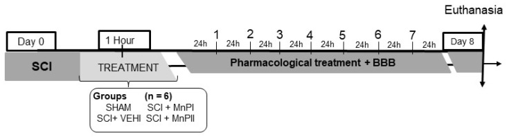
There is increasing interest in identifying drugs that can prevent or delay neurological complications following spinal cord injury, thus expanding the therapeutic window for other potential neuroprotective agents. In this context, manganese porphyrins (MnPs) have shown high antioxidant and anti-inflammatory potential in various experimental disease models, including stroke, cancer, diabetes, ischemia, and radiotherapy. However, they have been little evaluated in spinal cord injuries. This study aimed to assess the therapeutic potential of the manganese porphyrins [MnTE-2-PyP]5+ (MnPI) and [MnT(5-Br-3-E-Py)P]5+ (MnPII) in acute compressive spinal cord trauma in rats. Twenty-four animals were used (six animals/group). Following general inhalation anesthesia, acute compressive spinal cord trauma was induced in all groups except for the negative control (SHAM). Treatment commenced 60 min post-trauma, with animals receiving treatment for seven days at 24 h intervals. While no improvement in motor capacity was observed, MnPs effectively blocked the increase in oxidative stress and endoplasmic reticulum (ER) stress mediators caused by trauma, maintaining the protein expression levels of Hifα, 8-OHdG and MDA, as well as the expression of the genes Grp78, Chop, Ho1, and Perk, similar to those of the control group. Moreover, there was an increase in protein expression of SOD1, Cat, and GPX1, along with a restoration of SOD and CAT enzymatic activity. Additionally, MnPs improved the expression of IL-6, neurotrophic markers, and apoptotic factors. In conclusion, treatment with MnPs attenuated the oxidative stress and ER stress caused by acute compressive spinal cord trauma and restored spinal expression of neurotrophic mediators.

Keywords: apoptosis; endoplasmic reticulum stress; neuroprotection; oxidative stress.


Conflict of interest statement

The authors declare no conflicts of interest.

Figures

Figure A1
Figure A1
Photographic documentation of the experimental spinal cord injury surgical procedure in Wistar rats. (A) Retraction of epiaxial muscles and identification of spinous processes (arrow). (B) Dorsal lamina wear at T13 using a pneumatic drill (circle). (C) Visualization of the normal-appearing spinal cord following dorsal laminectomy (arrow). (D) Spinal cord injury induction via insertion of a Fogarty catheter into the spinal canal (arrow). Source: personal archive.
Figure 1
Figure 1
Representative diagram of the experimental schedule.
Figure 2
Figure 2
Motor function evaluation. Median daily scores of Wistar rats undergoing laminectomy (SHAM), acute compressive spinal cord injury (SCI + VEHI), and acute compressive spinal cord injury treated with MnPI and MnPII F (3, 21) = 47.30. (**** p < 0.0001).
Figure 3
Figure 3
Photomicrograph of a Wistar rat spinal cord cross-section stained with hematoxylin and eosin (H&E). (A) Morphologically normal spinal cord from the SHAM group, showing distinct white matter (WM) and gray matter (GM). The arrow indicates the ependymal canal. (B) Altered spinal cord morphology in the SCI + VEHI group, exhibiting pronounced multifocal areas of malacia in the white matter and portions of the gray matter (asterisk) dorsal to the ependymal canal. (C1) Axonal degeneration in a segment adjacent to the injury epicenter (highlight); (C2) neuronal necrosis and chromatolysis (highlight); (C3) gliosis (highlight); (C4) axonal degeneration within an area of malacia (highlight). 40× magnification.
Figure 4
Figure 4
Effects of MnPs treatment on the expression of cellular oxidative damage markers in the spinal cord of rats subjected to trauma. (AL) Photomicrographs of the immunolabeling of 8-OHdG (AD), MDA (EH), and HIF1α (IL) in the spinal cord of rats from the SHAM (A,E,I), SCI + VEHI (B,F,J), SCI + MnPI (C,G,K), and SCI + MnPII (D,H,L) groups. 40× magnification. (Hematoxylin; bar = 50 µm). (MO) Area of immunolabeling of 8-OHdG (M) F (3, 16) = 50.82, MDA (N) F (3, 16) = 201.3 and HIF1α (O) F (3, 16) = 155.6; (P) relative gene expression (fold change) of Hif1α F (3, 16) = 13.58 and Nrf2 F (3, 16) = 2.38 in the spinal cord of rats submitted to trauma, treated or not with MnPI and MnPII (mean ± SEM; * p < 0.05; ** p < 0.01; *** p < 0.001; **** p < 0.0001; # p < 0.05 test t).
Figure 5
Figure 5
Effects of MnPs treatment on the expression of antioxidant mediators in the spinal cord of rats subjected to trauma. (AL) Photomicrographs of CAT (AD), GPX1 (EH), and SOD 1 (IL) immunolabeling in the spinal cord of rats from the SHAM (A,E,I), SCI + VEHI (B,F,J), SCI + MnPI (C,G,K), and SCI + MnPII (D,H,L) groups. 40× magnification. (Hematoxylin; Bar = 50 µm). (MO) Area of immunolabeling of CAT (M) F (3, 16) = 67.91, GPX1 (N) F (3, 16) = 13.68 and SOD 1 (O) F (3, 16) = 21.30; (P) relative gene expression (fold change) of Cat F (3, 11) = 1.64, Gpx1 F (3, 16) = 2.95, and Sod1 F (3, 17) = 6.16; (Q) Enzymatic Activitys in the spinal cord of rats subjected to trauma, treated or not with MnPI and MnPII (mean ± SEM; * p < 0.05; ** p < 0.01; *** p < 0.001; **** p < 0.0001).
Figure 6
Figure 6
Effects of MnPs treatment on the expression of UPR mediators and endoplasmic reticulum stress in the spinal cord of rats subjected to trauma. (AH) Photomicrographs of GRP78 (AD), CHOP (E-H) immunolabeling in the spinal cord of rats from the SHAM (A,E), SCI + VEHI (B,F), SCI + MnPI (C,G), and SCI + MnPII (D,H) groups. 40× magnification. (Hematoxylin; Bar = 50 µm). (I,J) Area of GRP78 immunolabeling (I) F (3, 16) = 155.9, CHOP (J) F (3, 16) = 83.20; (K) relative gene expression (fold change) of Grp78 F (4, 25) = 2.41, Chop F (4, 16) = 1.93, Perk F (3, 16) = 1.83, and Ho1 F (3, 16) = 28.64 in the spinal cord of rats submitted to trauma, treated or not with MnPI and MnPII (mean ± SEM; * p < 0.05; *** p < 0.001; **** p < 0.0001).
Figure 7
Figure 7
Effects of MnPs treatment on the expression of immunological and apoptotic mediators in the spinal cord of rats subjected to trauma. (AH) Photomicrographs of IL-6 (AD), NeuN (EH) immunolabeling in the spinal cord of rats from the SHAM (A,E), SCI + VEHI (B,F), SCI + MnPI (C,G), and SCI + MnPII (D,H) groups. 40× magnification. (Hematoxylin; Bar = 50 µm). (I,J) Area of immunolabeling of IL-6 (I) F (3, 16) = 46.31, NeuN (J) F (3, 16) = 73.58; relative gene expression (fold change) of Gdnf F (3, 14) = 4.67, Ngf F (3, 15) = 2.71, Casp3 F (3, 14) = 4.77, and Casp9 (K) F (3, 15) = 6.35 in the spinal cord of rats subjected to trauma, treated or not with MnPI and MnPII (mean ± SEM; * p < 0.05; ** p < 0.01; **** p < 0.0001).

References

    1. Hu R., Zhou J., Lou C., Luo C., Lin J., Wang X., Li X., Bian X., Li Y., Wan Q., et al. Glial scar and neuroregeneration: Histological, functional, and magnetic resonance imaging analysis in chronic spinal cord injury. J. Neurosurg. Spine. 2010;13:169–180. doi: 10.3171/2010.3.SPINE09190. - DOI - PubMed
    1. Anjum A., Yazid M.D., Fauzi Daud M., Idris J., Ng A.M.H., Selvi Naicker A., Ismail O.H.R., Athi Kumar R.K., Lokanathan Y. Spinal Cord Injury: Pathophysiology, Multimolecular Interactions, and Underlying Recovery Mechanisms. Int. J. Mol. Sci. 2020;20:7533. doi: 10.3390/ijms21207533. - DOI - PMC - PubMed
    1. Bergman R., Lanz O., Shell L. Acute spinal cord trauma: Mechanisms and clinical syndromes. Vet. Med. 2000;95:846–849.
    1. Khorasanizadeh M., Yousefifard M., Eskian M., Lu Y., Chalangari M., Harrop J.S., Jazayeri S.B., Seyedpour S., Khodaei B., Hosseini M., et al. Neurological recovery following traumatic spinal cord injury: A systematic review and meta-analysis. J. Neurosurg. 2019;30:683–699. doi: 10.3171/2018.10.SPINE18802. - DOI - PubMed
    1. Dumont R.J., Okonkwo D.O., Verma S., Hurlbert R.J., Boulos P.T., Ellegala D.B., Dumont A.S. Acute spinal cord injury, part I: Pathophysiologic mechanisms. Clin. Neuropharmacol. 2001;24:254–264. doi: 10.1097/00002826-200109000-00002. - DOI - PubMed

LinkOut - more resources