. 2025 May 12;15(5):710.
doi: 10.3390/biom15050710.

A Novel Ashwagandha (Withania somnifera) Formulation Mitigates Sleep Deprivation-Induced Cognitive Impairment and Oxidative Stress in a Rat Model

Affiliations

A Novel Ashwagandha (Withania somnifera) Formulation Mitigates Sleep Deprivation-Induced Cognitive Impairment and Oxidative Stress in a Rat Model

Besir Er et al. Biomolecules. .

Abstract

Ashwagandha (Withania somnifera) is a well-known adaptogenic herb traditionally used to enhance sleep quality and mitigate stress-induced cognitive decline. This study investigated the effects of different doses of ashwagandha root extract (AE) formulations on cognitive function, oxidative stress, and neuronal plasticity in a rat model of sleep deprivation (SD). Forty-nine rats were randomly assigned to seven groups: control, wide platform (WP), SD, SD + A1 (15 mg/kg AE 1.5%), SD + A2 (30 mg/kg AE 1.5%), SD + A3 (5.5 mg/kg AE 8.0%), and SD + A4 (11 mg/kg AE 8.0%). The extract was administered orally for four weeks. SD induced via a modified wide platform model significantly impaired spatial memory, increased oxidative stress, and suppressed GABA receptor activity. Treatment with all AE doses, except 15 mg/kg AE 1.5%, considerably reduced serum corticosterone (12% for SD + A2, 15% for SD + A3, and 32% for SD + A4), CRH (11% for SD + A2, 14% for SD + A3, and 17% for SD + A4), ACTH (22% for SD + A2, 26% for SD + A3, and 38% for SD + A4), and MDA levels (31% for SD + A2, 34% for SD + A3, and 46% for SD + A4) (p < 0.05). All doses improved antioxidant enzyme activity and memory performance, while AE 8.0% doses notably increased serotonin (19% for SD + A3 and 33% for SD + A4) and dopamine levels (40% for SD + A3 and 50% for SD + A4). Moreover, AE treatment enhanced markers of neuronal plasticity and partially improved GABAergic function. These findings suggest that AE formulations, particularly at higher concentrations, exert neuroprotective effects against SD-induced cognitive impairment by modulating oxidative stress, neurotransmitter balance, and neuroplasticity, indicating their potential application in managing stress-related neurological disorders.

Keywords: GABAergic pathway; ashwagandha; memory; sleep deprivation; stress; withanolides.


Conflict of interest statement

A.M.M. and M.P. are employees of OmniActive Health Technologies, which funded this research. The funders had no role in the design, execution, or analysis of this study. Independent validation of data integrity was conducted to ensure unbiased results. The other authors declare no conflicts of interest.

Figures

Figure 1
Figure 1
Experimental procedures.
Figure 2
Figure 2
Sleep deprivation (SD) platform images for rats.
Figure 3
Figure 3
Effects of different ashwagandha doses on corticosterone (A), CRH (B), ACTH (C), serotonin (D), and dopamine (E) in sleep-deprived rats. Data were analyzed using one-way ANOVA followed by Tukey’s post hoc test for multiple comparisons. Different superscript letters (a–e) indicate statistically significant differences between groups (p < 0.05). The groups not sharing the same letter are significantly different. The error lines indicate SEM, and symbols show individual values. CRH: corticotropin-releasing hormone, ACTH: adrenocorticotropic hormone, C: control, WP: wide platform, SD: sleep deprivation, SD + A1: SD + ashwagandha 1.5% (15 mg/kg), SD + A2: SD + ashwagandha 1.5% (30 mg/kg), SD + A3: SD + ashwagandha 8% (5.5 mg/kg), SD + A4: SD + ashwagandha 8% (11 mg/kg).
Figure 4
Figure 4
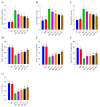
Effects of different ashwagandha doses on oxidative stress markers in sleep-deprived rats. (A) serum MDA, (B) liver MDA, (C) brain MDA, (D) brain superoxide dismutase (SOD), (E) brain catalase (CAT), (F) brain glutathione peroxidase (GSH-Px), and (G) total antioxidant capacity (TAC). Data were analyzed using one-way ANOVA followed by Tukey’s post hoc test for multiple comparisons. Different superscript letters (a–e) indicate statistically significant differences between groups (p < 0.05). The groups not sharing the same letter are significantly different. The error lines indicate SEM, and symbols show individual values. MDA: malondialdehyde, SOD: superoxide dismutase, CAT: catalase, GSH-Px: glutathione peroxidase, TAC: total antioxidant capacity. C: control, WP: wide platform, SD: sleep deprivation, SD + A1: SD + ashwagandha 1.5% (15 mg/kg), SD + A2: SD + ashwagandha 1.5% (30 mg/kg), SD + A3: SD + ashwagandha 8% (5.5 mg/kg), SD + A4: SD + ashwagandha 8% (11 mg/kg).
Figure 5
Figure 5
Effects of different ashwagandha doses on entries to target quadrant (A), probe trial (B), and spatial learning memory acquisition phase (SMAP) (C) in sleep-deprived rats. One-way ANOVA was used for group differences, and Tukey post hoc analysis for multiple comparisons. Kruskal–Wallis and Mann–Whitney U tests were used to compare the probe trials. Different superscripts (a–d) indicate the mean differences between the groups (p < 0.05). The groups not sharing the same letter are significantly different. The error lines indicate SEM for entries to the target quadrant and 95% confidence interval for the probe trial. Symbols show individual values. C: control, WP: wide platform, SD: sleep deprivation, SD + A1: SD + ashwagandha 1.5% (15 mg/kg), SD + A2: SD + ashwagandha 1.5% (30 mg/kg), SD + A3: SD + ashwagandha 8% (5.5 mg/kg), SD + A4: SD + ashwagandha 8% (11 mg/kg).
Figure 6
Figure 6
Effects of different ashwagandha doses on brain tissue NCAM (A), ICAM-1 (B), BDNF (C), NGF (D), and GAP-43 (E) protein levels and representative Western blot bands ((F) and Figure S1) in sleep-deprived rats. Western blot analysis was performed with incorporated β-actin to ensure equal protein loading. Representative bands are shown in panel (F). Data were analyzed using one-way ANOVA followed by Tukey’s post hoc test for multiple comparisons. Different superscript letters (a–g) indicate statistically significant differences between groups (p < 0.05). The groups not sharing the same letter are significantly different. The error lines indicate SEM. NCAM: neural cell adhesion molecule, ICAM: intercellular adhesion molecule-1, BDNF: brain-derived neurotrophic factor, NGF: nerve growth factor. C: control, WP: wide platform, SD: sleep deprivation, SD + A1: SD + ashwagandha 1.5% (15 mg/kg), SD + A2: SD + ashwagandha 1.5% (30 mg/kg), SD + A3: SD + ashwagandha 8% (5.5 mg/kg), SD + A4: SD + ashwagandha 8% (11 mg/kg). The original Western blot image can be found in the Supplementary Materials.
Figure 7
Figure 7
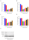
Effects of different ashwagandha doses on brain tissue GABAAR2 (A), GABABR1 (B), GABABR2 (C), and 5-HT1A (D) protein levels and representative Western blot bands ((E) and Figure S2) in sleep-deprived rats. Western blot analysis was performed with incorporated β-actin to ensure equal protein loading. Representative bands are shown in panel E. Data were analyzed using one-way ANOVA followed by Tukey’s post hoc test for multiple comparisons. Different superscript letters (a–f) indicate statistically significant differences between groups (p < 0.05). The groups not sharing the same letter are significantly different. The error lines indicate SEM. GABAA: gamma-aminobutyric acid type A receptor subunit alpha, GAP-43: growth-associated protein-43. C: control, WP: wide platform, SD: sleep deprivation, SD + A1: SD + ashwagandha 1.5% (15 mg/kg), SD + A2: SD + ashwagandha 1.5% (30 mg/kg), SD + A3: SD + ashwagandha 8% (5.5 mg/kg), SD + A4: SD + ashwagandha 8% (11 mg/kg). The original Western blot image can be found in the Supplementary Materials.

References

    1. Gohari A., Baumann B., Jen R., Ayas N. Sleep Deficiency: Epidemiology and Effects. Clin. Chest Med. 2022;43:189–198. doi: 10.1016/j.ccm.2022.02.001. - DOI - PubMed
    1. Krause A.J., Simon E.B., Mander B.A., Greer S.M., Saletin J.M., Goldstein-Piekarski A.N., Walker M.P. The Sleep-Deprived Human Brain. Nat. Rev. Neurosci. 2017;18:404–418. doi: 10.1038/nrn.2017.55. - DOI - PMC - PubMed
    1. Alrousan G., Hassan A., Pillai A.A., Atrooz F., Salim S. Early Life Sleep Deprivation and Brain Development: Insights From Human and Animal Studies. Front. Neurosci. 2022;16:833786. doi: 10.3389/fnins.2022.833786. - DOI - PMC - PubMed
    1. Prpar Mihevc S., Majdič G. Canine Cognitive Dysfunction and Alzheimer’s Disease—Two Facets of the Same Disease? Front. Neurosci. 2019;13:604. doi: 10.3389/fnins.2019.00604. - DOI - PMC - PubMed
    1. Taylor T.L., Fernandez E.J., Handley K.N., Hazel S.J. Non-Pharmacological Interventions for the Treatment of Canine Cognitive Dysfunction: A Scoping Review. Appl. Anim. Behav. Sci. 2023;269:106097. doi: 10.1016/j.applanim.2023.106097. - DOI

LinkOut - more resources