

Evaluation of Retinal and Optic Nerve Parameters in Recovered COVID-19 Patients: Potential Neurodegenerative Impact on the Ganglion Cell Layer
- PMID: 40428188
- PMCID: PMC12110219
- DOI: 10.3390/diagnostics15101195
Evaluation of Retinal and Optic Nerve Parameters in Recovered COVID-19 Patients: Potential Neurodegenerative Impact on the Ganglion Cell Layer
Abstract
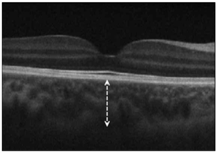
Background/Objectives: This study aimed to analyze optic nerve parameters, retinal nerve fiber layer thickness (RNFLT), ganglion cell layer thickness (GCLT), and subfoveal choroidal thickness (ChT) in patients who have recovered from coronavirus disease 2019 (COVID-19). Methods: This comparative study included 78 recovered COVID-19 patients (16 men, 62 women) and 56 age- and sex-matched healthy controls (18 men, 38 women). COVID-19 was confirmed in all patients, either through the detection of viral RNA in nasopharyngeal swabs via reverse transcriptase polymerase chain reaction or by serological testing for SARS-CoV-2 antibodies. Spectral-domain optical coherence tomography (SD-OCT) was used to assess optic nerve parameters, RNFLT, GCLT, and ChT. Results: The mean age was 35.0 ± 8.3 years in the COVID-19 group and 31.5 ± 8.3 years in the control group, with no statistically significant differences in age or sex distribution between groups (p = 0.41 and p = 0.16, respectively). Optic nerve parameters and RNFLT (overall and across the four peripapillary quadrants) did not differ significantly between the COVID-19 and control groups. However, the mean ganglion cell-inner plexiform layer (GC-IPL) thickness was significantly reduced in all quadrants in the COVID-19 group compared to the controls. No significant difference was observed in mean subfoveal ChT between groups. Conclusions: A significant reduction in ganglion GCLT was observed in recovered COVID-19 patients compared to healthy controls, suggesting a potential neurodegenerative effect of the disease on the optic nerve.
Keywords: COVID-19; optic coherence tomography; optic nerve; retinal ganglion cells; retinal nerve fiber layer.
Conflict of interest statement
The authors declare no conflicts of interest.
Figures
Similar articles
-
Optic nerve and macular optical coherence tomography in recovered COVID-19 patients.Eur J Ophthalmol. 2022 Jan;32(1):628-636. doi: 10.1177/11206721211001019. Epub 2021 Mar 15. Eur J Ophthalmol. 2022. PMID: 33719624
-
Assessment of Retinal Neurodegeneration and Choroidal Thickness in COVID-19 Patients Using Swept-Source OCT Technology.Klin Monbl Augenheilkd. 2021 Oct;238(10):1092-1097. doi: 10.1055/a-1340-0066. Epub 2021 Apr 14. Klin Monbl Augenheilkd. 2021. PMID: 33853186 English.
-
Optic Nerve Head Parameters and Peripapillary Retinal Nerve Fiber Layer Thickness in Patients with Coronavirus Disease 2019.Ocul Immunol Inflamm. 2022 Jul;30(5):1035-1038. doi: 10.1080/09273948.2020.1850800. Epub 2021 Feb 19. Ocul Immunol Inflamm. 2022. PMID: 33606593
-
Evaluation of retinal nerve fiber layer thickness and choroidal thickness in pseudoexfoliative glaucoma and pseudoexfoliative syndrome.Postgrad Med. 2016 May;128(4):444-8. doi: 10.1080/00325481.2016.1170579. Epub 2016 Apr 4. Postgrad Med. 2016. PMID: 27007173
-
Retinal nerve fibre layer and ganglion cell layer changes in children who recovered from COVID-19: a cohort study.Arch Dis Child. 2022 Feb;107(2):175-179. doi: 10.1136/archdischild-2021-321803. Epub 2021 Aug 2. Arch Dis Child. 2022. PMID: 34340983
Cited by
-
Optical Coherence Tomography (OCT) Findings in Post-COVID-19 Healthcare Workers.J Imaging. 2025 Jun 12;11(6):195. doi: 10.3390/jimaging11060195. J Imaging. 2025. PMID: 40558794 Free PMC article.
References
Grants and funding
LinkOut - more resources
Full Text Sources
Miscellaneous
