

Cytoimmunological Profile of Lower Airways in Post-COVID-19 Syndrome (PCS): Predictive Value of Bronchoalveolar Lavage
- PMID: 40429360
- PMCID: PMC12112653
- DOI: 10.3390/jcm14103361
Cytoimmunological Profile of Lower Airways in Post-COVID-19 Syndrome (PCS): Predictive Value of Bronchoalveolar Lavage
Abstract
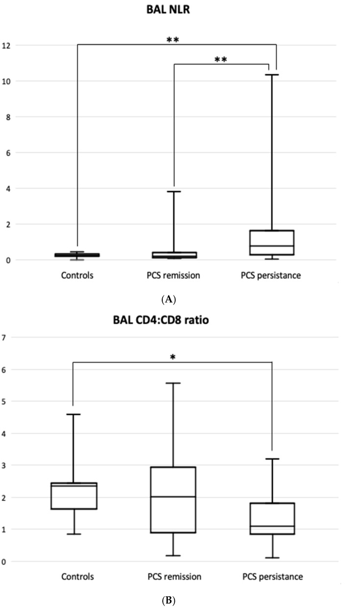
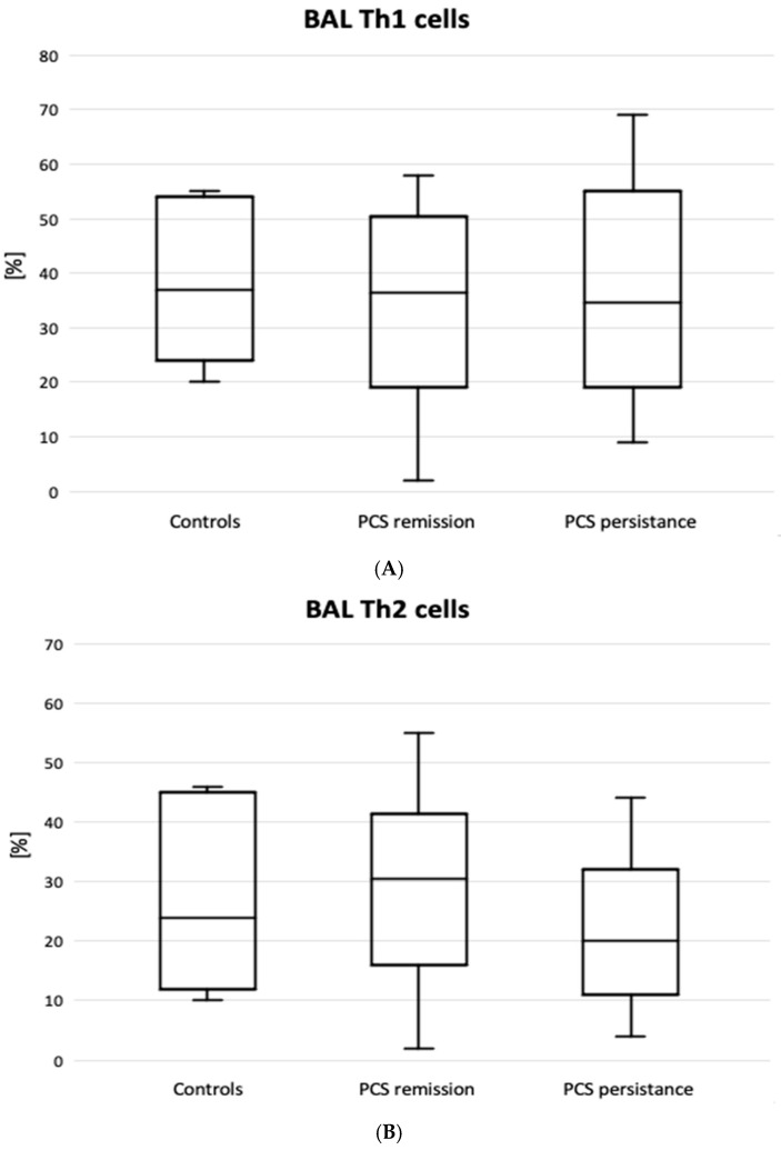
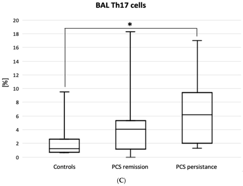
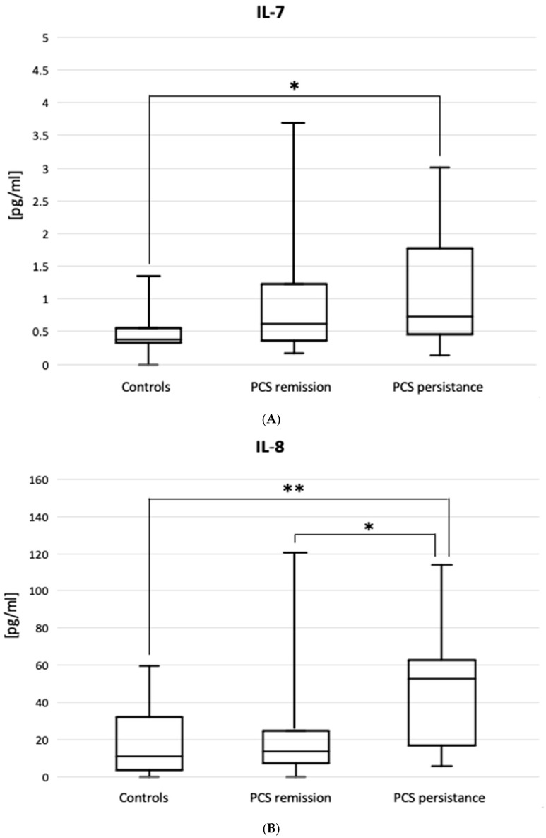
Background: It has yet to be determined whether the immunocytological profile of the bronchoalveolar lavage (BAL) in respiratory post-COVID syndrome (PCS) reflects the risk of persistent interstitial lung disease (ILD), including pulmonary fibrosis. In this study, we aimed to assess the prognostic value of the BAL cytoimmunologic profile in PCS-related ILD. Materials and Methods: We enrolled 58 non-smoking patients with a history of COVID-19 and new-onset ILD, divided into PCS remission and PCS persistence groups based on clinical data, including repeated computed tomography and pulmonary function tests. We phenotyped BAL major T cell subsets, immune checkpoints (including programmed cell death-1, PD1), and markers of Th1/Th2/Th17 polarization. Results: The PCS groups compared to the control showed increased total cell, lymphocyte, and neutrophil counts and a high BAL neutrophil:lymphocyte ratio (NLR). PCS persistence compared to the controls presented an increased neutrophil count (26 [17-36] vs. 2.6 [1.9-5.4] 103/mL, median [Q1-Q3], p < 0.001) and percentage, BAL NLR (0.77 [0.26-1.63] vs. 0.21 [0.17-0.31], p < 0.0001), CD8+PD1+ cell percentage (43.5 [34-60.5] vs. 24.5 [22-44]%, p = 0.045), and a decreased CD4:CD8 ratio. A high percentage of CD4+CD196+CD183 cells (relevant to Th17 activity, 6.2 [2.0-9.4] vs. 1.2 [0.7-2.7]%, p = 0.02) and increased BAL supernatant elevated IL-8 levels (62.5 [16-243] vs. 10.9 [3.44-32] pg/mL, p = 0.002) were found in the PCS persistence vs. control groups. In the total PCS group, predicted values of Vital Capacity (VC) [16-243] and Diffusing Lung Capacity for CO (DLCO) correlated negatively with BAL NLR; VC correlated negatively with BAL CD8+PD1+; and DLCO correlated positively with the CD4:CD8 ratio. Conclusions: Worse prognosis in PCS is associated with higher BAL NLR, BAL neutrophilia, an elevated percentage of CD8+PD1+ lymphocytes, and a decline in the CD4:CD8 ratio. Th17 cells and IL-8 participate in lung PCS persistence.
Keywords: T exhausted cells; Th17 cells; bronchoalveolar lavage; interstitial lung disease; lung lymphocytes; lung neutrophils; post-COVID syndrome.
Conflict of interest statement
The authors declare that they have no competing interests.
Figures
References
-
- World Health Organization COVID-19 Epidemiological Update—24 December 2024. [(accessed on 21 April 2025)]. Available online: https://www.who.int/publications/m/item/covid-19-epidemiological-update-....
-
- Fernandez-de-Las-Peñas C., Notarte K.I., Macasaet R., Velasco J.V., Catahay J.A., Ver A.T., Chung W., Valera-Calero J.A., Navarro-Santana M. Persistence of post-COVID symptoms in the general population two years after SARS-CoV-2 infection: A systematic review and meta-analysis. J. Infect. 2024;88:77–88. doi: 10.1016/j.jinf.2023.12.004. - DOI - PubMed
-
- Soriano J.B., Murthy S., Marshall J.C., Relan P., Diaz J.V. WHO Clinical Case Definition Working Group on Post-COVID-19 Condition. A clinical case definition of post-COVID-19 condition by a Delphi consensus. Lancet Infect. Dis. 2022;22:e102–e107. doi: 10.1016/S1473-3099(21)00703-9. - DOI - PMC - PubMed
Grants and funding
LinkOut - more resources
Full Text Sources
Research Materials
