

Do Onodi Cells Influence the Onset of Sphenoiditis? A Multicentric Cross-Sectional Study
- PMID: 40429504
- PMCID: PMC12112168
- DOI: 10.3390/jcm14103508
Do Onodi Cells Influence the Onset of Sphenoiditis? A Multicentric Cross-Sectional Study
Abstract
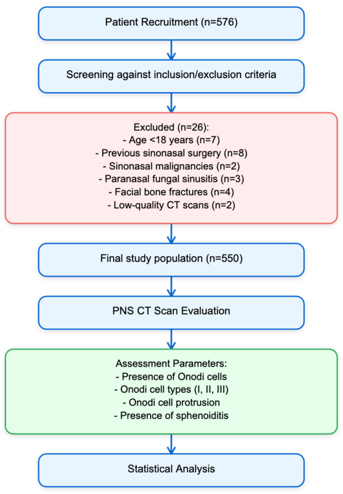
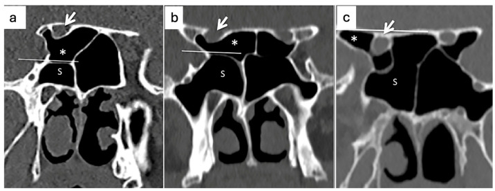
Background: Sphenoiditis poses diagnostic and treatment challenges due to its complex anatomy and potential for serious complications. Anatomic variations, such as Onodi cells, could play a role in the onset and spreading of inflammation. The diagnosis and treatment of sphenoiditis can be more difficult if Onodi cells are present, especially due to their proximity to delicate vital tissues. Objectives: The purpose of this study was to look at the frequency, features, and relationship between Onodi cells and sphenoiditis. Methods: This multicentric study comprised 550 people who received sinonasal CT imaging. The Thimmaiah classification was used to assess the presence and features of Onodi cells, and radiographic results were used to diagnose sphenoiditis. We conducted univariate and multivariate logistic regression to evaluate the relationships between sphenoiditis and Onodi cells. Results: The prevalence of Onodi cells was 32.40%, with a higher prevalence on the right side (18.40%) compared to the left side (8.40%). The multivariable analysis revealed a significant correlation between right-side Type II Onodi cells and a higher incidence of sphenoiditis (OR = 6.81, 95% CI: 1.14-38.97, p = 0.029). In the univariable analysis (OR = 3.00, 95% CI: 1.15-6.96, p = 0.015), but not in the multivariable analysis, the presence of Type I Onodi cells on the left side was significantly associated with sphenoiditis. Conclusions: There may be a link between a higher incidence of sphenoiditis and the presence of Type II Onodi cells on the right side. In order to validate these findings and clarify the underlying processes of this connection, more prospective research is required.
Keywords: Onodi cells; anatomical landmarks; paranasal sinuses; sphenoiditis; tomography.
Conflict of interest statement
The authors declare no conflicts of interest. The funders had no role in the design of the study; in the collection, analyses, or interpretation of data; in the writing of the manuscript; or in the decision to publish the results.
Figures
Similar articles
-
The role of Onodi cells in sphenoiditis: results of multiplanar reconstruction of computed tomography scanning.Braz J Otorhinolaryngol. 2017 Jan-Feb;83(1):88-93. doi: 10.1016/j.bjorl.2016.01.011. Epub 2016 Apr 20. Braz J Otorhinolaryngol. 2017. PMID: 27161189 Free PMC article.
-
Is there a relationship between Onodi cell and optic canal?Eur Arch Otorhinolaryngol. 2019 Apr;276(4):1057-1064. doi: 10.1007/s00405-019-05284-0. Epub 2019 Jan 7. Eur Arch Otorhinolaryngol. 2019. PMID: 30617426
-
The prevalence and pattern of pneumatization of Onodi cell in Thai patients.J Med Assoc Thai. 2011 Sep;94(9):1122-6. J Med Assoc Thai. 2011. PMID: 21970203
-
Identification of Onodi cell and new classification of sphenoid sinus for endoscopic sinus surgery.Int Forum Allergy Rhinol. 2015 Nov;5(11):1068-76. doi: 10.1002/alr.21567. Epub 2015 Jun 10. Int Forum Allergy Rhinol. 2015. PMID: 26097234 Review.
-
Sphenoid sinuses: pneumatisation and anatomical variants-what the radiologist needs to know and report to avoid intraoperative complications.Surg Radiol Anat. 2020 Sep;42(9):1013-1024. doi: 10.1007/s00276-020-02490-y. Epub 2020 May 11. Surg Radiol Anat. 2020. PMID: 32394118 Review.
Cited by
-
A new CBCT-based classification of posterior extramural ethmoid cells.Surg Radiol Anat. 2025 Jul 16;47(1):173. doi: 10.1007/s00276-025-03686-w. Surg Radiol Anat. 2025. PMID: 40670633
References
-
- Onodi A. Der Sehnerv and Die NebenOhlen der Nase. Alfred Holder; Wien, Germany: 1907. The optic nerve and the accessory sinuses of the nose; pp. 1–6.
-
- Chipczyńska B., Kosowski H., Skrzat J. Sphenoid sinus and sphenoid bone fractures in patients with isolated orbital injuries. Med. Sci. Monit. 2016;22:4327–4332.
