Case Reports
. 2025 May 19;14(10):3547.
doi: 10.3390/jcm14103547.

Multimodality Imaging Leading the Way to a Prompt Diagnosis and Management of Transthyretin Amyloidosis

Affiliations
Case Reports

Multimodality Imaging Leading the Way to a Prompt Diagnosis and Management of Transthyretin Amyloidosis

Anca Bălinișteanu et al. J Clin Med. .

Abstract

Background/Objectives: A 43-year-old male presented with neurological symptoms and asymptomatic cardiac dysfunction, left ventricular hypertrophy, and impaired global longitudinal strain with apical sparing, associated with elevated NT-proBNP. Methods: Multimodality imaging (bone scintigraphy and cardiac magnetic resonance) revealed cardiac amyloid deposition. Genetic testing confirmed variant transthyretin amyloidosis (ATTR) with mixed phenotype. Results: Treatment with tafamidis 20 mg for stage I polyneuropathy, available at that moment, was initiated with good neurological outcome. Three years later, cardiac function deteriorated, following a moderate COVID-19 infection, with heart failure symptoms and reduced ventricular and atrial functions. For progressive ATTR cardiomyopathy, we intensified therapy to tafamidis free acid 61 mg, associated with SGLT2 inhibitor, spironolactone, and furosemide with subsequent improvements of symptoms and stabilization of imaging findings. Conclusions: This case emphasizes the importance of multimodal imaging in early detection, monitoring, and guiding individualized management in ATTR cardiomyopathy.

Keywords: ATTR cardiomyopathy; COVID-19; cardiac amyloidosis; multimodality imaging; transthyretin amyloidosis.


Conflict of interest statement

The authors declare no conflict of interest.

Figures
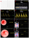
Figure 1
Figure 1
ECG and transthoracic echocardiographic images. (A) ECG- sinus rhythm, right bundle branch block, and pseudo-infarct pattern in V1–V3; (B) concentric left ventricular (LV) hypertrophy (IVS, interventricular septum of 14 mm, LVPW, LV posterior wall of 15 mm, LV mass of 130 g/m2); (C) restrictive filling pattern, with E/A of 2.1, DTE of 85 msec, E/E′ mean of 15, E = peak of early filling velocity, A = peak of late atrial filling velocity, DTE = mitral flow deceleration time, E′ = early diastolic tissue velocity; (D) worsening of diastolic dysfunction, restrictive filling pattern, with E/A of 2.4, E/E′ mean of 20; (E) bull’s eye plot by 2D speckle tracking echocardiography (STE) with significantly reduced global longitudinal strain (GLS) (−10.6%) and global altered deformation mainly at the basal and mid-ventricular segments, with a typical “apical sparing” pattern; (F) 2D STE at the level of the left atrium (LA) showing significantly decreased LA contractile function (Lact) and also of LA reservoir function (LAr); (G) bull’s eye plot by 2D speckle tracking echocardiography (STE) with additional significant reduction in global longitudinal strain (GLS) (−6.6%) and global altered deformation mainly at the basal and mid-ventricular segments, with a typical “apical sparing” pattern; (H) 2D STE at the level of the left atrium (LA) showing additional significantly reduction in LA contractile function (Lact = 1%) and also in LA reservoir function (Lar = 3%).
Figure 2
Figure 2
Bone scintigraphy with 99mTc-DPD and single-photon emission computerized tomography (SPECT) imaging. (A) Bone scintigraphy with positive result defined as grade 3 = high myocardial uptake, greater than the bone. (B) SPECT confirming intra-myocardial uptake in both right and left ventricles (yellow arrows).
Figure 3
Figure 3
Anatomo-pathological evaluation: salivary gland and abdominal fat pad biopsies. (A) Amyloid deposits in adipose tissue showing yellow-green birefringence in polarized light—Congo red stain, polarized light microscopy 4×. (B) Small peri-glandular and perivascular amyloid deposits in a minor salivary gland. Congo red stain, 4×. (C) Amyloid deposits showing typical yellow-green birefringence in polarized light—Congo red stain, polarized light microscopy 4×.
Figure 4
Figure 4
Cardiac magnetic resonance images. (A) Short-axis view showing left ventricular hypertrophy (IVS = 13.9 mm, AWT = 13 mm, LWT = 14.8 mm, PWT = 14 mm). (B) Extracellular volume (ECV) bull’s eye plot evaluated by cardiac magnetic resonance, using a 16-segment model, displaying a progressive decrease of the ECV from base to apex, explaining the substrate of apical sparing deformation pattern. (C) Short axis views at basal (bSAX), midventricular (mSAX) and apical (aSAX) levels showing concentric left ventricle hypertrophy in SSFP cine images (the first column), with diffuse elevation in native T1 mapping (the second column), transmural hyperenhancement, more pronounced at the basal and midventricular levels, with a dark blood pool in LGE images (the third column) and diffuse reduction in post-contrast T1 mapping (forth column). SSFP: Steady-state free precession; LGE: late gadolinium enhancement.

References

    1. Maurer M.S., Schwartz J.H., Gundapaneni B., Elliot P.M., Merlini G., Waddington-Cruz M., Kristen A.V., Grogan M., Witteles R., Damy T., et al. Tafamidis treatment for patients with transthyretin amyloid cardiomyopathy. N. Eng. J. Med. 2018;379:1007–1016. doi: 10.1056/NEJMoa1805689. - DOI - PubMed
    1. Rimbas R.C., Balinisteanu A., Magda S.L., Visoiu S.I., Beganu E., Nicula A.I., Vinereanu D. New advanced imaging parameters and biomarkers—A step forward in the diagnosis and prognosis of TTR cardiomyopathy. J. Clin. Med. 2022;11:2360. doi: 10.3390/jcm11092360. - DOI - PMC - PubMed
    1. Licordari R., Trimarchi G., Teresi LRestelli D., Lofrumento F., Perna A., Campisi M., de Gregorio C., Grimaldi P., Calabrò D., Costa F., et al. Cardiac Magnetic Resonance in HCM Phenocopies: From Diagnosis to Risk Stratification and Therapeutic Management. J. Clin. Med. 2023;12:3481. doi: 10.3390/jcm12103481. - DOI - PMC - PubMed
    1. De Gregorio C., Trimarchi G., Faro D.C., De Gaetano F., Campisi M., Losi V., Liso V., Tamburino C., Di Bella G., Monte I.P. Myocardial Work Appraisal in Transthyretin Cardiac Amyloidosis and Nonobstructive Hypertrophic Cardiomyopathy. Am. J. Cardiol. 2023;208:173–179. doi: 10.1016/j.amjcard.2023.09.055. - DOI - PubMed
    1. Pugliatti P., Trimarchi G., Barocelli F., Pizzino F., Di Spigno F., Tedeschi A., Piccione M.C., Irrera P., Aschieri D., Niccoli G., et al. Advancing Cardiac Amyloidosis Care Through Insights from Cardiopulmonary Exercise Testing. J. Clin. Med. 2024;13:7285. doi: 10.3390/jcm13237285. - DOI - PMC - PubMed

Publication types

LinkOut - more resources