Review
. 2025 May 20;14(10):3576.
doi: 10.3390/jcm14103576.

The Role of Cardiovascular Magnetic Resonance Imaging in Athletic Individuals-A Narrative Review

Affiliations
Review

The Role of Cardiovascular Magnetic Resonance Imaging in Athletic Individuals-A Narrative Review

Neil Grech et al. J Clin Med. .

Abstract

Cardiovascular magnetic resonance imaging (MRI) is an advanced cardiac imaging modality that is often required when evaluating athletic individuals. Unrestricted imaging planes, excellent spatial resolution, and a lack of ionising radiation are some of the benefits of this modality. Cardiac MRI has been established as the gold standard imaging modality for morphological assessment, volumetric analysis, and tissue characterisation. Cardiac MRI without any doubt is an excellent diagnostic tool when evaluating athletes with symptoms or those individuals exhibiting equivocal findings at screening. It is also useful for athletes who fall within the grey zone and is especially important among athletes with a suspected or confirmed diagnosis. Cardiac MRI plays a strategic role when adopting a shared decision-making model in athletes with heart disease, tailoring and personalising medical care to the condition and the athlete's wishes. The aim of this review is to provide a comprehensive yet practical overview of the role of cardiac MRI when evaluating athletes in clinic.

Keywords: athlete; cardiac MRI; cardiomyopathy; fibrosis; sudden cardiac death.


Conflict of interest statement

The authors declare no conflict of interest.

Figures

Figure 1
Figure 1
Commonly adopted cardiac MRI sequences when evaluating athletes.
Figure 2
Figure 2
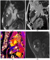
A mother presents to clinic for screening following the sudden death of her son who was a long-distance runner. Autopsy confirmed hypertrophic cardiomyopathy (HCM) secondary to a pathogenic TNNT2 variant. The victim’s uncle was also diagnosed with HCM in the interim and referred for transplantation. The mother was a carrier for the TNNT2 variant. The ECG was abnormal; echocardiography was normal. CMR identified mid-wall fibrosis in the lateral wall, despite the absence of left ventricular hypertrophy.
Figure 3
Figure 3
Caucasian male soccer player presenting with inferolateral T-wave inversion on ECG. SSFP cine imaging (A) showing apical hypertrophy, with evidence of apical fibrosis on post-contrast imaging (B,C).
Figure 4
Figure 4
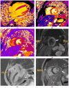
A 21 year old male that presented with chest pain during a pre-participation evaluation to join the armed forces. ECG showing biphasic ST segments in leads V1-V3, with T-wave inversion in II/III/aVF. CT coronary angiogram ruled out epicardial coronary disease. A stress echocardiogram ruled out dynamic LVOT obstruction. Echocardiogram confirmed the presence of hypertrophic cardiomyopathy, secondary to a likely pathogenic MYBPC3 variant. Cardiac MRI (3T scanner) showing septal hypertrophy (arrow) (A), extensive replacement (arrow) (B), and interstitial fibrosis in the septal segments (arrow) (C) (T1 map using a MOLLI 5b(3b)3b [3T]). Perfusion imaging (D) also identifying an extensive perfusion defect (arrow) in the hypertrophied segments, confirming microvascular dysfunction.
Figure 5
Figure 5
A 21 year old Caucasian male found to have lateral T-wave inversion on pre-recruitment ECG to join the military. Echocardiography showed dilated left and right ventricles, a bicuspid aortic valve (AoV), and possibly an atrial septal defect (ASD). Cardiac MRI confirmed the presence of dilated ventricles (A) exceeding reference ranges for male athletes. A bicuspid AoV (Sievers Classification Type 0) was also confirmed (B), without regurgitation. Phase-contrast flow imaging confirmed the presence of an ASD (C). Post-contrast imaging showed no replacement fibrosis.
Figure 6
Figure 6
A 21 year old endurance athlete, referred for screening as her mother was diagnosed with arrhythmogenic cardiomyopathy secondary to a Desmoplakin pathogenic variant. The athlete was also gene-positive, with a normal ECG and echocardiogram. Cardiac MRI identified a ring-like subepicardial scar on post-contrast imaging (A), with a diffusely elevated T1 (B), resulting in a diagnosis of non-dilated left ventricular cardiomyopathy.
Figure 7
Figure 7
An 18 year old male Caucasian competitive swimmer who was seen because of a very long PR interval in the context of non-cardiogenic syncope. Echocardiography revealed a dilated LV. This was confirmed on CMR, though perfectly within normal athletic specific reference ranges. Post-contrast imaging showed posterior RV insertion-point fibrosis.
Figure 8
Figure 8
A 19 year old male soccer player who presented with dyspnoea, found to have increased LV trabeculation on echocardiogram. Cardiac MRI showed prominent apical hypertrabeculation, with preserved thickness of the non-compacted layer. There was preserved LV function, with a normal LV size and absence of regional wall motion abnormalities and fibrosis. The findings were attributed to athletic remodelling.
Figure 9
Figure 9
A 42 year old Caucasian male, engaging in power sport (recreational) for 20 years, presented with a profound drop in exercise tolerance. Murmur heard on physical examination. T-wave inversion in the inferior leads noted. Supraventricular arrhythmias during an exercise test. Mitral valve (MV) prolapse with moderate regurgitation noted on echocardiogram. Cardiac MRI showed a dilated LV, an LV ejection fraction of 58%, and mitral annular disjunction (arrow) (A). There was prolapse of the posterior MV leaflet with moderate regurgitation (B). Post-contrast imaging identified a subepicardial scar in the lateral wall (arrow) (C,D), consistent with a diagnosis of malignant MV prolapse. He was subsequently referred for MV surgery.
Figure 10
Figure 10
A 24 year old long-distance runner reported a drop in exercise tolerance. A murmur was heard at auscultation. Cardiac MRI showed a bicuspid aortic valve (Sievers Classification Type 0) (A), with eccentric closure and moderate regurgitation (B).
Figure 11
Figure 11
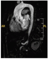
In a 34 year old Caucasian male soccer player, a routine screening echocardiogram identified a dilated proximal ascending aorta. He had a normal blood pressure; genetics was negative. A 3D aortogram on cardiac MRI confirmed the presence of a dilated aortic root and proximal ascending aorta, measuring 43 mm.
Figure 12
Figure 12
A 22 year old Caucasian male soccer player who presented for a routine screening echocardiogram. A dilated right ventricle (RV) led to a cardiac MRI. This confirmed a dilated RV (A), exceeding athletic reference ranges. MRI also identified a sinus venosus defect and anomalous pulmonary venous drainage (B), with a Qp:Qs > 1.5, reaching surgical thresholds.
Figure 13
Figure 13
A 52 year old North African male soccer player who had presented with acute coronary syndrome, requiring PCI to LAD 10 years before. He was referred for risk stratification. A cardiac MRI showed evidence of an LAD infarct (50% thickness) (arrows) (A,B) with preserved LV ejection fraction.
Figure 14
Figure 14
A 21 year old Caucasian male who played soccer professionally at club level. He presented with Troponin-positive chest pain. Invasive coronary angiography was normal. A cardiac MRI showed a mildly reduced LV ejection fraction, together with a transmural LAD infarct involving the anterior and lateral wall (A,B). A bubble echocardiographic study confirmed the presence of a large patent foramen ovale (C). This was corrected percutaneously, though later again presenting with another MINOCA presentation, complicated with a large LV thrombus (D).
Figure 15
Figure 15
A 17 year old Caucasian male who played handball at club level presented with recurrent myocarditis. His father, a former endurance athlete, presented with an out of hospital cardiac arrest, was implanted with a defibrillator, and was genetically positive for a likely pathogenic DSG2 variant. Cardiac MRI in the patient in question showed evidence of an extensive subepicardial high signal extending into the anterior and lateral wall (arrows), suggestive of persistent replacement fibrosis (A,B). He unfortunately refused to undergo genetic testing.
Figure 16
Figure 16
A 15 year old Caucasian male who played basketball at club level, presenting with chest pain, fever, and Troponin elevation. Cardiac MRI showed evidence of myocardial injury (A,B) on T1 mapping, with myocardial oedema on T2 mapping (C) and STIR (D) imaging (arrows). Post-contrast imaging showed evidence of extracellular volume expansion, with subepicardial late-gadolinium enhancement in the anterior and lateral wall (E,F) (arrows). The findings satisfied Lake Louise Criteria for myocarditis.

Similar articles

References

    1. Sharma S., Merghani A., Mont L. Exercise and the heart: The good, the bad, and the ugly. Eur. Heart J. 2015;36:1445–1453. doi: 10.1093/eurheartj/ehv090. - DOI - PubMed
    1. Kokkinos P., Myers J., Faselis C., Panagiotakos D.B., Doumas M., Pittaras A., Manolis A., Kokkinos J.P., Karasik P., Greenberg M., et al. Exercise Capacity and Mortality in Older Men. Circulation. 2010;122:790–797. doi: 10.1161/CIRCULATIONAHA.110.938852. - DOI - PubMed
    1. WHO . Guidelines on Physical Activity and Sedentary Behaviour. World Health Organization; Geneva, Switzerland: 2020. [(accessed on 24 January 2025)]. Available online: https://www.ncbi.nlm.nih.gov/books/NBK566046/
    1. Dvorak J., Grimm K., Schmied C., Junge A. Development and implementation of a standardized precompetition medical assessment of international elite football players-2006 FIFA world cup Germany. Clin. J. Sport Med. 2009;19:316–321. doi: 10.1097/JSM.0b013e3181b21b6e. - DOI - PubMed
    1. Ljungqvist A., Jenoure P.J., Engebretsen L., Alonso J.M., Bahr R., Clough A.F., de Bondt G., Dvorak J., Maloley R., Matheson G., et al. The International Olympic Committee (IOC) Consensus Statement on Periodic Health Evaluation of Elite Athletes, March 2009. Clin. J. Sport Med. 2009;43:631–643. doi: 10.1097/jsm.0b013e3181b7332c. - DOI - PubMed

LinkOut - more resources