. 2025 May 8;26(10):4505.
doi: 10.3390/ijms26104505.

Exploring the Histopathological Features of Thrombus-Associated Localized Amyloid Deposition: Comprehensive Analysis Employing Immunohistochemistry and Proteomics

Affiliations

Exploring the Histopathological Features of Thrombus-Associated Localized Amyloid Deposition: Comprehensive Analysis Employing Immunohistochemistry and Proteomics

Shojiro Ichimata et al. Int J Mol Sci. .

Abstract

Amyloid deposition has been reported to localize within thrombi; however, its pathological characteristics, particularly its precursor proteins, remain poorly understood. This study aimed to elucidate the pathological features of thrombus-associated amyloid deposition by immunohistochemistry combined with proteomic analyses using liquid chromatography-tandem mass spectrometry with laser microdissection. Our findings revealed that thrombus-associated amyloid deposits within the thrombus and vessel wall primarily comprised apolipoprotein A-I, with a mixture of amyloid fibrils derived from amyloidogenic proteins, including transthyretin and lactoferrin. Given that these proteins are present in the blood, our results support a previous hypothesis that proteins denatured during thrombus aging are a source of amyloid. Furthermore, phagocytes were infiltrated around the intramural and extravascular deposits rather than around the amyloid deposits within the thrombus. Therefore, amyloid deposits generated within the thrombus may be transported from regions with limited blood flow to the vessel wall and surrounding tissues, where blood flow is present, during thrombus processing. These deposits were primarily removed by phagocytic cells. Our results suggest that a facilitative effect on deposition occurs via a cross-seeding mechanism between amyloid fibrils and that phagocytes can remove amyloid deposits. These findings help elucidate the pathogenesis of localized amyloidosis.

Keywords: apolipoprotein A-I; atherosclerosis; lactoferrin; localized amyloidosis; phagocyte; proteomics; thrombus; transthyretin.


Conflict of interest statement

The authors declare no conflicts of interest.

Figures

Figure 1
Figure 1
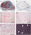
Representative pathological findings in aortic aneurysm (Case 2 in Table 2). (ac) Elastica–Masson staining; (dh) phenol Congo red (pCR) staining under bright-field (d,e,h) and polarized light (f,g) observation. (i) Panel a indicates intrathrombus amyloid deposition, whereas (ii) indicates intramural amyloid deposition. Medial elastic fibers are disrupted at amyloid deposition site within wall ((c), arrow). Scale bar = 5 mm (a,h); 2 mm (c,e,g); 200 μm (b,d,f).
Figure 2
Figure 2
Representative pathological findings in internal carotid artery (Case 8 in Table 2). (a) Hematoxylin–eosin staining; (b) Elastica–Masson staining; (cg) pCR staining under bright-field (ce) and polarized light (f,g) observation. (ac) FHT formation is observed. (dg) Intrathrombus area and border/vessel wall area contain amyloid deposition. Scale bar = 2 mm (ac); 200 μm (dg).
Figure 3
Figure 3
Representative pathological findings of arteriovenous fistula-related aneurysm in forearm approximately 20 years after thrombus occlusion (Case 18). (a) Elastica–Masson staining; (be) pCR staining under bright-field (be) and polarized light (f) observation. (a,b) Amyloid deposition is observed in thrombus (i), border area to vessel wall (ii), and granuloma area (iii). Panels (ce) are higher-magnification views of amyloid deposition foci in (i), (ii), and (iii), respectively. Notably, phagocyte infiltration is inevident around amyloid deposits in thrombus (c), but it is observed within vessel wall (d). (e,f) Amyloid deposition is identified within cytoplasm of macrophages and multinucleated giant cells in granuloma area (arrows). Scale bar = 5 mm (a,b); 200 μm (c,d); 100 μm (e,f).
Figure 4
Figure 4
Representative pathological findings showing congophilic state at varying degrees among amyloid deposits (Case 4). (a,b) pCR staining under bright-field (a) and polarized light (b) observation. (a) Within arterial wall, amyloid deposits are present, one area showing prominent inflammatory cell infiltrate, including multinucleated giant cells (arrow), and another area exhibiting less phagocyte infiltration (arrowhead). Under bright-field observation, congophilic state did not clearly differ between two lesions. (b) Under polarized light, apple-green birefringence was weak in former deposition area but was clear in latter. Scale bar = 200 μm (a,b).
Figure 5
Figure 5
Representative proteomics results. (a) Proteins identified in intrathrombus amyloid deposition in Case 19; (b) proteins identified in amyloid deposition found in border to vessel wall in Case 18. * emPAI stands for exponentially modified protein abundance index used to estimate relative protein quantification in mass spectrometry-based proteomics analysis. Igκ, immunoglobulin kappa light chain; SAP, serum amyloid P-component.
Figure 6
Figure 6
Representative IHC findings in amyloid deposits (Case 4). (a,b) pCR staining under bright-field (a) and polarized light (b) observation. IHC for apolipoprotein A-I (ApoAI, (c)); lactoferrin (Lac, (d)); transthyretin (TTR, (e)); and immunoglobulin κ light chain (Igκ, (f)). Arrows indicate the amyloid deposits. Amyloid deposits in this case (a,b) are moderately positive for ApoAI (c) and Lac (d), weakly positive for TTR (e), and negative for Igκ (f). (e) TTR immunoreactivity in thrombus-associated amyloid deposits is significantly weaker than that observed in patients with systemic ATTR amyloidosis (Case 1) that underwent IHC at the same time as the control with IHC-grade 3+ (inset). Scale bar = 200 μm (af).
Figure 7
Figure 7
Schematic representation of hypothesized progression and resorption process of thrombus-associated amyloids. (−) Absent; (+) Present to a mild degree; (++) Abundantly present.
Figure 8
Figure 8
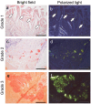
Representative microphotographs of thrombus-associated amyloid deposition evaluated using grading system employed in this study. (af) pCR staining under bright-field (a,c,e) and polarized light (b,d,f) observation. (a,b) Grade 1+, focal and tiny amyloid deposition (arrow); (c,d) Grade 2+, multifocal deposition with nodular deposits; (e,f) Grade 3+, multifocal deposition with some massive deposits. Scale bar = 200 μm (af).

Similar articles

References

    1. Buxbaum J.N., Eisenberg D.S., Fandrich M., McPhail E.D., Merlini G., Saraiva M.J.M., Sekijima Y., Westermark P. Amyloid nomenclature 2024: Update, novel proteins, and recommendations by the International Society of Amyloidosis (ISA) Nomenclature Committee. Amyloid. 2024;31:249–256. doi: 10.1080/13506129.2024.2405948. - DOI - PubMed
    1. Westermark G.T., Westermark P. Localized amyloids important in diseases outside the brain--lessons from the islets of Langerhans and the thoracic aorta. FEBS J. 2011;278:3918–3929. doi: 10.1111/j.1742-4658.2011.08298.x. - DOI - PubMed
    1. Riba M., Del Valle J., Auge E., Vilaplana J., Pelegri C. From corpora amylacea to wasteosomes: History and perspectives. Ageing Res. Rev. 2021;72:101484. doi: 10.1016/j.arr.2021.101484. - DOI - PubMed
    1. Ichimata S., Hata Y., Yoshinaga T., Katoh N., Kametani F., Yazaki M., Sekijima Y., Nishida N. Amyloid-forming corpora amylacea and spheroid-type amyloid deposition: Comprehensive analysis using immunohistochemistry, proteomics, and a literature review. Int. J. Mol. Sci. 2024;25:4040. doi: 10.3390/ijms25074040. - DOI - PMC - PubMed
    1. Castellani R.J., Shanes E.D., McCord M., Reish N.J., Flanagan M.E., Mesulam M.M., Jamshidi P. Neuropathology of anti-amyloid-beta immunotherapy: A case report. J. Alzheimer’s Dis. 2023;93:803–813. doi: 10.3233/JAD-221305. - DOI - PubMed

LinkOut - more resources