. 2025 May 10;26(10):4566.
doi: 10.3390/ijms26104566.

Liensinine Prevents Acute Myocardial Ischemic Injury via Inhibiting the Inflammation Response Mediated by the Wnt/β-Catenin Signaling Pathway

Affiliations

Liensinine Prevents Acute Myocardial Ischemic Injury via Inhibiting the Inflammation Response Mediated by the Wnt/β-Catenin Signaling Pathway

En Ma et al. Int J Mol Sci. .

Abstract

Myocardial infarction (MI) is characterized by the sudden reduction in myocardial blood flow and remains the leading cause of death worldwide. Because MI causes irreversible damage to the heart, discovering drugs that can limit the extent of ischemic damage is crucial. Liensinine (LSN) is a natural alkaloid that has exhibited beneficial effects in various cardiovascular diseases, including MI; however, its molecular mechanisms of action remain largely unelucidated. In this study, we constructed murine models of MI to examine the potential beneficial effects and mechanisms of LSN in myocardial ischemic injury. Murine models of MI in wild-type and cardiomyocyte-specific β-catenin knockout mice were used to explore the role of LSN and Wnt/β-catenin signaling in MI-induced cardiac injuries and inflammatory responses. The administration of LSN markedly improved cardiac function and decreased the extent of ischemic damage and infarct size following MI. LSN not only prevented excessive inflammatory responses but also inhibited the aberrant activation of Wnt/β-catenin signaling, two factors that are critically involved in the exacerbation of MI-induced injury. Our findings provide important new mechanistic insight into the beneficial effect of LSN in MI-induced cardiac injury and suggest the therapeutic potential of LSN as a novel drug in the treatment of MI.

Keywords: Wnt/β-catenin; inflammation; liensinine; myocardial infarction.


Conflict of interest statement

The authors declare no conflicts of interest.

Figures

Figure 1
Figure 1
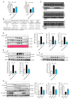
LSN prevents MI-induced cardiac dysfunction and remodeling in mice. (A) Echocardiographic assessment of ejection fraction (left) and fractional shortening (right) at 4 weeks post-myocardial infarction in mice administered with PBS or LSN. n = 8 or more. (B) M-mode echocardiography at 4 weeks post-myocardial infarction in mice administered with PBS or LSN. (C) Table of cardiac function parameters in Sham, MI + PBS, and MI + LSN at 4 weeks post-MI. Data are presented as mean ± SD. n = 8 or more for each group. LVID;d: Left Ventricular Internal Dimension, diastolic; LVID;s: Left Ventricular Internal Dimension, systolic. (D) Masson’s trichrome staining showing ischemic hearts (top, scale bar = 1000 µm) and enlarged area of local left ventricular (LV) border region of ischemic hearts 4 weeks post-MI (bottom, scale bar = 100 μm). (E) Representative immunoblots (upper) and quantification (lower) of serum ANP, BNP, and CTnI in mice administered with PBS or LSN at 3 weeks post-MI. n = 7 or more. (F) ANP, BNP, β-MHC, Collagen I, and Collagen III mRNA levels in heart tissue of mice administered with PBS or LSN at 1 week post-MI. n = 5 or more. Data are presented as mean ± SEM along with individual data points; * p < 0.05, ** p < 0.01, *** p < 0.001 vs. MI + PBS.
Figure 2
Figure 2
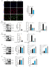
LSN prevents MI-induced early cardiac inflammatory responses. (A) TNF-α, IL-1β, and IL-6 mRNA levels in heart tissue of mice administered with PBS or LSN at 1 week post-MI. n = 5 or more. (B) ELISA analysis of serum IFN-γ, IL-1β, IL-6, and TNF-α levels in mice administered with PBS or LSN at 1 week post-MI. n = 9 or more. (C) Representative immunoblots (upper) and quantification (lower) of P-P65 and P-iκB expression in the heart of mice administered with PBS or LSN at 1 week post-MI. n = 4 or more. (D) Immunofluorescence staining (upper) and quantification (lower) of P65 foci (red), cardiomyocyte-specific α-actinin 2 (Actn2, green), and DAPI (blue) in heart tissue of mice treated as in (C). n = 4 or more. Scale bar, 16 μm. Data are presented as mean ± SEM along with individual data points; * p < 0.05, ** p < 0.01, *** p < 0.001 vs. MI + PBS.
Figure 3
Figure 3
β-catenin deletion prevents MI-induced cardiac injury and inflammatory response. (A) Echocardiographic assessment of ejection fraction (left) and fractional shortening (right) at 1 week post-MI in β-catenin fl/fl and β-catenin KO mice. n = 6 or more. (B) Table of cardiac function parameters in β-catenin fl/fl and β-catenin KO mice at 1 week post-MI. n = 6 or more for each group. LVID;d: Left Ventricular Internal Dimension; diastolic; LVID;s: Left Ventricular Internal Dimension; systolic. (C) M-mode echocardiography at 1 week post-MI in β-catenin fl/fl and β-catenin KO mice. (D) Representative immunoblots (left) and quantification (right) of serum ANP, BNP, and cTnI in β-catenin fl/fl and β-catenin KO mice at 1 week post-MI. n = 3 or more. (E) Representative immunoblots (upper) and quantification (lower) of total P-P65 and P-iκB expression in the heart of β-catenin fl/fl and β-catenin KO mice at 1 week post-MI. n = 3 or more. (F) Representative immunoblots (upper) and quantification (lower) of nuclear P65 expression in the heart of β-catenin fl/fl and β-catenin KO mice at 1 week post-MI. n = 3 or more. (G) Representative immunoblots (left) and quantification (right) of n/nuclear β-catenin, P65, and c/cytoplasmic phospho-β-catenin expression in the heart of mice administered with PBS or LSN at 1 week post-MI. n = 4 or more. Data are presented as mean ± SEM along with individual data points; * p < 0.05, ** p < 0.01, *** p < 0.001.
Figure 4
Figure 4
LSN prevents MI-induced proinflammatory response through inhibiting Wnt/β-catenin signaling. (A) Immunofluorescence staining (left) and quantification (right) of P65 foci (red), CTnT (green), and DAPI (blue) in H9C2 cardiomyocytes induced by TNF-α following pretreatment with LSN for 12 h. n = 4. Scale bar, 50 μm. (B) Representative immunoblots (left) and quantification (right) of total β-catenin, P-P65, and P-iκB expression following siRNA-mediated knockdown of β-catenin for 48 h, and subsequent treatment with TNF-α for 12 h in H9C2 cardiomyocytes. n = 3 or more. (C) Representative immunoblots (left) and quantification (right) of nuclear β-catenin and P65 expression following siRNA-mediated knockdown of β-catenin for 48 h, and subsequent treatment with TNF-α for 12 h in H9C2 cardiomyocytes. n = 3 or more. (D) Representative immunoblots (left) and quantification (right) of total β-catenin, P-P65, and P-iκB expression following TNF-α or LiCl treatment for 12 h in H9C2 cardiomyocytes. n = 3 or more. (E) Representative immunoblots (left) and quantification (right) of nuclear β-catenin and P65 expression following TNF-α or LiCl treatment for 12 h in H9C2 cardiomyocytes. n = 3 or more. Data are presented as mean ± SEM along with individual data points; * p < 0.05, ** p < 0.01, *** p < 0.001.

Similar articles

References

    1. Wu X., Reboll M.R., Korf-Klingebiel M., Wollert K.C. Angiogenesis after acute myocardial infarction. Cardiovasc. Res. 2021;117:1257–1273. doi: 10.1093/cvr/cvaa287. - DOI - PubMed
    1. Neri M., Fineschi V., Di Paolo M., Pomara C., Riezzo I., Turillazzi E., Cerretani D. Cardiac oxidative stress and inflammatory cytokines response after myocardial infarction. Curr. Vasc. Pharmacol. 2015;13:26–36. doi: 10.2174/15701611113119990003. - DOI - PubMed
    1. Yap J., Irei J., Lozano-Gerona J., Vanapruks S., Bishop T., Boisvert W.A. Macrophages in cardiac remodelling after myocardial infarction. Nat. Rev. Cardiol. 2023;20:373–385. doi: 10.1038/s41569-022-00823-5. - DOI - PubMed
    1. Zhang Q., Wang L., Wang S., Cheng H., Xu L., Pei G., Wang Y., Fu C., Jiang Y., He C., et al. Signaling pathways and targeted therapy for myocardial infarction. Signal Transduct. Target. Ther. 2022;7:78. doi: 10.1038/s41392-022-00925-z. - DOI - PMC - PubMed
    1. Barandon L., Casassus F., Leroux L., Moreau C., Allières C., Lamazière J.M., Dufourcq P., Couffinhal T., Duplàa C. Secreted frizzled-related protein-1 improves postinfarction scar formation through a modulation of inflammatory response. Arter. Thromb. Vasc. Biol. 2011;31:e80–e87. doi: 10.1161/ATVBAHA.111.232280. - DOI - PubMed

MeSH terms

LinkOut - more resources