. 2025 May 10;26(10):4589.
doi: 10.3390/ijms26104589.

Early Zinc Supplementation Enhances Epididymal Sperm Glycosylation, Endocrine Activity, and Antioxidant Activity in Rats Exposed to Cadmium

Affiliations

Early Zinc Supplementation Enhances Epididymal Sperm Glycosylation, Endocrine Activity, and Antioxidant Activity in Rats Exposed to Cadmium

Sergio Marín de Jesús et al. Int J Mol Sci. .

Abstract

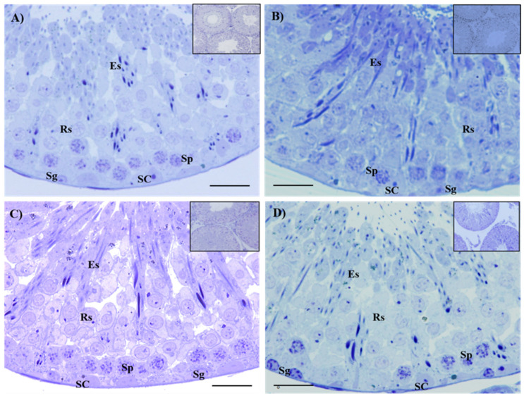
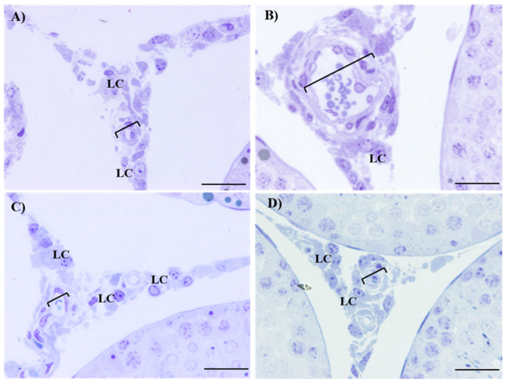
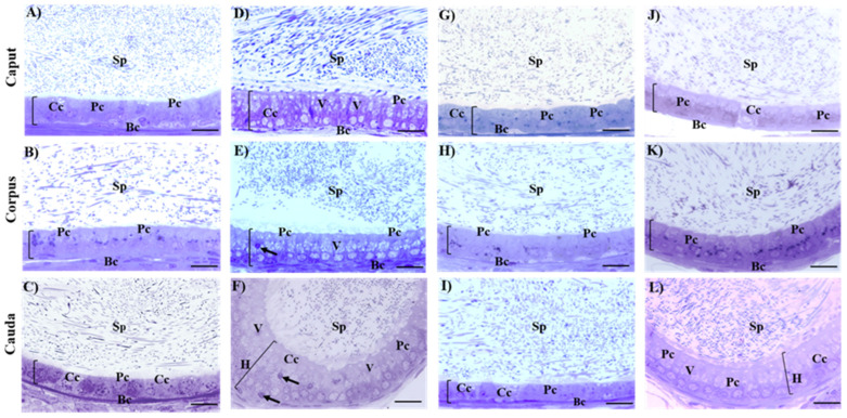
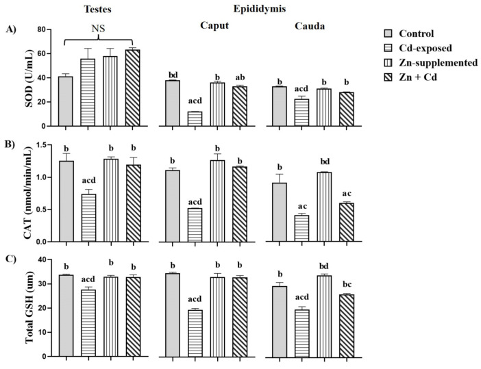
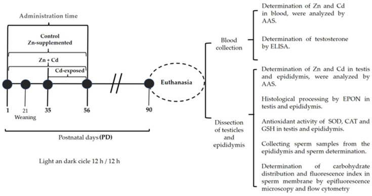
Sperm maturation involves changes in plasma membrane glycosylation for fertilization. Cadmium (Cd) exerts a negative effect by disrupting testicular and epididymal function, altering antioxidant activity. Zinc (Zn) is an essential element known for its antioxidant properties, role in testosterone synthesis, and support of spermatogenesis. However, its effect on sperm membrane glycosylation, as well as endocrine and antioxidant activity, after exposure to Cd has remained unexplored. This study evaluated the impact of Zn on epididymal sperm glycosylation, endocrine activity, and antioxidant activity in Cd-exposed rats. Four groups of male Wistar rats were analyzed: control, Cd-exposed, Zn-supplemented, and Zn + Cd groups. On postnatal day 90, tissues and blood were collected for Zn and Cd quantification, testosterone levels, antioxidant activity, histological analysis, and sperm quality. The results showed that Cd concentration increased significantly, reduced testosterone levels, modified antioxidant activity, and caused structural damage in the epididymis. The Cd-exposed group showed disrupted glycosylation and distribution patterns and reduced sperm quality. The Zn + Cd group showed lower Cd accumulation, preserved testosterone levels, restored antioxidant activity, and preserved glycosylation patterns and sperm quality. This study highlights the protective role of Zn in mitigating Cd-induced reproductive toxicity, probably through the competitive inhibition of Cd uptake and antioxidant support, thereby preserving fertility.

Keywords: cadmium; glycosylation; sperm maturation; spermatogenesis; zinc.


Conflict of interest statement

The authors declare no conflicts of interest.

Figures

Figure 1
Figure 1
Histological sections of seminiferous tubules in the testes show the presence of cell types involved in spermatogenesis. The control is shown in (A), while the Cd-exposed is shown in (B), the Zn-supplemented in (C), and the Zn + Cd in (D) groups. The histological images contained the following cell types: spermatogonia (Sg), spermatocytes (Sp), round spermatids (Rs), elongated spermatids (Es), and Sertoli cells (SC); toluidine blue, 60×. Bar 30 µm.
Figure 2
Figure 2
Histological sections of the testes interstitium. Leydig cells and some blood vessels with endothelial damage due to Cd treatment are observed as shown in (B) in comparison with the control (A), the Zn-supplemented (C), and the Zn + Cd (D) groups. Leydig cell (LC); bracketed bar indicates (blood vessels); toluidine blue, 60×. Bar 30 µm.
Figure 3
Figure 3
Histological sections of the epididymal regions. The characteristic cell types of the epididymal epithelium are seen, providing a luminal environment for the protection and maturation of sperm. Cd exposure caused an increase in epithelial height, vacuolization, and hyperplasia. In the Zn + Cd group, Zn protected the caput and corpus regions and reduced damage to the cauda. Control: (A) caput, (B) corpus, (C) cauda; Cd-exposured: (D) caput, (E) corpus, (F) cauda; Zn-supplemented: (G) caput, (H) corpus, (I) cauda; and Zn + Cd: (J) caput, (K) corpus, (L) cauda groups. Principal cell (Pc); basal cell (Bc); clear cell (Cc); sperm (Sp); hyperplasia (H); epithelial height (bar in brackets); vacuolization (V); dense vesicles (arrows); toluidine blue, 60×. Bar 20 µm.
Figure 4
Figure 4
Antioxidant activity of the testes and epididymis. The activity of SOD, CAT, and GSH was altered in both the testes and the epididymis with Cd-exposure. However, in the Zn + Cd, Zn-supplementation favored the antioxidant activity in both organs. SOD (A), CAT (B), and GSH (C). Data expressed as means ± SEM for all analyzed groups (n = 7/treatment group). a p < 0.05 indicates differences vs. the control group; b p < 0.05 indicates differences vs. the Cd-exposed group; c p < 0.05 indicates differences vs. the Zn-supplemented group; d p < 0.05 indicates differences vs. the Zn + Cd group. No significant difference (NS).
Figure 5
Figure 5
The photomicrograph shows the distribution patterns of fluorescence of N-acetylglucosamine and/or sialic acid in sperm. The sperm showed different patterns of fluorescence along their plasma membranes. Pattern 1 (P1): fluorescence throughout the spermatozoon; Pattern 2 (P2): fluorescence in the head; Pattern 3 (P3): fluorescence in the head and midbody; Pattern 4 (P4): sperm without fluorescence. Fluorescence microscopy, 20×.
Figure 6
Figure 6
Percentages of distribution of fluorescence patterns of N-acetylglucosamine and/or sialic acid in the membrane of sperm in the three epididymal regions. The fluorescence distribution of Pattern 2 predominated in all regions, among treatments, the Cd-exposed group showed a decrease in the distribution of Patterns 1, 2, and 3 and an increase in Pattern 4. In the Zn + Cd group, Zn supplementation favored the distribution of N-acetylglucosamine and/or sialic acid. Data are expressed as means ± SEM for all analyzed groups (n = 7/treatment group). a p < 0.05 indicates differences vs. the control group; b p < 0.05 indicates differences vs. the Cd-exposed group; c p < 0.05 indicates differences vs. the Zn-supplemented group; d p < 0.05 indicates differences vs. the Zn + Cd group.
Figure 7
Figure 7
Fluorescence index of N-acetylglucosamine and/or sialic acid in the sperm of the three epididymal regions. Cd-exposed group decreased the presence of N-acetylglucosamine and/or sialic acid. However, in the Zn + Cd- group, Zn supplementation favored the presence of carbohydrates. Data expressed as means ± SEM for all analyzed groups (n = 7/treatment group). a p < 0.05 indicates differences vs. the control group; b p < 0.05 indicates differences vs. the Cd-exposed group; c p < 0.05 indicates differences vs. the Zn-supplemented group; d p < 0.05 indicates differences vs. the Zn + Cd group.
Figure 8
Figure 8
Micrographs of the distribution patterns of mannose fluorescence in sperm. The sperm showed different fluorescence patterns along their plasma membrane. Pattern 1 (P1): fluorescence in the head and main body; Pattern 2 (P2): fluorescence in the head; Pattern 3 (P3): fluorescence in the head and intermediate body; Pattern 4 (P4): sperm without fluorescence. Fluorescence microscopy, 20×.
Figure 9
Figure 9
Percentages of distribution patterns of mannose fluorescence in the sperm membrane in the three epididymal regions. The fluorescence distribution of Pattern 2 was predominant in the epididymal regions, and between treatments, the group treated with Cd decreased the distribution of Patterns 1, 2, and 3 and increased Pattern 4. However, in the Zn + Cd group, Zn supplementation favored the distribution of mannose. Data expressed as means ± SEM for all analyzed groups (n = 7/treatment group). a p < 0.05 indicates differences vs. the control group; b p < 0.05 indicates differences vs. the Cd group; c p < 0.05 indicates differences vs. the Zn-supplemented group; d p < 0.05 indicates differences vs. the Zn + Cd group.
Figure 10
Figure 10
Fluorescence index of mannose in the sperm of the three epididymal regions. The Cd-exposed group decreased the presence of mannose. However, in the Zn + Cd group, Zn supplementation favored the presence of the carbohydrate. Data expressed as means ± SEM for all analyzed groups (n = 7/treatment group). a p < 0.05 indicates differences vs. the control group; b p < 0.05 indicates differences vs. the Cd-exposed group; c p < 0.05 indicates differences vs. the Zn-supplemented group; d p < 0.05 indicates differences vs. the Zn + Cd group.
Figure 11
Figure 11
Experimental design and sample processing. Four experimental groups of Wistar rats were established: control (saline), Cd-exposed (CdCl2 0.5 mg/kg body weight), Zn-supplemented (ZnCl2 1 mg/kg body weight), and Zn + Cd (CdCl2 + ZnCl2). The treatments were administered intraperitoneally on the indicated days. After 90 days, the animals were euthanized to collect blood, testis, and epididymis samples. The following were analyzed: Cd and Zn metals via AAS; testosterone concentration via ELISA; histological processing via EPON; oxidative stress via ELISA (SOD, CAT, and GSH); sperm parameters and carbohydrates via epifluorescence and flow cytometry. Body weight (BW), postnatal day (PD), intraperitoneal (IP), atomic absorption spectrophotometry (AAS), enzyme-linked immunosorbent assay (ELISA), epoxy resin (EPON), superoxide dismutase (SOD), catalase (CAT), glutathione (GSH).

Similar articles

References

    1. Gervasi M.G., Visconti P.E. Molecular changes and signaling events occurring in spermatozoa during epididymal maturation. Andrology. 2017;5:204–218. doi: 10.1111/andr.12320. - DOI - PMC - PubMed
    1. Barrachina F., Battistone M.A., Castillo J., Mallofré C., Jodar M., Breton S., Oliva R. Sperm acquire epididymis-derived proteins through epididymosomes. Hum. Reprod. 2022;37:651–668. doi: 10.1093/humrep/deac015. - DOI - PMC - PubMed
    1. Dacheux J.L., Dacheux F. New insights into epididymal function in relation to sperm maturation. Reproduction. 2013;147:R27–R42. doi: 10.1530/REP-13-0420. - DOI - PubMed
    1. Camargo M., Intasqui P., Bertolla R.P. Understanding the seminal plasma proteome and its role in male fertility. Basic Clin. Androl. 2018;28:6. doi: 10.1186/s12610-018-0071-5. - DOI - PMC - PubMed
    1. Vickram S., Rohini K., Srinivasan S., Nancy Veenakumari D., Archana K., Anbarasu K., Jeyanthi P., Thanigaivel S., Gulothungan G., Rajendiran N., et al. Role of Zinc (Zn) in Human Reproduction: A Journey from Initial Spermatogenesis to Childbirth. Int. J. Mol. Sci. 2021;22:2188. doi: 10.3390/ijms22042188. - DOI - PMC - PubMed

LinkOut - more resources