. 2025 May 13;26(10):4660.
doi: 10.3390/ijms26104660.

Computational Search for Inhibitors of SOD1 Mutant Infectivity as Potential Therapeutics for ALS Disease

Affiliations

Computational Search for Inhibitors of SOD1 Mutant Infectivity as Potential Therapeutics for ALS Disease

Marco Carnaroli et al. Int J Mol Sci. .

Abstract

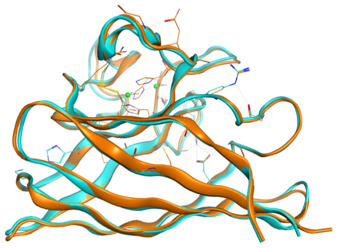
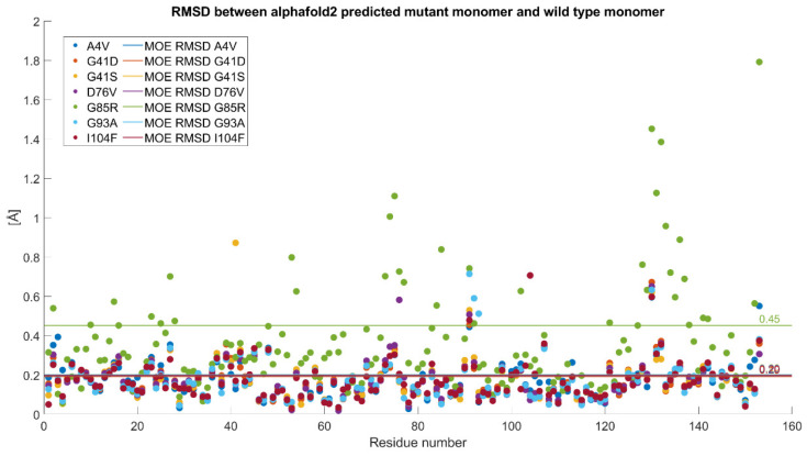
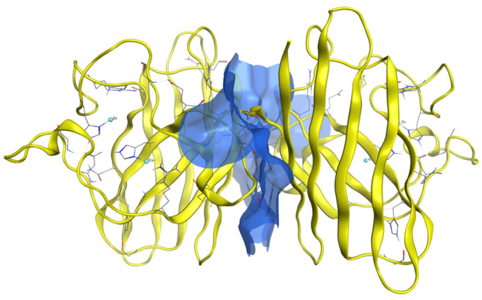
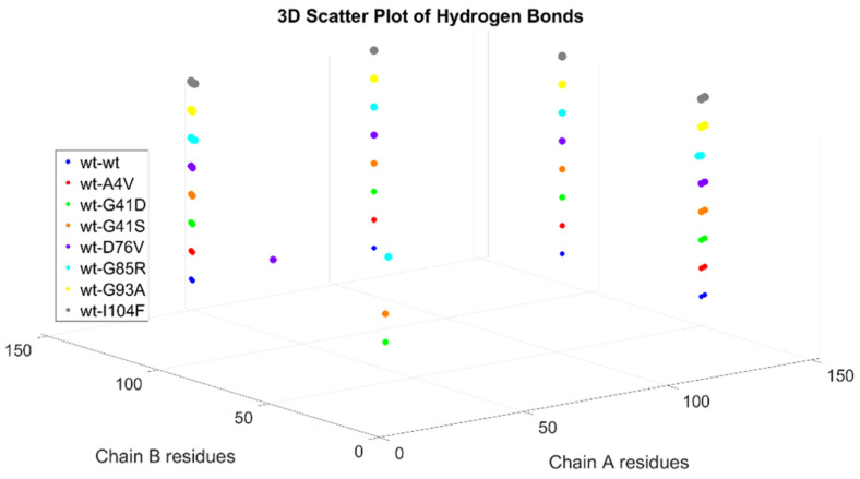
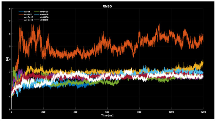
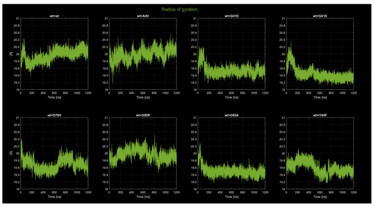
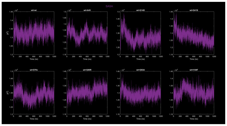
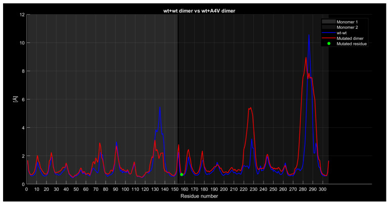
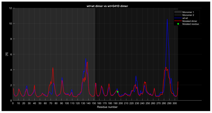
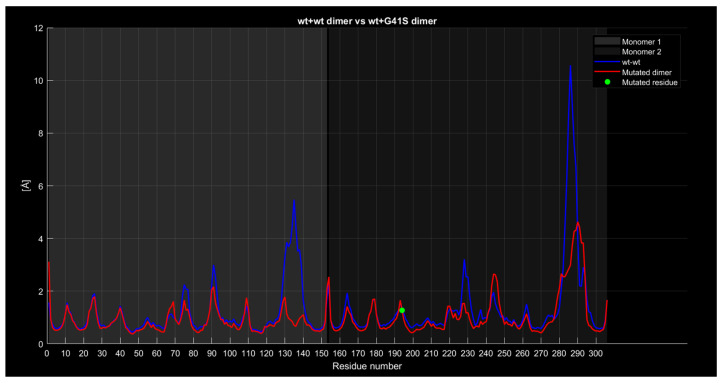
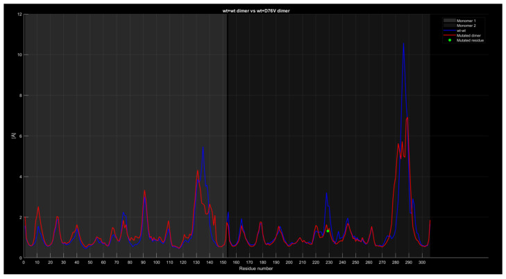
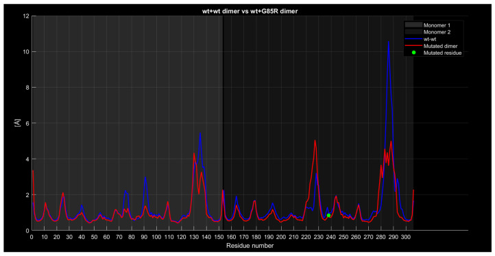
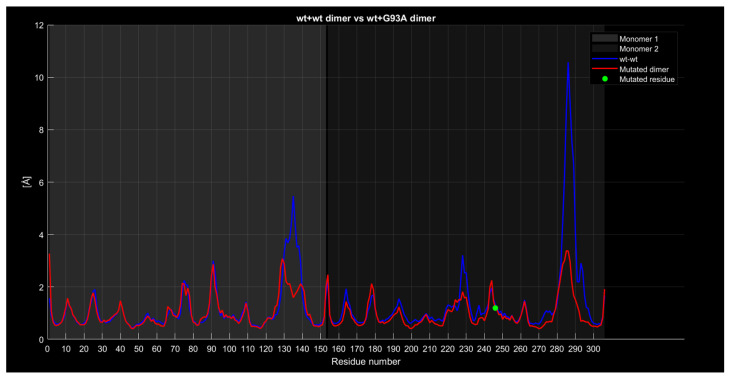
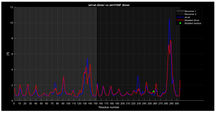
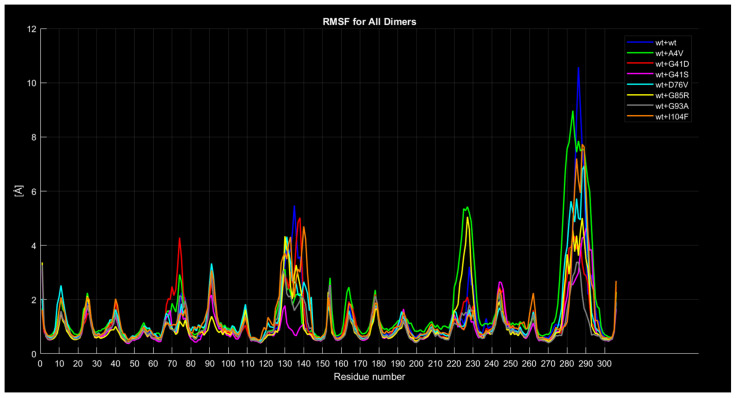
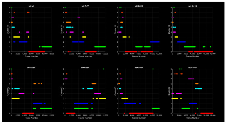
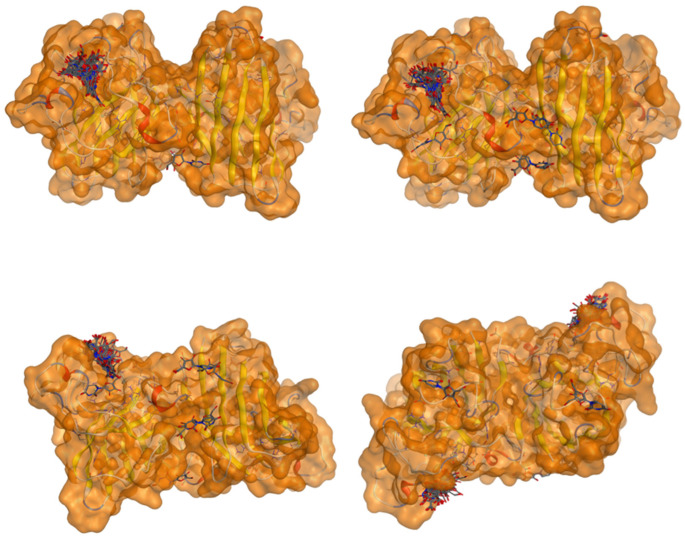
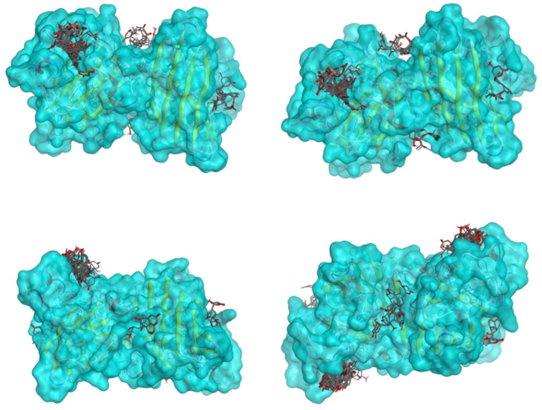
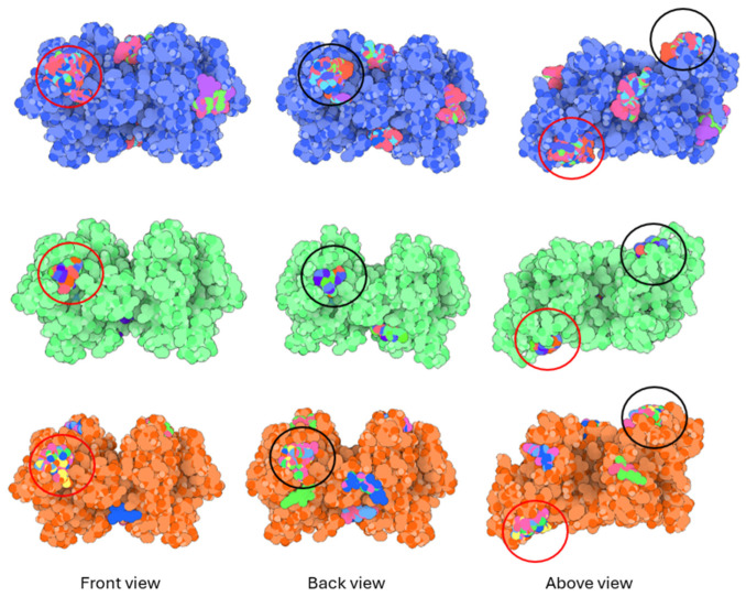
Familial amyotrophic lateral sclerosis (ALS) is a progressive neurodegenerative disease characterized by the selective degeneration of motor neurons. Among the main genetic causes of ALS, over 200 mutations have been identified in the Cu/Zn superoxide dismutase (SOD1) protein, a dimeric metalloenzyme essential for converting superoxides from cellular respiration into less toxic products. Point mutations in SOD1 monomers can induce protein misfolding, which spreads to wild-type monomers through a prion-like mechanism, leading to dysfunctions that contribute to the development of the disease. Understanding the structural and functional differences between the wild-type protein and its mutated variants, as well as developing drugs capable of inhibiting the propagation of misfolding, is crucial for identifying new therapeutic strategies. In this work, seven SOD1 mutations (A4V, G41D, G41S, D76V, G85R, G93A, and I104F) were selected, and three-dimensional models of SOD1 dimers composed of one wild-type monomer and one mutated monomer were generated, along with a control dimer consisting solely of wild-type monomers. Molecular dynamics simulations were conducted to investigate conformational differences between the dimers. Additionally, molecular docking was performed using a library of ligands to identify compounds with high affinity for the mutated dimers. The study reveals some differences in the mutated dimers following molecular dynamics simulations and in the docking of the selected ligands with the various dimers.

Keywords: ALS mutations; SOD1 inhibitors; docking; molecular dynamics.


Conflict of interest statement

The authors declare no conflicts of interest.

Figures

Figure 1
Figure 1
The monomer derived from PDB entry “3ecu” is shown in light blue, superimposed on the monomer derived from “7wwt” in orange, with the metal ions depicted in green.
Figure 2
Figure 2
Comparison between the RMSD of the various mutants (shown as dots in the legend) based on their positions in the amino acid sequence. The horizontal lines, following the same color scheme, represent the RMSD values calculated for each mutant.
Figure 3
Figure 3
A generic SOD1 dimer shown in yellow with the surface which includes the contacts between the two monomers highlighted in blue.
Figure 4
Figure 4
Illustration of the pairs of residues from chain A and chain B that are involved in the hydrogen bonds at the interface of the various dimers.
Figure 5
Figure 5
Time evolution of RMSD values for all eight dimers analyzed.
Figure 6
Figure 6
Time evolution of the radius of gyration for all eight dimers.
Figure 7
Figure 7
Time evolution of SASA for all eight dimers.
Figure 8
Figure 8
The wt+A4V dimer RMSF compared to the wild-type dimer.
Figure 9
Figure 9
The wt+G41D dimer’s RMSF compared to the wild-type dimer.
Figure 10
Figure 10
The wt+G41S dimer RMSF compared to the wild-type dimer.
Figure 11
Figure 11
The wt+D76V dimer RMSF compared to the wild-type dimer.
Figure 12
Figure 12
The wt+G85R dimer’s RMSF compared to the wt+wt dimer.
Figure 13
Figure 13
The wt+G93A dimer’s RMSF compared to the wt+wt dimer.
Figure 14
Figure 14
The wt+I104F dimer’s RMSF compared to the wt+wt dimer.
Figure 15
Figure 15
Mutant RMSF curves compared to the wild type curve.
Figure 16
Figure 16
Evolution of cluster population over time. Each trajectory frame is assigned to a specific cluster, represented by a distinct color. Note that although the same color scheme is used, a cluster of a given color in one protein does not correspond to the cluster of the same color in another protein.
Figure 17
Figure 17
Superposition of the wt+wt dimer’s representative structures, with each color corresponding to a specific cluster.
Figure 18
Figure 18
Ramachandran plot of protein residues for all the dimers. Each blue point represents the pair of φ and ψ values for a specific generic residue, while the colored points represent the same but for a mutated residue.
Figure 19
Figure 19
The same as in Figure 18, but in this case, the dimer structures are derived from cluster 0 after the conformational clustering.
Figure 20
Figure 20
Three-dimensional representation of dimer wt+wt in pink with ligand 22 docked multiple (100) times.
Figure 21
Figure 21
Three-dimensional representation of dimer wt+wt in orange with ligand 36 docked multiple (100) times.
Figure 22
Figure 22
Three-dimensional representation of dimer wt+wt in light blue with ligand 38 docked multiple (100) times.
Figure 23
Figure 23
Graphical representation of ligands 37, 24, and 34 bonded to the wt+wt dimer. Each line represents the wt+wt dimer bonded 100 times to a specific ligand in three perspective views. The two binding regions shared between them are circled in red and black.

Similar articles

References

    1. Brotman R.G., Moreno-Escobar M.C., Joseph J., Munakomi S., Pawar G. Amyotrophic Lateral Sclerosis. StatPearls; Treasure Island, FL, USA: 2025. - PubMed
    1. Rosen D.R., Siddique T., Patterson D., Figlewicz D.A., Sapp P., Hentati A., Donaldson D., Goto J., O’Regan J.P., Deng H.X., et al. Mutations in Cu/Zn superoxide dismutase gene are associated with familial amyotrophic lateral sclerosis. Nature. 1993;362:59–62. doi: 10.1038/362059a0. - DOI - PubMed
    1. Berdyński M., Miszta P., Safranow K., Andersen P.M., Morita M., Filipek S., Żekanowski C., Kuźma-Kozakiewicz M. SOD1 mutations associated with amyotrophic lateral sclerosis: Analysis of variant severity. Nature. 2022;12:103. doi: 10.1038/s41598-021-03891-8. - DOI - PMC - PubMed
    1. Bernard E., Pegat A., Svahn J., Bouhour F., Leblanc P., Millecamps S., Thobois S., Guissart C., Lumbroso S., Mouzat K. Clinical and molecular landscape of ALS patients with SOD1 mutations: Novel pathogenic variants and novel phenotypes. A single ALS center study. Int. J. Mol. Sci. 2020;21:6807. doi: 10.3390/ijms21186807. - DOI - PMC - PubMed
    1. ALSoD. [(accessed on 1 April 2024)]. Available online: https://alsod.ac.uk/output/gene.php/SOD1.

MeSH terms

LinkOut - more resources