. 2025 May 14;26(10):4696.
doi: 10.3390/ijms26104696.

Biosynthesized Calcium Peroxide Nanoparticles as a Multifunctional Platform for Liver Cancer Therapy

Affiliations

Biosynthesized Calcium Peroxide Nanoparticles as a Multifunctional Platform for Liver Cancer Therapy

Sen Wu et al. Int J Mol Sci. .

Abstract

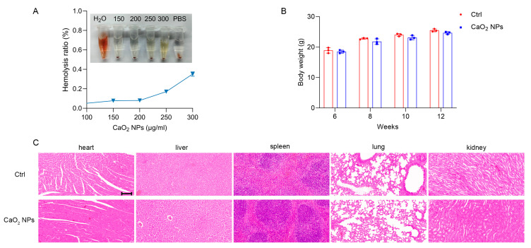
To overcome the limitations associated with chemically synthesized nanoparticles in cancer therapy, researchers have increasingly focused on developing nanoparticles with superior biocompatibility and prolonged tumor retention using biosynthetic methods. In this study, we first identified the presence of calcium peroxide nanoparticles (CaO2 NPs) in the blood of individuals who had ingested calcium gluconate. Furthermore, the dropwise addition of calcium gluconate to human serum resulted in the spontaneous self-assembly of CaO2 NPs. Next, following tail vein injection of fluorescently labeled CaO2 NPs into subcutaneous tumor-bearing nude mice, we observed that the nanoparticles exhibited prolonged accumulation at the tumor sites compared to other organs through visible-light imaging. Immunofluorescence staining demonstrated that CaO2 NPs co-localized with vesicular transport-associated proteins, such as PV-1 and Caveolin-1, as well as the albumin-binding-associated protein SPARC, suggesting that their transport from tumor blood vessels to the tumor site is mediated by Caveolin-1- and SPARC-dependent active transport pathways. Additionally, the analysis of various organs in normal mice injected with CaO2 NPs at concentrations significantly higher than the experimental dose showed no apparent organ damage. Hemolysis assays indicated that hemolysis occurred only at calcium concentrations of 300 µg/mL, whereas the experimental concentration remained well below this threshold with no detectable hemolytic activity. In a subcutaneous tumor-bearing nude mouse model, treatment with docetaxel-loaded CaO2 NPs showed a 68.5% reduction in tumor volume compared to free docetaxel (DTX) alone. These novel biosynthetic CaO2 NPs demonstrated excellent biocompatibility, prolonged retention at the tumor site, safety, and drug-loading capability.

Keywords: CaO2 NPs; biocompatibility; biosynthetic methods; drug loading; prolonged retention.


Conflict of interest statement

The authors declare that they have no known competing financial interests or personal relationships that could have appeared to influence the work reported in this paper.

Figures

Scheme 1
Scheme 1
The biosynthesis of CaO2 NP nanocarriers for the treatment of liver cancer.
Figure 1
Figure 1
Characterization analysis of CaO2 NPs. (A) Representative TEM image of CaO2 NPs isolated from human blood, scale bar = 1 μm. (B) Representative TEM image of CaO2 NPs biosynthesized by calcium gluconate and human serum in vitro, scale bar = 500 nm. (C) High-resolution transmission electron microscopy of CaO2 NPs (lattice: 0.28 nm), scale bar = 10 nm. (D) Size distribution of CaO2 NPs in PBS as characterized by DLS. (E) Negative CaO2 NP surface charge at −12.75 mV in phosphate buffer. (F) Dot blot assay to detect albumin in isolated CaO2 NPs. Human serum albumin (HSA) served as positive control, calcium gluconate used as negative control.
Figure 2
Figure 2
Tumor targeting of CaO2 NPs. (A) Absorbance spectra of ICG and ICG-CaO2 NPs. (B) Excitation spectra of ICG and ICG-CaO2 NPs. (C) FTIR spectra of ICG, CaO2 NPs, and ICG-CaO2 NPs. (D) Representative in vitro fluorescence images of tumors and major organs at 1 h after intravenous treatment of tumor-bearing mice with ICG-CaO2 NPs (10 mg/kg calcium, n = 3 mice). (E) Representative in vitro fluorescence images of tumors and major organs at 72 h after intravenous treatment of tumor-bearing mice with ICG-CaO2 NPs (10 mg/kg calcium, n = 3 mice).
Figure 3
Figure 3
CaO2 NPs enter tumor tissues through active pathways. Representative immunofluorescence images of tumor sections from the tumor-bearing mice treated with CaO2 NPs (10 mg/kg calcium). (A) Tumor sections stained with anti-PV-1 antibodies (green), CaO2 NPs (red), and DAPI (blue), and the merged image (yellow). Scale bar = 20 μm. (B) Tumor sections were stained with anti-Caveolin-1 antibodies (green), CaO2 NPs (red), and DAPI (blue). Scale bar = 20 μm. (C) Tumor sections stained with anti-SPARC antibodies (green), CaO2 NPs (red), and DAPI (blue), and the merged image (yellow). Scale bar = 20 μm. (D) Live-cell imaging showing lysosomes (red) and calcium ions (green) in MHCC 97-H cells incubated with 150 mg/L CaO2 NPs; scale bar = 10 μm.
Figure 4
Figure 4
CaO2 NPs as nanocarriers promote apoptosis in liver cancer cells. (A) The Cell Counting Kit (CCK-8) assay of MHCC 97-H cells. Group (1): the cells were treated with CaO2 NPs (150 mg/L), DTX (37.5 mg/L), or DTX@CaO2 NPs (containing 37.5 mg/L DTX) and tested after incubation for 48 h. Group (2): The cells were treated with CaO2 NPs (300 mg/L), DTX (75 mg/L), or DTX@CaO2 NPs (containing 75 mg/L DTX) and tested after incubation for 48 h. The bars represent the results from three independent experiments (n = 3); *** p < 0.001. (B) The flow cytometry analysis using Annexin V/PI staining. MHCC 97-H cells were treated with 150 mg/L CaO2 NPs, 37.5 mg/L DTX, or DTX@CaO2 NPs (containing 37.5 mg/L DTX) for 24 h. (C) The quantitative analysis of the apoptotic cells in B (n = 3); * p < 0.033, *** p < 0.001.
Figure 5
Figure 5
CaO2 NPs as drug delivery nanocarriers for liver tumor treatment. (A) Influence of different treatment methods on volume size of nude mouse transplanted tumor: (a) control group (inject normal saline); (b) inject CaO2 NPs (10 mg/kg calcium); (c) inject 3 mg/kg DTX; (d) inject DTX@CaO2 NPs (3 mg/kg DTX). (B) Tumor volume on day 28 after different treatments (n = 3 mice). ** p < 0.002, *** p < 0.001.
Figure 6
Figure 6
The biosafety assessment of CaO2 NPs. (A) The hemolysis rate of CaO2 NPs. PBS served as the positive control, while deionized water served as the negative control. (B) The body weights of the BALB/c mice treated with CaO2 NPs (blue) or saline vehicle control (red) for 6 weeks. The mice were treated with CaO2 NPs (10 mg/kg calcium) by intravenous injection once per week, and their weights were noted every two weeks. (C) The histological analysis of the BALB/c mice heart, liver, spleen, lungs, and kidneys (CaO2 NPs (20 mg/kg calcium) treatment by tail vein for 6 weeks). Representative images are shown (n = 3). Scale bar = 100 μm.

References

    1. Sung H., Ferlay J., Siegel R.L., Laversanne M., Soerjomataram I., Jemal A., Bray F. Global Cancer Statistics 2020: GLOBOCAN Estimates of Incidence and Mortality Worldwide for 36 Cancers in 185 Countries. CA Cancer J. Clin. 2021;71:209–249. doi: 10.3322/caac.21660. - DOI - PubMed
    1. Yao X., Xie R., Cao Y., Tang J., Men Y., Peng H., Yang W. Simvastatin induced ferroptosis for triple-negative breast cancer therapy. J. Nanobiotechnol. 2021;19:311. doi: 10.1186/s12951-021-01058-1. - DOI - PMC - PubMed
    1. Suliman I.H., Kim K., Chen W., Kim Y., Moon J.H., Son S., Nam J. Metal-Based Nanoparticles for Cancer Metalloimmunotherapy. Pharmaceutics. 2023;15:2003. doi: 10.3390/pharmaceutics15072003. - DOI - PMC - PubMed
    1. Chandoliya R., Sharma S., Sharma V., Joshi R., Sivanesan I. Titanium Dioxide Nanoparticle: A Comprehensive Review on Synthesis, Applications and Toxicity. Plants. 2024;13:2964. doi: 10.3390/plants13212964. - DOI - PMC - PubMed
    1. Nam K.T., Wartena R., Yoo P.J., Liau F.W., Lee Y.J., Chiang Y.M., Hammond P.T., Belcher A.M. Stamped microbattery electrodes based on self-assembled M13 viruses. Proc. Natl. Acad. Sci. USA. 2008;105:17227–17231. doi: 10.1073/pnas.0711620105. - DOI - PMC - PubMed

LinkOut - more resources