. 2025 May 16;26(10):4799.
doi: 10.3390/ijms26104799.

On the Potential Role of Phytate Against Neurodegeneration: It Protects Against Fe3+-Catalyzed Degradation of Dopamine and Ascorbate and Against Fe3+-Induced Protein Aggregation

Affiliations

On the Potential Role of Phytate Against Neurodegeneration: It Protects Against Fe3+-Catalyzed Degradation of Dopamine and Ascorbate and Against Fe3+-Induced Protein Aggregation

Samantha Rebeca Godoy et al. Int J Mol Sci. .

Abstract

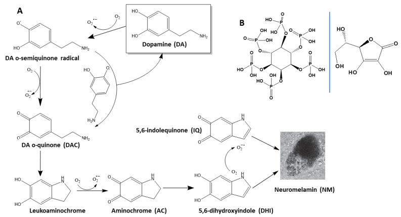
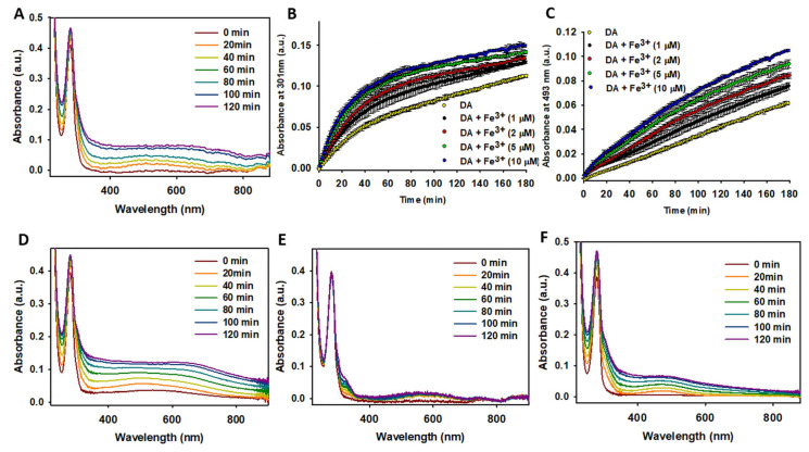
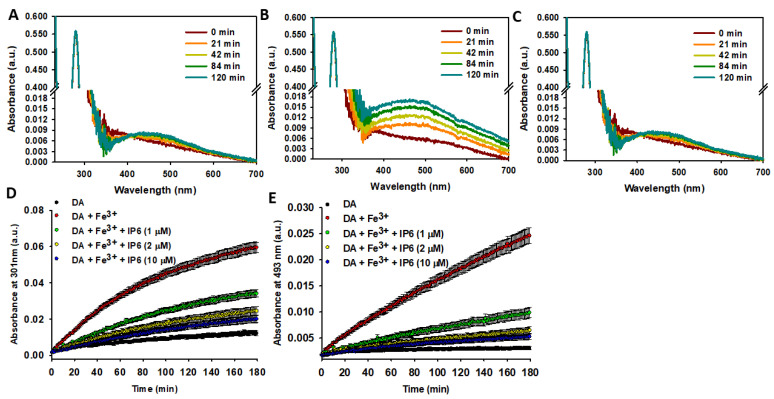
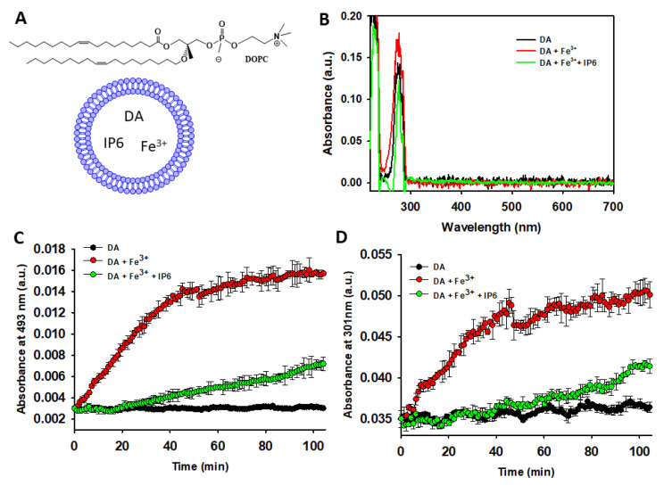
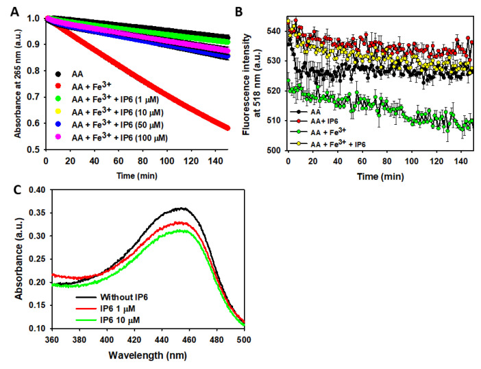
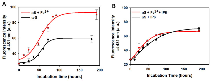
Myo-inositol-1,2,3,4,5,6-hexakisphosphate (IP6) is commonly found in plant-derived foods and has important pharmacological properties against many pathologies. One of them appears to be neurodegeneration, which is notably stimulated by dysregulated metal metabolism. Consequently, we explore the role of IP6 in mitigating neurodegenerative events catalyzed by dysregulated free iron. More precisely, we performed spectrophotometric measurements in aqueous solutions to investigate the ability of IP6 to chelate Fe3+ and inhibit its role in catalyzing the oxidative degradation of dopamine and ascorbic acid, two key molecules in neuronal redox systems. Our results demonstrate that IP6 effectively prevents the formation of harmful intermediates, such as neuromelanin and reactive oxygen species, which are linked to neuronal damage. Additionally, we assessed the effect of IP6 on Fe3+-induced protein aggregation, focusing on α-synuclein, which is closely associated with Parkinson's disease. Our data reveal that IP6 accelerates the conversion of toxic α-synuclein oligomers into less harmful amyloid fibrils, thereby reducing their neurotoxic potential. Our findings highlight the dual function of IP6 as a potent Fe3+ chelator and modulator of protein aggregation pathways, reinforcing its potential as a neuroprotective agent. Consequently, IP6 offers promising therapeutic potential for mitigating the progression of neurodegenerative disorders such as Parkinson's and Alzheimer's diseases.

Keywords: ascorbic acid; dopamine; phytic acid; α-synuclein.


Conflict of interest statement

The authors declare no conflicts of interest.

Figures

Figure 1
Figure 1
Oxidation pathway of DA to form NM and structural formula of IP6 and AA. (A) Chemical structures of the products generated during DA oxidation. The non-enzymatic degradation of DA produces superoxide radicals and quinones, which subsequently aggregate to form insoluble neuromelamin (NM). (B) Chemical structures of myo-inositol-1,2,3,4,5,6-hexakisphosphate (phytate; IP6; left) and ascorbic acid (AA; right).
Figure 2
Figure 2
Study of the effect of Fe3+ on DA degradation. (A) Temporal variation of UV-Vis absorbance spectra for a solution containing DA (100 μM) in the presence of Fe3+ (5 μM). (B,C) Temporal absorbance variation at 301 nm (B) and at 493 nm (C) of a solution containing DA (100 μM) in the absence (yellow dots) and presence of Fe3+ at different concentrations: black dots (1 μM), red dots (2 μM), green dots (5 μM), and blue dots (10 μM). (D) Temporal variation of UV-Vis spectra of a solution containing DA (100 μM) in the presence of Fe3+ (5 μM) and H2O2 (50 μM). (E) Temporal variation of UV-Vis spectra of a solution containing DA (100 μM) in the presence of Fe3+ (5 μM) and tris(2-carboxyethyl)phosphine (TCEP; 50 μM). (F) Temporal variation of UV-Vis spectra of a solution containing DA (100 μM) in the presence of Fe3+ (5 μM) and EDTA (50 μM). All solutions were prepared in 10 mM phosphate (pH 7.4) containing 75 mM NaCl, and spectra were recorded at 37 °C. Statistics: Data in panels (B,C) (n = 3) were analyzed using two-way repeated measures ANOVA (factors: time and conditions) and one-way ANOVAs for specific comparisons. Significant differences were observed between DA alone and DA + Fe3+ (≥1 μM Fe3+ accelerated DA degradation; p < 0.05).
Figure 3
Figure 3
Effect of IP6 on Fe3+-catalyzed degradation of DA. (AC) Temporal variation of UV-Vis spectra for a solution containing DA (100 μM) incubated at 37 °C in 20 mM MES buffer (pH 6.0) with 75 mM NaCl: (A) without Fe3+ nor IP6; (B) with Fe3+ (10 μM); (C) with Fe3+ (10 μM) and IP6 (1 μM). (D) Temporal variation of absorbance at 301 nm for a solution containing DA (100 μM) alone (black dots) or with Fe3+ (10 μM) and varying IP6 concentrations (0, 1, 2, and 10 μM). (E) Temporal variation of absorbance at 493 nm under the same conditions described in panel (D). Statistics: Data in panels (D,E) (n = 3) were analyzed using a two-way repeated measures ANOVA (factors: time and conditions) to assess the effect of IP6 on Fe3+-driven DA oxidation. Fe3+ significantly increased DA oxidation compared to DA alone (p < 0.05). IP6 (≥1 μM) significantly inhibited Fe3+-induced DA degradation (p < 0.05 vs. Fe3+ alone). Increasing IP6 from 1 μM to 2 or 10 μM further enhanced inhibition (p < 0.05).
Figure 4
Figure 4
Studying the ability of IP6 to inhibit Fe3+-catalyzed DA degradation inside of SUVs. (A) Chemical structure of 1,2-dioleoyl-sn-glycero-3-phosphocholine (DOPC) and graphical representation of SUVs containing encapsulated DA, IP6, and Fe3+. Encapsulated DA and Fe3+ within the aqueous core remain solvated. IP6 is likely bound to cationic DOPC in the absence of Fe3+, while the soluble Fe3+-IP6 complex would predominate in Fe3+-containing conditions. Non-encapsulated molecules were removed via dialysis, ensuring DA, Fe3+, and IP6 remain isolated from the SUV outer hydrophilic layer. (B) UV-Vis spectra of disaggregated DOPC-SUVs (using 1% Triton X-100) assembled in 20 mM MES buffer (pH 6.0) with 75 mM NaCl and 1 mM DA (black), 1 mM DA and Fe3+ (10 μM) (red), or 1 mM DA, 10 μM Fe3+, and 50 μM IP6 (green). (C) Temporal variation of the absorbance at 493 nm of a solution containing DA (1 mM) in a 20 mM MES buffer (pH 6.0) that also contained 75 mM NaCl. The data were collected in the absence (black dots) and in the presence of Fe3+ (10 μM) with or without IP6 (50 μM). In this study, DA, Fe3+, and IP6 were encapsulated in DOPC-SUVs. (D) The same as described in panel C, but the absorbance was collected at 301 nm. Statistics: Data shown in panels (C,D) (n = 3) were analyzed via two-way repeated measures ANOVA (factors: time and conditions). DA and Fe3+ (encapsulated) vs. DA alone (encapsulated): Fe3+ significantly increased DA oxidation (p < 0.05). Fe3+ + DA + IP6 (encapsulated) vs. DA + Fe3+ (encapsulated): IP6 markedly reduced Fe3+-induced DA oxidation (p < 0.05). DA + Fe3+ + IP6 vs. control (DA alone): we did not observe significant difference in DA degradation.
Figure 5
Figure 5
Effect of Fe3+ and IP6 on AA degradation and ROS formation/scavenging. (A) UV-Vis study of the time-dependent AA (70 μM) degradation at 25 °C, monitored by absorbance decay at 265 nm when AA was alone (black), in the presence of Fe3+ (2.5 μM) (red), in the presence of Fe3+ (2.5 μM) and IP6 (1 μM) (green), in the presence of Fe3+ (2.5 μM) and IP6 (10 μM) (yellow), in the presence of Fe3+ (2.5 μM) and IP6 (50 μM) (blue), and in the presence of Fe3+ (2.5 μM) and IP6 (100 μM) (purple). (B) Time-dependent ROS formation (via fluorescein (10 μM) fluorescence decay at λexc 490 nm) of a solution prepared in 10 mM sodium phosphate buffer containing 150 mM NaCl (pH 7.4) and (i) AA (70 μM) alone (black); (ii) AA (70 μM) and Fe3+ (2.5 μM) (green); (iii) AA (70 μM) and IP6 (50 μM) (red); and (iv) AA (70 μM), Fe3+ (2.5 μM), and IP6 (50 μM) (yellow). In both panels, the data points are the means from all the replicas, and the error bars represent the standard deviation from the different independent measurements. (C) UV–Vis spectra of the neocuproine-Cu+ complex formed from HO·-mediated salicylic acid hydroxylation in the absence (black) or in the presence of either 1 μM IP6 (red) or 10 μM IP6 (green). All the experiments were carried out in triplicate. Statistics: Kinetic data shown in panels (A,B) (n = 3) were analyzed by two-way repeated measures ANOVA (factors: time and conditions). The differences between the UV-Vis spectra of panel C were studied using the one-way ANOVA (with Tukey or Dunnett post hoc tests). Panel (A): Fe3+ significantly accelerated AA degradation vs. AA alone (p < 0.05). IP6 (≥1 μM) markedly inhibited Fe3+-catalyzed AA oxidation (p < 0.05 vs. Fe3+ alone). No dose-dependent improvement observed at higher IP6 concentrations (p > 0.05). Panel (B): Fe3+ increased ROS production (reflected by a faster fluorescein decay rate, p < 0.05 vs. AA alone). IP6 (50 μM) reversed Fe3+-induced ROS generation (p < 0.05 vs. Fe3+; no difference vs. AA alone). IP6 alone did not affect fluorescein decay (p > 0.05 vs. AA alone). Panel (C): IP6 (1–10 μM) significantly reduced HO· levels (p < 0.05 vs. control). No difference between 1 μM and 10 μM IP6 (p > 0.05), suggesting saturation at low doses.
Figure 6
Figure 6
Effect of IP6 on the kinetics of αS amyloid fibril formation. (A) Time-dependent variation in fluorescence emission at 481 nm (λexc 440 nm) of solutions containing 120 μM monomeric αS prepared in 20 mM phosphate buffer (pH 7.4) with 150 mM NaCl, either without (black line) or with Fe3+ (2 μM) (red line). Solutions were incubated at 37 °C while shaking, and aliquots were taken at various incubation times and diluted to an αS concentration of 10 μM prior to fluorescence measurement. (B) Time-dependent variation in fluorescence emission at 481 nm (λexc 440 nm) of solutions containing 120 μM monomeric αS in 20 mM phosphate buffer (pH 7.4) with 150 mM NaCl and IP6 (20 μM), either without (black line) or with Fe3+ (2 μM) (red line). Solutions were incubated at 37 °C while shaking, and aliquots were taken at different incubation times and diluted up to an αS concentration of 10 μM before fluorescence measurement. Statistics: Data shown in the panels (n = 3) were analyzed using a two-way ANOVA (factors: Fe3+ and IP6) on aggregation endpoints (e.g., plateau Tht fluorescence intensity). Fe3+ effect without IP6: in the absence of IP6, Fe3+ significantly increased the final fibril yield compared to αS incubated without Fe3+ (p < 0.05). Fe3+ effect with IP6: in the presence of IP6, there was no significant difference between the +Fe3+ and −Fe3+ samples (p > 0.05), indicating that IP6 completely abolished the Fe3+-induced enhancement of αS aggregation. IP6 effect: the presence of IP6 resulted in a significantly lower Tht fluorescence plateau (indicating fewer amyloid fibrils formed) compared to the corresponding conditions without IP6 (p < 0.05, IP6 vs. no IP6).

Similar articles

References

    1. Feizollahi E., Mirmahdi R.S., Zoghi A., Zijlstra R.T., Roopesh M.S., Vasanthan T. Review of the beneficial and anti-nutritional qualities of phytic acid, and procedures for removing it from food products. Food Res. Int. 2021;143:110284. doi: 10.1016/j.foodres.2021.110284. - DOI - PubMed
    1. Chen Y., Yuan W., Xu Q., Reddy M.B. Neuroprotection of phytic acid in Parkinson’s and Alzheimer’s disease. J. Funct. Foods. 2023;110:105856. doi: 10.1016/j.jff.2023.105856. - DOI
    1. Silva V.M., Putti F.F., White P.J., Reis A.R.D. Phytic acid accumulation in plants: Biosynthesis pathway regulation and role in human diet. Plant Physiol. Biochem. 2021;164:132–146. doi: 10.1016/j.plaphy.2021.04.035. - DOI - PubMed
    1. Schlemmer U., Frølich W., Prieto R.M., Grases F. Phytate in foods and significance for humans: Food sources, intake, processing, bioavailability, protective role and analysis. Mol. Nutr. Food Res. 2009;53((Suppl. 2)):S330–S375. doi: 10.1002/mnfr.200900099. - DOI - PubMed
    1. Kumar A., Singh B., Raigond P., Sahu C., Mishra U.N., Sharma S., Lal M.K. Phytic acid: Blessing in disguise, a prime compound required for both plant and human nutrition. Food Res. Int. 2021;142:110193. doi: 10.1016/j.foodres.2021.110193. - DOI - PubMed

MeSH terms

LinkOut - more resources