Review
. 2025 May 17;26(10):4819.
doi: 10.3390/ijms26104819.

Recent Advances in Nano-Drug Delivery Strategies for Chalcogen-Based Therapeutic Agents in Cancer Phototherapy

Affiliations
Review

Recent Advances in Nano-Drug Delivery Strategies for Chalcogen-Based Therapeutic Agents in Cancer Phototherapy

Subhrakant Jena et al. Int J Mol Sci. .

Abstract

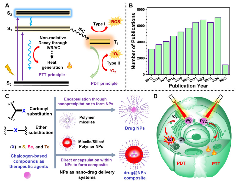
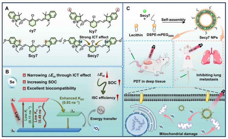
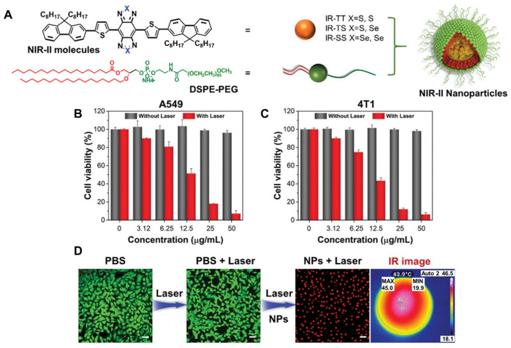
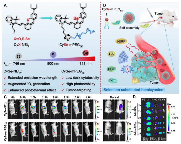
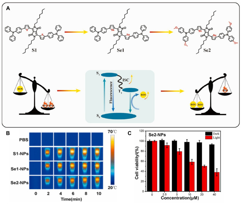
Chalcogen-containing therapeutic agents (TAs), which include sulfur (S), selenium (Se), and tellurium (Te) atoms, have recently emerged as a promising class of photosensitizers (PSs) and photothermal agents (PTAs) for cancer phototherapy. The incorporation of heavier chalcogens into organic chromophores leads to visible-to-near-infrared (VIS-NIR) light absorption, efficient triplet harvesting, and adequate heat and energy transfer efficiency, all of which are paramount for photodynamic therapy (PDT) and photothermal therapy (PTT). However, chalcogen-based PSs/PTAs suffer from photostability, bioavailability, and targeted delivery issues, which minimize their PDT/PTT performances. Nevertheless, significant progress in the rational design of nanoencapsulation strategies has been achieved to overcome the challenges of chalcogen-based TAs for effective phototherapeutic cancer treatment. This review highlights the recent advances (within the last five years) in nano-drug delivery approaches adapted for chalcogen-substituted PSs/PTAs for PDT, PTT, or synergistic PDT/PTT, integrating imaging and treatment. The PSs/PTAs described in this review are classified into three classes: (i) sulfur, (ii) selenium, and (iii) tellurium-containing TAs used in phototherapy applications. This review offers a comprehensive perspective on the design of chalcogen-substituted photosensitizers (PSs) and photothermal agents (PTAs), covering spectroscopic and computational characterization, nanoformulation strategies, and their roles in enhancing reactive oxygen species (ROS) generation and photothermal conversion efficiency for improved in vitro and in vivo performance. We hope this work will encourage further research into nanotechnological strategies designed to enhance the phototherapeutic efficacy of chalcogen-containing therapeutic agents.

Keywords: cancer therapy; chalcogen; nano-drug delivery; photodynamic therapy; photothermal therapy; reactive oxygen species; triplet harvesting.


Conflict of interest statement

The authors declare no conflicts of interest.

Figures

Scheme 1
Scheme 1
(A) A simplified Jablonski energy diagram representing the main photophysical and photochemical processes involved in PDT and PTT. ISC: intersystem crossing, IVR: intramolecular vibrational energy redistribution, and VC: vibrational cooling. (B) Increase in the number of publications from 2015 to present, obtained from the search for phototherapy (with keywords PDT and PTT) in the Web of Science portal (https://www.webofscience.com/wos/woscc/basic-search, accessed on 12 March 2025). (C) Illustration of the design and nanoformulation strategies for chalcogen–based conventional organic chromophores. (D) Schematic representation of the cell apoptosis process induced by PDT/PTT upon irradiation of nanoformulated TAs.
Figure 1
Figure 1
(A) Illustration of the preparation and applications of TSQ-NPs. (B) Fluorescence microscope image of TSQ-NPs on labeled KB cells showing subcellular accumulation of TSQ. The low and high magnification images of KB cells utilize blue excitation for Mito–Tracker green (green emission) and green excitation for TSQ (red emission). The scale bar indicates 20 μm. (C) TSQ-NPs induced an in vitro cytotoxic effect (20 μg/mL) against KB cells upon irradiation using red light (6 W, λ = 630 nm) for 5 min. * Data from ≥2 replicates. ns, not significant. Adapted with permission from Ref. [68]. Copyright 2024, American Chemical Society.
Figure 2
Figure 2
(A) Molecular structures of PDI−4CHA-X, where X = S. (B) Schematic representation of encapsulation strategy within silica nanocapsules (SNCs). (C,D) In vitro cell viability studies of 4T1 cancer cells incubated with PDI−4CHA−3S@SNCs at different concentrations with or without 808 nm and 1064 nm laser irradiation for 5 min (1.0 W cm−2). Adapted with permission from Ref. [69]. Copyright 2023, Elsevier.
Figure 3
Figure 3
Illustration of the synthetic pathways of SNP from ONP and the self-assembly process to produce SNP NPs. It further demonstrates the irradiation of SNP NPs using 690 nm laser light to produce ROS with photothermal effects. The figure also depicts the relaxation processes responsible for PDT (type–I and type–II) and PTT (heat generation) and outlines the key involved photophysical properties. Adapted with permission from Ref. [70]. Copyright 2025, Elsevier.
Figure 4
Figure 4
(A) Molecular structure of the Os–based PS rac–[Os(phen)2(IP–4T)](Cl)2 (ML18J03) and schematic representations of lipid formulations of ML18J03 using DSPE-mPEG2000 micelles (mic–ML18J03) and PEG–modified DPPC liposomes (lipo–ML18J03). (B) Min–max plots showing the interassay distribution of EC50 values of ML18J03 and lipid-formulated ML18J03 in SK–MEL–28 human melanoma cells upon irradiation using (i) Visible–light, (ii) Green–light, and (iii) Red–light, and (iv) Relationship between excitation light and statistical significance between variances demonstrating that the most reduced interassay variability is for visible–light excitation of lipid-formulated ML18J03. * Data from 2 replicates. ns, not significant. (C) Normalized transient absorption decays measured at 660 nm observation wavelength. Adapted with permission from Ref. [89]. Copyright 2022, John Wiley & Sons, Inc.
Figure 5
Figure 5
(A) Graphical representation of CR dye NP formation for tumor–targeting PTT. In vivo CR NP-mediated PTT. (B) IR thermal images, and (C) respective temperature changes at the tumor sites upon 808 nm (1.0 W cm−2) laser irradiation of Colon26 tumor-bearing mouse model treated with PBS or CR NPs at different time points (n = 5). (D) Tumor growth profiles of four groups during the treatment course. Data from ≥3 replicates. ns, not significant. *** p < 0.001. Adapted with permission under the terms of the CC-BY license from Ref. [92]. Copyright 2024, the authors, published by Springer Nature.
Figure 6
Figure 6
Schematic demonstration of π–bridge engineered NIR–II AIEgens with multimodal phototheranostic performance. (A) Illustration of π–bridge manipulation strategy for constructing NIR–II AIEgens. (B) Illustration of the nanofabrication process of BT-NS to form BT-NS NPs and its application in FLI/PAI/PTI trimodal imaging–guided synergistic phototherapy. (C) ROS production ability of BT-NS NPs (5 μM) upon 660 nm laser irradiation (0.3 W cm−2). (D) Temperature change in BT-NS NPs at varied concentration in aqueous solutions upon 660 nm laser irradiation (0.3 W cm−2). Adapted with permission from Ref. [93]. Copyright 2024, John Wiley & Sons, Inc.
Figure 7
Figure 7
(A) Illustration of type–I PDT processes of Se6-NPs and type–II PDT processes of chlorin e6 (Ce6)-NPs: Se6-NPs exhibited better PDT treatment than Ce6-NPs in either hypoxia or normoxia conditions by generating dual radicals (OH and O2•−). The proposed radical generation processes were also shown in the case of Se6-NPs in an aqueous solution (top right corner). (B) Chemical structures and NPs of Se6, Se5, and amphiphilic copolymer DSPE–PEG. Cell viability studies of 4T1 cells treated with (C) Se6-NPs and (D) Ce6-NPs at different concentrations upon 488 nm light irradiation (20 mW cm−2) for 30 min under normoxia or hypoxia conditions (n = 6, mean ± SD). (E) Picture showing the accumulation of Se6-NPs for more than 48 h in the tumor sites of a tail vein-injected tumor-bearing mouse model. Adapted under the terms of the CC–BY license from Ref. [100]. Copyright 2023, the authors, published by John Wiley & Sons, Inc.
Figure 8
Figure 8
(A) The chemical structure of the investigated cy7 derivatives. (B) Se facilitated relevant photophysical parameters to enhance the ISC process, leading to 1O2 generation. (C) Illustration of self–assembly process for Secy7 NPs production and demonstration of in vivo PDT in a mouse model. Reproduced with permission from Ref. [103]. Copyright 2024, John Wiley & Sons, Inc.
Figure 9
Figure 9
(A) Schematic illustration of the preparation process of the NIR–II NPs. Cytotoxicity of (B) A549 cells and (C) 4T1 cells incubated with IR–SS NPs of various concentrations with or without 1064 nm laser irradiation (1 W cm−2). (D) Live/dead confocal laser scanning images of A549 cells after multiple treatments. Green fluorescence calcein AM and red fluorescence PI represent live and dead cells, respectively. The scale bar represents 50 μm. Adapted with permission from Ref. [24]. Copyright 2020, John Wiley & Sons, Inc.
Figure 10
Figure 10
(A) Molecular structures of CyX–NEt2 (X = O, S, Se) and CySe–mPEG5K, designed through the copper(I)–catalyzed click reaction of CySe–NEt2 and a PEG chain. (B) Schematic demonstration of tumor-targeting imaging–guided multimodal phototherapy. (C) In vivo fluorescence imaging in tail vein–injected tumor-bearing mice treated with CySe–NEt2 and CySe–mPEG5K (100 mM, 100 mL in PBS buffer). (D) In vivo biodistribution fluorescence images of tail vein-injected tumor-bearing mice treated with CySe–NEt2 and CySemPEG5K for 1 h, 2 h, and 3 h. Adapted with permission under the terms of the CC–BY–NC license from the Ref. [107]. Copyright 2023, the authors, published by Royal Society of Chemistry.
Figure 11
Figure 11
(A) Illustration of a rational design of diketopyrrolopyrrole–based compounds for efficient phototherapy. (B) IR thermal images of the 5 μM S1-NPs, Se1-NPs, and Se2-NPs under 635 nm laser irradiation at 0.3 W cm−2 for different times. (C) Relative viability of A549 cells treated with Se2-NPs at various doses under 635 nm laser irradiation or not. Adapted with permission from Ref. [109]. Copyright 2024, Elsevier.
Figure 12
Figure 12
(A) Chemical structure of chalcogen atom–substituted CR dyes (CR-X, X = O, S, Se, and Te). (B) Absorption spectra of CRO, CRS, CRSe, and CRTe; (C) computed HOMO−LUMO energy gap of CRO, CRS, CRSe, and CRTe. (D) Nanoformulation strategy of CRTe. (E) Photothermal stability of CRTe NPs during four cycles of 1064 nm light irradiation. (F) Relative viability of 4T1 cells treated with various concentrations of CRTe NPs without or with 1064 nm laser irradiation (0.7 W cm−2). (G) Fluorescence microscope images of CRTe-NP-treated 4T1 cells co-stained with calcein–AM (green color, live cells) and propidium iodide (red color, dead cells). Adapted with permission from Ref. [27]. Copyright 2024, American Chemical Society.
Figure 13
Figure 13
(A) Schematic illustration of the preparation of PPy–Te NPs; 4T1 cell viability when treated with NPs II–VII. (B) Cell viability in the dark and (C) upon 808-nm laser irradiation. Adapted with permission from Ref. [125]. Copyright 2020, John Wiley & Sons, Inc.
Figure 14
Figure 14
(A) Schematic illustration of the preparation process of F127/BODIPY-X (X = O, S, Se, and Te) NPs. In vitro study of U87 cells exposed to F127/BODIPY-X and 660 nm laser irradiation for 10 min via DCFH–DA. (B) Fluorescence microscopy images, scale bar: 100 μm, and (C) flow cytometry results of intracellular ROS generation. (D) IR thermal images of mice bearing U87 tumor cells treated with PBS or F127/BODIPY-Te NPs under irradiation of a 660 nm laser (1.0 W cm−2). Adapted from Ref. [26]. Copyright 2024, Royal Society of Chemistry.

Similar articles

References

    1. Murray S.G., Hartley F.R. Coordination chemistry of thioethers, selenoethers, and telluroethers in transition-metal complexes. Chem. Rev. 1981;81:365–414. doi: 10.1021/cr00044a003. - DOI
    1. Ma Y.-Z., Yu L., Zhou Q., Fu W. Dinuclear ytterbium(iii) benzamidinate complexes with bridging s32−, se22− and te22− ligands: Synthesis, structure and magnetic properties. Dalton Trans. 2024;53:8118–8123. doi: 10.1039/D4DT00724G. - DOI - PubMed
    1. Kaur M., Rob A., Caton-Williams J., Huang Z. Biochalcogen Chemistry: The Biological Chemistry of Sulfur, Selenium, and Tellurium. Volume 1152. American Chemical Society; Washington, DC, USA: 2013. Biochemistry of nucleic acids functionalized with sulfur, selenium, and tellurium: Roles of the single-atom substitution; pp. 89–126.
    1. Yang S., Zhao G., Gao Y., Sun Y., Zhang G., Fan X., Li Y., Li Y. In-solution direct oxidative coupling for the integration of sulfur/selenium into DNA-encoded chemical libraries. Chem. Sci. 2022;13:2604–2613. doi: 10.1039/D1SC06268A. - DOI - PMC - PubMed
    1. Chen X., Li B. How nature incorporates sulfur and selenium into bioactive natural products. Curr. Opin. Chem. Biol. 2023;76:102377. doi: 10.1016/j.cbpa.2023.102377. - DOI - PMC - PubMed

LinkOut - more resources