. 2025 May 18;26(10):4827.
doi: 10.3390/ijms26104827.

Gap Junctional Interaction of Endothelial Progenitor Cells (EPC) with Endothelial Cells Induces Angiogenic Network Formation In Vitro

Affiliations

Gap Junctional Interaction of Endothelial Progenitor Cells (EPC) with Endothelial Cells Induces Angiogenic Network Formation In Vitro

Christina Buchberger et al. Int J Mol Sci. .

Abstract

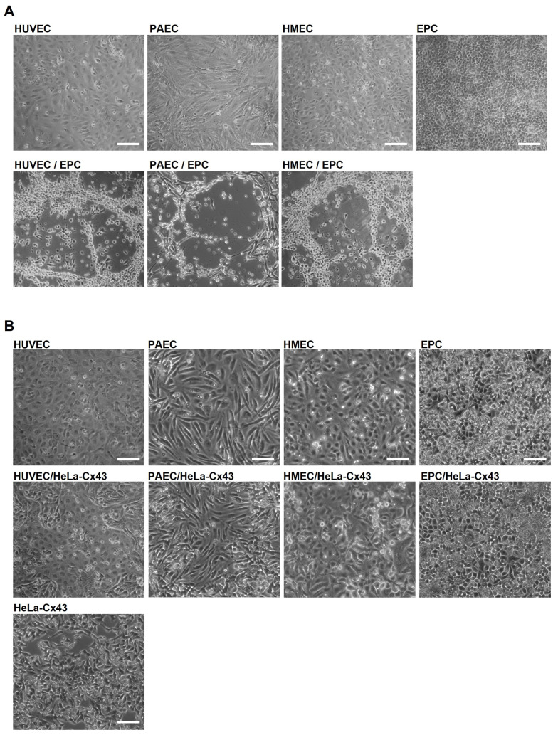
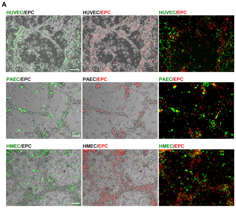
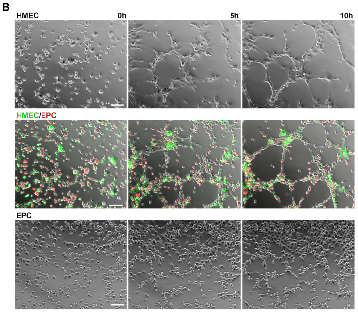
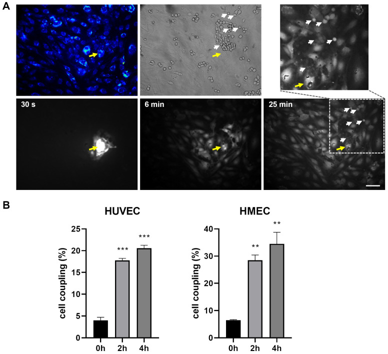
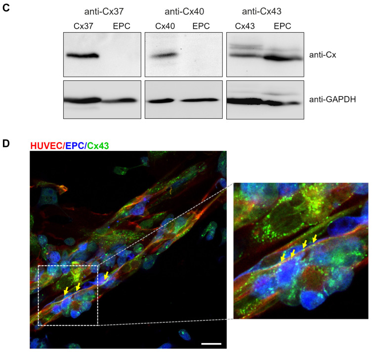
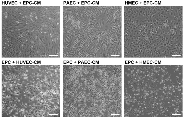
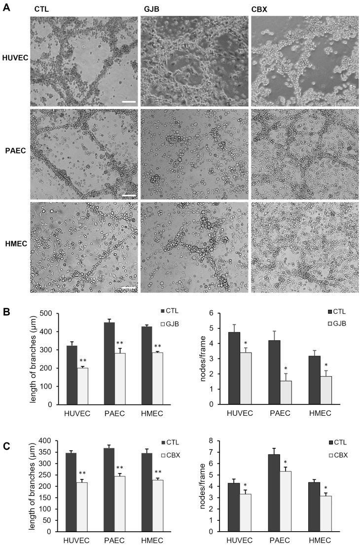
Endothelial progenitor cells (EPC) are considered to support neovascularization and endothelial repair by being incorporated into newly formed or injured vessels and by improving vascularization in a paracrine manner by secreting proangiogenic factors. Here, we studied the role of gap junctional communication between EPC and endothelial cells in long-term co-cultures in vitro. The cultivation of endothelial cells together with mouse embryonic EPC (E 7.5) induced the spontaneous formation of angiogenic networks after 3-6 days consisting of both cell types, but not in the respective monocultures, whereas their respective cultivation on a basement matrix induced the formation of tube-like structures, as expected. The angiogenic network formation could not be mimicked by the incubation of endothelial cells with supernatants of EPC only. We therefore hypothesized that direct interaction and cell-cell communication is required to induce the angiogenic network formation in co-cultures with endothelial cells. Expression analysis demonstrated expression of the gap junctional protein connexin 43 (Cx43) in EPC. Moreover, dye injection studies as well as FACS analysis identified gap junctional communication between endothelial cells and EPC. The inhibition of gap junctions by pharmacological blockers significantly reduced the angiogenic network formation, confirming that gap junctional communication between both cell types is required for this process.

Keywords: angiogenesis; connexin; endothelial cells; endothelial progenitor cells; gap junction.


Conflict of interest statement

The authors declare no conflicts of interest. The funders had no role in the design of the study; in the collection, analyses, or interpretation of data; in the writing of the manuscript; or in the decision to publish the results.

Figures

Figure 1
Figure 1
Co-cultivation of EC with EPC induces angiogenic network formation on uncoated cell dishes. (A) Representative images of HUVEC, PAEC and HMEC co-cultured with EPC or cultured as monolayers. EC/EPC co-cultivation induced an angiogenic network formation typically after 3–6 days (n = 7). Pictures were taken after 4 days. Scale bar: 100 µm. (B) Representative images of HUVEC, PAEC, HMEC and EPC cultivated as monolayers or co-cultured with HeLa-Cx43 and as control HeLa-Cx43 as monolayer. No angiogenic networks could be observed after 3–6 days in co-culture with HeLa-Cx43 cells. Representative pictures were taken after 4 days (n = 4). Scale bar: 100 µm.
Figure 2
Figure 2
Angiogenic networks are formed by both cell types. (A) Angiogenic networks were formed on uncoated dishes in co-cultures of EC with EPC. Representative images of co-cultures of HUVEC, PAEC or HMEC, pre-labeled with the fluorescence dye PKH67 (green), and EPC fluorescently labeled with PKH26 (red) after 6 days (n = 4–6). Scale bar: 100 µm. (B) Angiogenic tube formation of EC, EPC and EC/EPC co-cultures on an angiogenic basement matrix (Geltrex). Representative images of HMEC, EPC or HMEC in co-culture with EPC at different time points (0 h, 5 h, 10 h) are shown (n = 6). Scale bar: 100 µm.
Figure 2
Figure 2
Angiogenic networks are formed by both cell types. (A) Angiogenic networks were formed on uncoated dishes in co-cultures of EC with EPC. Representative images of co-cultures of HUVEC, PAEC or HMEC, pre-labeled with the fluorescence dye PKH67 (green), and EPC fluorescently labeled with PKH26 (red) after 6 days (n = 4–6). Scale bar: 100 µm. (B) Angiogenic tube formation of EC, EPC and EC/EPC co-cultures on an angiogenic basement matrix (Geltrex). Representative images of HMEC, EPC or HMEC in co-culture with EPC at different time points (0 h, 5 h, 10 h) are shown (n = 6). Scale bar: 100 µm.
Figure 3
Figure 3
EC communicate with EPC via functional gap junction channels containing Cx43. (A) Representative images demonstrating the gap junctional dye transfer from HUVEC to EPC. HUVEC monolayers were stained with the fluorescent dye CMTMR (blue; top left) and then co-cultivated with EPC (n = 3 independent cell cultures). After 28 h of cocultivation, the fluorescent gap junction permeable dye Alexa Fluor 488 was injected into a single HUVEC cell (yellow arrow, injected cell not visible in the transmission channel). The diffusion of the fluorescent dye into surrounding HUVEC and EPC via gap junctions after 6 and 25 min is shown. A magnified image section marked by dotted lines depicts the fluorescent dye propagation into EPC (white arrows) after 25 min. The transmission picture (top middle) shows the distribution of unstained EPC (white arrows) on the HUVEC monolayer. Scale bar: 100 µm. (B) Gap junctional coupling of EPC with EC: HUVEC (left) and HMEC (right). HUVEC or HMEC were stained with the gap junction permeable dye calcein (0.04 µM, green fluorescence) and co-cultivated with PKH26-labeled EPC (red fluorescence) for 2 h and 4 h. Subsequently, the amount of green fluorescence in red-labelled EPC, indicating the gap junctional transfer of calcein, was quantified by FACS analysis. Data are represented as percentage of cell coupling, n = 2 (0 h), n = 4 (2 h, 4 h) independent cell cultures, (***) p < 0.001, (**) p < 0.01 versus each time point. (C) Western blot analysis of the expression of Cx37, Cx40 and Cx43 in EPC. HeLa cells expressing Cx37, Cx40 or Cx43 were used as positive control. Equal loading was confirmed by detection of GAPDH. (D) Immunofluorescence stainings of Cx43 in HUVEC/EPC co-cultures after 6 days. To distinguish EPC from HUVEC, GFP-transfected EPC (blue) were used. The co-cultures were stained for the endothelial specific cell marker CD31 (red) to specifically label HUVEC. Cx43 (green) was stained with a polyclonal antibody against Cx43. The right image shows a magnified section of the left image (dotted line) showing the membrane localization of Cx43 between both cell types (yellow arrows). Scale bar: 20 µm.
Figure 3
Figure 3
EC communicate with EPC via functional gap junction channels containing Cx43. (A) Representative images demonstrating the gap junctional dye transfer from HUVEC to EPC. HUVEC monolayers were stained with the fluorescent dye CMTMR (blue; top left) and then co-cultivated with EPC (n = 3 independent cell cultures). After 28 h of cocultivation, the fluorescent gap junction permeable dye Alexa Fluor 488 was injected into a single HUVEC cell (yellow arrow, injected cell not visible in the transmission channel). The diffusion of the fluorescent dye into surrounding HUVEC and EPC via gap junctions after 6 and 25 min is shown. A magnified image section marked by dotted lines depicts the fluorescent dye propagation into EPC (white arrows) after 25 min. The transmission picture (top middle) shows the distribution of unstained EPC (white arrows) on the HUVEC monolayer. Scale bar: 100 µm. (B) Gap junctional coupling of EPC with EC: HUVEC (left) and HMEC (right). HUVEC or HMEC were stained with the gap junction permeable dye calcein (0.04 µM, green fluorescence) and co-cultivated with PKH26-labeled EPC (red fluorescence) for 2 h and 4 h. Subsequently, the amount of green fluorescence in red-labelled EPC, indicating the gap junctional transfer of calcein, was quantified by FACS analysis. Data are represented as percentage of cell coupling, n = 2 (0 h), n = 4 (2 h, 4 h) independent cell cultures, (***) p < 0.001, (**) p < 0.01 versus each time point. (C) Western blot analysis of the expression of Cx37, Cx40 and Cx43 in EPC. HeLa cells expressing Cx37, Cx40 or Cx43 were used as positive control. Equal loading was confirmed by detection of GAPDH. (D) Immunofluorescence stainings of Cx43 in HUVEC/EPC co-cultures after 6 days. To distinguish EPC from HUVEC, GFP-transfected EPC (blue) were used. The co-cultures were stained for the endothelial specific cell marker CD31 (red) to specifically label HUVEC. Cx43 (green) was stained with a polyclonal antibody against Cx43. The right image shows a magnified section of the left image (dotted line) showing the membrane localization of Cx43 between both cell types (yellow arrows). Scale bar: 20 µm.
Figure 4
Figure 4
The angiogenic network formation is not induced by the incubation of EC with conditioned media of EPC. Supernatants of EPC (EPC-CM) grown in endothelial cell culture medium for 48 h were used to cultivate HUVEC, PAEC or HMEC for 5 days. Vice versa, EPC were cultured in conditioned media of HUVEC (HUVEC-CM), PAEC (PAEC-CM) or HMEC (HMEC-CM) for 5 days. The conditioned medium was changed every 2 days. Representative images demonstrate that the angiogenic network formation is not induced by angiogenic growth factors in the conditioned medium secreted by EPC (n = 4 independent cell cultures). Scale bar: 100 µm.
Figure 5
Figure 5
Angiogenic network formation is reduced by the inhibition of gap junctions. (A) Representative images of co-cultures of HUVEC, PAEC or HMEC with EPC which were treated with different gap junction blockers (GJB, CBX) or with the solvents alone as mock control (CTL) for 6 days. Blockers (GJB: 1 mM heptanol and 2.5 µM meclofenamic acid; CBX: 10 µM carbenoxolone) were added every second day. Scale bar: 100 µm. (B,C) The pharmacological inhibition of gap junctions by the treatment with GJB (B) or CBX (C) significantly reduced the length of branches and the number of branching points (nodes/frame) in angiogenic networks of HUVEC/EPC, PAEC/EPC and HMEC/EPC co-cultures (HUVEC/EPC: GJB: n = 4; CBX: n = 4; PAEC/EPC: GJB: n = 6; CBX: n = 9; HMEC/EPC: GJB: n = 6; CBX: n = 7; * p < 0.05, ** p < 0.01; GJB or CBX vs. CTL with the same number of n, respectively).

Similar articles

References

    1. Ratliff B.B., Ghaly T., Brudnicki P., Yasuda K., Rajdev M., Bank M., Mares J., Hatzopoulos A.K., Goligorsky M.S. Endothelial Progenitors Encapsulated in Bioartificial Niches Are Insulated from Systemic Cytotoxicity and Are Angiogenesis Competent. Am. J. Physiol. Renal Physiol. 2010;299:F178–F186. doi: 10.1152/ajprenal.00102.2010. - DOI - PMC - PubMed
    1. Shin H.S., Thakore A., Tada Y., Pedroza A.J., Ikeda G., Chen I.Y., Chan D., Jaatinen K.J., Yajima S., Pfrender E.M., et al. Angiogenic Stem Cell Delivery Platform to Augment Post-Infarction Neovasculature and Reverse Ventricular Remodeling. Sci. Rep. 2022;12:17605. doi: 10.1038/s41598-022-21510-y. - DOI - PMC - PubMed
    1. Kupatt C., Horstkotte J., Vlastos G.A., Pfosser A., Lebherz C., Semisch M., Thalgott M., Buttner K., Browarzyk C., Mages J., et al. Embryonic Endothelial Progenitor Cells Expressing a Broad Range of Proangiogenic and Remodeling Factors Enhance Vascularization and Tissue Recovery in Acute and Chronic Ischemia. FASEB J. 2005;19:1576–1578. doi: 10.1096/fj.04-3282fje. - DOI - PubMed
    1. Vajkoczy P., Blum S., Lamparter M., Mailhammer R., Erber R., Engelhardt B., Vestweber D., Hatzopoulos A.K. Multistep Nature of Microvascular Recruitment of Ex Vivo-Expanded Embryonic Endothelial Progenitor Cells during Tumor Angiogenesis. J. Exp. Med. 2003;197:1755–1765. doi: 10.1084/jem.20021659. - DOI - PMC - PubMed
    1. Brandes R.P., Ushio-Fukai M. Endothelial Progenitor Cells=Epc=Elemental Pernicious Complexity. Antioxid. Redox Signal. 2011;15:911–914. doi: 10.1089/ars.2010.3803. - DOI - PMC - PubMed

LinkOut - more resources