. 2025 May 19;26(10):4852.
doi: 10.3390/ijms26104852.

Serum RNA Profile Reflects Fluid Status and Atrophic Retinal Changes in Neovascular Age-Related Macular Degeneration

Affiliations

Serum RNA Profile Reflects Fluid Status and Atrophic Retinal Changes in Neovascular Age-Related Macular Degeneration

Hanna Heloterä et al. Int J Mol Sci. .

Abstract

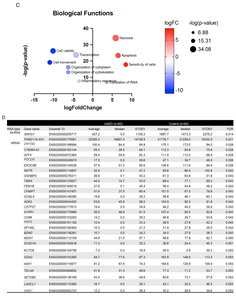
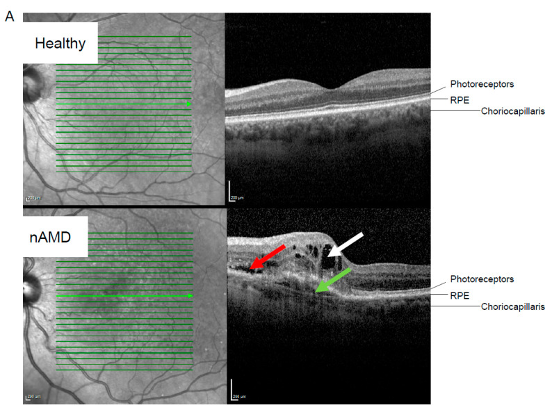
The increasing prevalence of age-related macular degeneration (AMD), a disease that can result in the loss of central vision, is an emerging problem worldwide due to aging societies. Growing patient numbers create a challenge for the healthcare system. Understanding the mechanisms of AMD pathogenesis will aid in early, personalized, and efficient intervention, helping to mitigate this issue. Current diagnostic methods rely on optical coherence tomography and angiography imaging, which identify existing damages, but do not provide information on the mechanisms behind them. In the present work, we demonstrate a difference in the serum RNA profile between neovascular AMD (nAMD) patients and controls. Moreover, the RNA profile of nAMD patients corresponded with anatomical changes in the retinal fluid compartments as well as atrophic changes of the retina. We followed two independent ways to control false positive leads, and when these approaches were combined, thioredoxin-related transmembrane protein 4 (TMX4) was observed to be differentially expressed by both approaches. This finding opens a new pathway in AMD studies, which are limited due to restricted access to live human target material and the limited value of animal models of human AMD.

Keywords: RNA sequencing; age-related macular degeneration; aging; differentially expressed RNAs; retina; serum RNA.


Conflict of interest statement

The authors declare no conflicts of interest.

Figures

Figure 1
Figure 1
Patient characteristics and mRNAs upregulated in nAMD samples. (A) Patient characteristics in the study population. (B) Most significantly altered biological functions between nAMD and control samples based on serum RNA analysis. Blue color indicates downregulation in nAMD and the stronger the color, the stronger the association. (C) Most significantly altered biological functions in pathway analysis. Blue indicates downregulation and orange color upregulation in nAMD. The color intensity indicates association strength. (D) RNAs significant after FDR adjustment. * One value is missing in the nAMD group. Abbreviations: nAMD, neovascular age-related macular degeneration; BMI, body mass index; FDR, false discovery rate.
Figure 1
Figure 1
Patient characteristics and mRNAs upregulated in nAMD samples. (A) Patient characteristics in the study population. (B) Most significantly altered biological functions between nAMD and control samples based on serum RNA analysis. Blue color indicates downregulation in nAMD and the stronger the color, the stronger the association. (C) Most significantly altered biological functions in pathway analysis. Blue indicates downregulation and orange color upregulation in nAMD. The color intensity indicates association strength. (D) RNAs significant after FDR adjustment. * One value is missing in the nAMD group. Abbreviations: nAMD, neovascular age-related macular degeneration; BMI, body mass index; FDR, false discovery rate.
Figure 2
Figure 2
RNA profile differences in the nAMD group based on retinal fluid status. (A) Representative optical tomography images from healthy and neovascular AMD (nAMD) cases. The white arrow indicates intraretinal fluid, the red arrow indicates subretinal fluid, and the green arrow indicates subretinal pigment epithelium (sub-RPE) fluid. (B) Altered RNAs in SRF vs. IRF analysis. Adjusted values from ANCOVA analysis are shown. (C) Highlights of the functions of the altered lncRNAs and encoded proteins by mRNAs. Abbreviations: nAMD, neovascular age-related macular degeneration; IRF, intraretinal fluid; SRF, subretinal fluid; lncRNA, long non-coding ribonucleic acid; mRNA, messenger ribonucleic acid; FTX, FTX transcript XIST regulator; NINJ2-AS1, NINJ2 antisense RNA 1; CRCP, CGRP receptor component; TUBGCP3, tubulin gamma complex component 3; CENPT, centromere protein T; TGM2, transglutaminase 2; ANGPT1, angiopoietin 1; TMX4, thioredoxin-related transmembrane protein 4; CEP162, centrosomal protein 162; CHMP6, charged multivesicular body protein 6; GPR65, G protein-coupled receptor 65; R3HDM1, R3H domain containing 1; STARD3, StAR-related lipid transfer domain containing 3; TMEM64, transmembrane protein 64; PREPL, prolyl endopeptidase-like; TAB3, TGF-beta activated kinase 1 (MAP3K7) binding protein 3; CDC14B, cell division cycle 14B; CDC42BPA, CDC42 binding protein kinase alpha; ARMCX3, armadillo repeat containing X-linked 3; CAMKK2, calcium/calmodulin-dependent protein kinase kinase 2; ZBTB37, zinc finger and BTB domain containing 37; SPATA13, spermatogenesis-associated 13 [29,30,31,32,33,34,35,36,37,38,39,40,41,42,43,44,45,46,47,48,49,50,51,52,53,54,55,56,57,58,59,60,61,62,63,64,65,66].
Figure 2
Figure 2
RNA profile differences in the nAMD group based on retinal fluid status. (A) Representative optical tomography images from healthy and neovascular AMD (nAMD) cases. The white arrow indicates intraretinal fluid, the red arrow indicates subretinal fluid, and the green arrow indicates subretinal pigment epithelium (sub-RPE) fluid. (B) Altered RNAs in SRF vs. IRF analysis. Adjusted values from ANCOVA analysis are shown. (C) Highlights of the functions of the altered lncRNAs and encoded proteins by mRNAs. Abbreviations: nAMD, neovascular age-related macular degeneration; IRF, intraretinal fluid; SRF, subretinal fluid; lncRNA, long non-coding ribonucleic acid; mRNA, messenger ribonucleic acid; FTX, FTX transcript XIST regulator; NINJ2-AS1, NINJ2 antisense RNA 1; CRCP, CGRP receptor component; TUBGCP3, tubulin gamma complex component 3; CENPT, centromere protein T; TGM2, transglutaminase 2; ANGPT1, angiopoietin 1; TMX4, thioredoxin-related transmembrane protein 4; CEP162, centrosomal protein 162; CHMP6, charged multivesicular body protein 6; GPR65, G protein-coupled receptor 65; R3HDM1, R3H domain containing 1; STARD3, StAR-related lipid transfer domain containing 3; TMEM64, transmembrane protein 64; PREPL, prolyl endopeptidase-like; TAB3, TGF-beta activated kinase 1 (MAP3K7) binding protein 3; CDC14B, cell division cycle 14B; CDC42BPA, CDC42 binding protein kinase alpha; ARMCX3, armadillo repeat containing X-linked 3; CAMKK2, calcium/calmodulin-dependent protein kinase kinase 2; ZBTB37, zinc finger and BTB domain containing 37; SPATA13, spermatogenesis-associated 13 [29,30,31,32,33,34,35,36,37,38,39,40,41,42,43,44,45,46,47,48,49,50,51,52,53,54,55,56,57,58,59,60,61,62,63,64,65,66].
Figure 3
Figure 3
RNA profile differences in the nAMD group during the atrophy analysis. (A) Optical coherent tomography images from healthy cases and various levels of atrophy. White arrows indicate outer retina damage, while the black arrow shows changes at the RPE level. Abbreviations: RPE = retinal pigment epithelium, iORA = incomplete outer retinal atrophy, cORA = complete outer retinal atrophy, iRORA = incomplete RPE and outer retinal atrophy, cRORA = complete RPE and outer retinal atrophy. (B) Altered RNAs in atrophy analysis. Adjusted values from the ANCOVA analysis are shown. (C) Highlights of the functions of the proteins differentially expressed by altered mRNAs. Abbreviations: nAMD, neovascular age-related macular degeneration; iORA, incomplete outer retinal atrophy; cORA, complete outer retinal atrophy cORA; iRORA, incomplete RPE and outer retinal atrophy; cRORA, complete RPE and outer retinal atrophy; CMTR1, cap methyltransferase 1; CD28, CD28 molecule; EIF6, eukaryotic translation initiation factor 6; CEBPG, CCAAT enhancer binding protein gamma; FLNB, filamin B; PIH1D1, PIH1 domain containing 1; DUSP16, dual specificity phosphatase 16; MIOS, meiosis regulator for oocyte development; RBM28, RNA binding motif protein 28; PPIL2, peptidylprolyl isomerase-like 2; EIF1AX, eukaryotic translation initiation factor 1A X-linked; ARHGEF7, Rho guanine nucleotide exchange factor 7 [16,67,68,69,70,71,72,73,74,75,76,77,78,79,80,81].
Figure 3
Figure 3
RNA profile differences in the nAMD group during the atrophy analysis. (A) Optical coherent tomography images from healthy cases and various levels of atrophy. White arrows indicate outer retina damage, while the black arrow shows changes at the RPE level. Abbreviations: RPE = retinal pigment epithelium, iORA = incomplete outer retinal atrophy, cORA = complete outer retinal atrophy, iRORA = incomplete RPE and outer retinal atrophy, cRORA = complete RPE and outer retinal atrophy. (B) Altered RNAs in atrophy analysis. Adjusted values from the ANCOVA analysis are shown. (C) Highlights of the functions of the proteins differentially expressed by altered mRNAs. Abbreviations: nAMD, neovascular age-related macular degeneration; iORA, incomplete outer retinal atrophy; cORA, complete outer retinal atrophy cORA; iRORA, incomplete RPE and outer retinal atrophy; cRORA, complete RPE and outer retinal atrophy; CMTR1, cap methyltransferase 1; CD28, CD28 molecule; EIF6, eukaryotic translation initiation factor 6; CEBPG, CCAAT enhancer binding protein gamma; FLNB, filamin B; PIH1D1, PIH1 domain containing 1; DUSP16, dual specificity phosphatase 16; MIOS, meiosis regulator for oocyte development; RBM28, RNA binding motif protein 28; PPIL2, peptidylprolyl isomerase-like 2; EIF1AX, eukaryotic translation initiation factor 1A X-linked; ARHGEF7, Rho guanine nucleotide exchange factor 7 [16,67,68,69,70,71,72,73,74,75,76,77,78,79,80,81].

Similar articles

References

    1. Kaarniranta K., Blasiak J., Liton P., Boulton M., Klionsky D.J., Sinha D. Autophagy in age-related macular degeneration. Autophagy. 2023;19:388–400. doi: 10.1080/15548627.2022.2069437. - DOI - PMC - PubMed
    1. Fleckenstein M., Schmitz-Valckenberg S., Chakravarthy U. Age-Related Macular Degeneration: A Review. JAMA. 2024;331:147–157. doi: 10.1001/jama.2023.26074. - DOI - PubMed
    1. Boopathiraj N., Wagner I.V., Dorairaj S.K., Miller D.D., Stewart M.W. Recent Updates on the Diagnosis and Management of Age-Related Macular Degeneration. Mayo Clin. Proc. Innov. Qual. Outcomes. 2024;8:364–374. doi: 10.1016/j.mayocpiqo.2024.05.003. - DOI - PMC - PubMed
    1. Helotera H., Kaarniranta K. A Linkage between Angiogenesis and Inflammation in Neovascular Age-Related Macular Degeneration. Cells. 2022;11:3453. doi: 10.3390/cells11213453. - DOI - PMC - PubMed
    1. Helotera H., Siintamo L., Kivinen N., Abrahamsson N., Aaltonen V., Kaarniranta K. Analysis of prognostic and predictive factors in neovascular age-related macular degeneration Kuopio cohort. Acta Ophthalmol. 2024;102:703–713. doi: 10.1111/aos.16681. - DOI - PubMed

LinkOut - more resources