. 2025 May 20;26(10):4892.
doi: 10.3390/ijms26104892.

Mass Spectrometric ITEM-FOUR Analysis Reveals Coding Single-Nucleotide Polymorphisms in Human Cardiac Troponin T That Evade Detection by Sandwich ELISAs Which Use Monoclonal Antibodies M7 and M11.7 from the Elecsys Troponin T® Assay

Affiliations

Mass Spectrometric ITEM-FOUR Analysis Reveals Coding Single-Nucleotide Polymorphisms in Human Cardiac Troponin T That Evade Detection by Sandwich ELISAs Which Use Monoclonal Antibodies M7 and M11.7 from the Elecsys Troponin T® Assay

Kristjan Kormann et al. Int J Mol Sci. .

Abstract

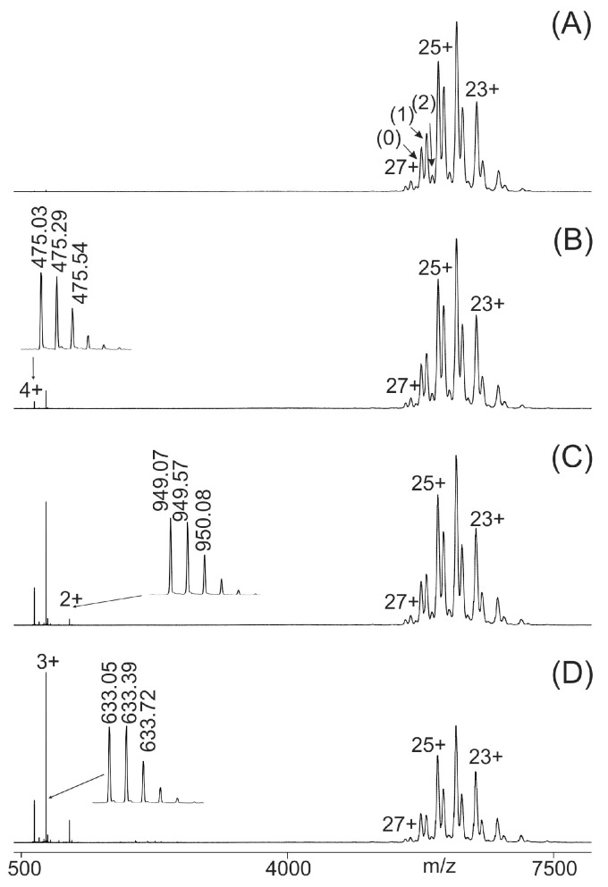
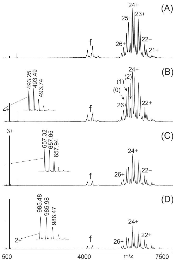
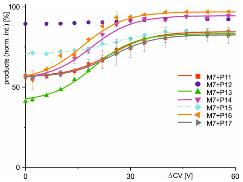
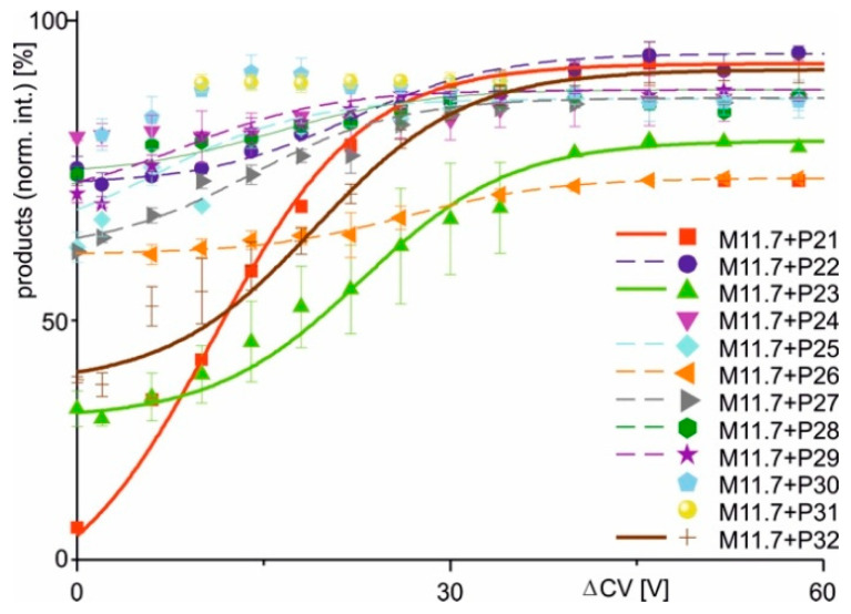
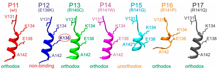
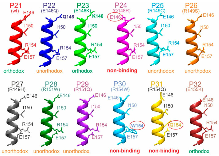
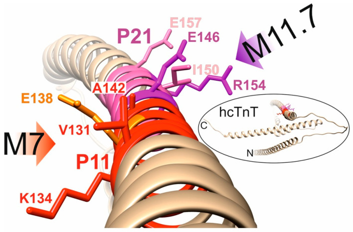
Immunoassays for cardiac troponin, such as the Elecsys® hs-TnT, have become the gold standard for myocardial infarction diagnostics. While various protein/chemical factors affecting the troponin complex and thus its diagnostic accuracy have been investigated, the role of coding single-nucleotide polymorphisms remains underexplored. To evaluate potential cSNP-induced interference with antibody binding in the Elecsys® hs-TnT immunoassay, we applied ITEM-FOUR, a mass spectrometry-based method that quantifies changes in antibody binding upon amino acid substitutions in epitope peptides. Candidate cSNPs were selected from the dbSNP database and were mapped to human cardiac troponin T by molecular modeling. Consuming micromolar antibody concentrations and microliter sample volumes, two wild-type and 17 cSNP-derived variant epitope peptides-six for monoclonal antibody M7 and eleven for monoclonal antibody M11.7-were investigated to reveal the binding motifs "V131-K134-E138-A142" for M7 and "E146-I150-R154-E157" for M11.7. Loss of binding to M11.7 was observed for substitutions Q148R (rs730880232), R154W (rs483352832), and R154Q (rs745632066), whereas the E138K (rs730881100) exchange disrupted binding of M7. Except for cSNP Q148R, they are associated with cardiomyopathies, placing affected individuals at risk of both underlying heart disease and false-negative hs-TnT assay results in cases of myocardial infarction. Our results highlight the need to account for cSNP-related interferences in antibody-based diagnostics. ITEM-FOUR offers a powerful approach for tackling this challenge, fostering next-generation assay development.

Keywords: ITEM-FOUR; human troponin T; immune complex analysis; myocardial infarction; nano-ESI mass spectrometry; single-amino-acid polymorphism; single-nucleotide polymorphism.


Conflict of interest statement

The authors declare no conflicts of interest.

Figures

Figure 1
Figure 1
Nano-ESI mass spectra of peptide 11 (LVSLKDRIERRRAER)–M7 antibody mixtures with increasing collision cell voltage differences (ΔCV). (A): 2 V, (B): 14 V, (C): 30 V, (D): 52 V. Charge states are given for the ion signals (right ion series) of the antibody (0) and the immune complexes (antibody plus one peptide (1) and antibody plus two peptides (2)). Charge states for peptide ion signals are given on the left. The inlets in (BD) show zoom views of the isotopically resolved peptide ion signals and their m/z values. The quadrupole was set to block transmission of ions < m/z 3850. The molar ratio of peptide to antibody was 2.1:1. Solvent: 200 mM ammonium acetate, pH 6.7.
Figure 2
Figure 2
Nano-ESI mass spectra of peptide 21 (AEQQRIRNEREKERQ)–M11.7 antibody mixtures with increasing collision cell voltage differences (ΔCV). (A): 2 V, (B): 14 V, (C): 30 V, (D): 52 V. Charge states are given for the ion signals (right ion series) of the antibody (0) and the immune complexes (antibody plus one peptide (1) and antibody plus two peptides (2)). Charge states for peptide ion signals are given on the left. The inlets in (B), (C), and (D) show zoom views of the isotopically resolved peptide ion signals and their m/z values. Multiply charged antibody fragment ions are labeled f. The quadrupole was set to block transmission of ions < m/z 3850. The molar ratio of peptide to antibody was 13: 1. Solvent: 200 mM ammonium acetate, pH 6.7.
Figure 3
Figure 3
Courses of normalized product ion intensities of M7 antibody–epitope peptide complexes as functions of collision cell voltage differences (ΔCV). The immune complex dissociations with peptides P11 (red square), P12 (violet dot), P13 (green triangle), P14 (violet triangle), P15 (light blue diamond), P16 (orange triangle), and P17 (gray triangle) are shown. Each data point is the mean of two independent measurements (see Table S3). Vertical bars indicate standard deviations. The sigmoidal shaped curves were fitted using a Boltzmann function. Solid lines indicate orthodox binding. Dotted lines indicate unorthodox binding. Curve parameters are given in Table 2.
Figure 4
Figure 4
Courses of normalized product ion intensities of M11.7 antibody–epitope peptide complexes as functions of collision cell voltage differences (ΔCV). The immune complex dissociations with peptides P21 (red square), P22 (violet dot), P23 (green triangle), P24 (violet triangle), P25 (light blue diamond), P26 (orange triangle), P27 (gray triangle), P28 (green hexagon), P29 (purple star), P30 (cyan pentagon), P31 (yellow dot), and P32 (brown cross) are shown. Each data point is the mean of two independent measurements (see Table S4). Vertical bars indicate standard deviations. The sigmoidal shaped curves were fitted using a Boltzmann function. Solid lines indicate orthodox binding. Dotted lines indicate unorthodox binding. Curve parameters are given in Table 2.
Figure 5
Figure 5
Ribbon cartoons of M7 epitope peptide backbone structure models. Alpha helices of P11 to P17 are shown. Selected amino acid residues are shown (stick model) and labeled (single-letter code). Wild-type (wt) and amino acid exchanges (point mutations) are indicated in parentheses. Amino acid numbering as in the full-length hcTnT protein. Binding modes with the M7 antibody are given at the bottom. Amino acid residues that prevent binding are circled. The “V-K-E-A” binding motif amino acid residues are shown.
Figure 6
Figure 6
Ribbon cartoons of M11.7 epitope peptide backbone structure models. Alpha helices of P21 to P32 are shown. Selected amino acid residues are shown (stick models) and labeled (single-letter code). Wild-type (wt) and amino acid exchanges (point mutations) are indicated in parentheses. Amino acid numbering as in the full-length hcTnT protein. Binding modes with the M11.7 antibody are given at the bottom. Amino acid residues that prevent binding are circled. The “E-I-R-E” binding motif amino acid residues are shown.
Figure 7
Figure 7
Ribbon cartoon of the hcTnT backbone structure model. The alpha helix that contains the M7 (red) and the M11.7 (pink) epitopes is shown. Selected amino acid residues are shown (stick models) and labeled (single-letter code). Amino acid residues E138 (orange), E146 (purple) and R154 (purple) are required for antibody binding. The orientations of antibody docking are indicated with red (M7) and purple (M11.7) arrows. A ribbon structure model of full-length hcTnT (UniProt accession no. P45379) is shown in the insert (circled). N- and C-termini are labeled.

Similar articles

References

    1. Dwivedi S., Purohit P., Misra R., Pareek P., Goel A., Khattri S., Pant K.K., Misra S., Sharma P. Diseases and Molecular Diagnostics: A Step Closer to Precision Medicine. Indian J. Clin. Biochem. 2017;32:374–398. doi: 10.1007/s12291-017-0688-8. - DOI - PMC - PubMed
    1. Jeremias A. The utility of troponin measurement to detect myocardial infarction: Review of the current findings. Vasc. Health Risk Manag. 2010;6:691–699. doi: 10.2147/VHRM.S5306. - DOI - PMC - PubMed
    1. Mingels A.M., Mills N.L., Mueller C. Cardiac troponin T and I: Back to basics. Eur. Heart J. Acute Cardiovasc. Care. 2023;12:631–632. doi: 10.1093/ehjacc/zuad084. - DOI - PMC - PubMed
    1. Mohammed A.A., Januzzi J.L., Jr. Clinical applications of highly sensitive troponin assays. Cardiol. Rev. 2010;18:12–19. doi: 10.1097/CRD.0b013e3181c42f96. - DOI - PubMed
    1. Mair J., Hammarsten O. Potential analytical interferences in cardiac troponin immunoassays. J. Lab. Precis. Med. 2023;8:12. doi: 10.21037/jlpm-22-65. - DOI

LinkOut - more resources