. 2025 May 21;26(10):4957.
doi: 10.3390/ijms26104957.

Single Cell RNA Sequencing of Papillary Cancer Mesenchymal Stem/Stromal Cells Reveals a Transcriptional Profile That Supports a Role for These Cells in Cancer Progression

Affiliations

Single Cell RNA Sequencing of Papillary Cancer Mesenchymal Stem/Stromal Cells Reveals a Transcriptional Profile That Supports a Role for These Cells in Cancer Progression

Danny Jandu et al. Int J Mol Sci. .

Abstract

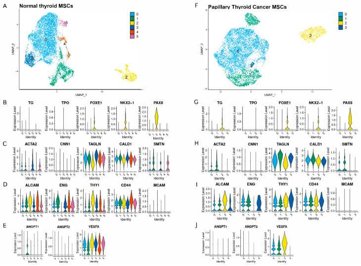
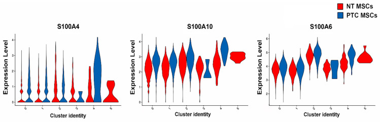
Papillary thyroid cancer (PTC) contains mesenchymal stem/stromal cells (MSCs), but their contribution to PTC progression is not clear. In this study, we compared the transcriptional signatures of normal thyroid (NT) and PTC-derived MSCs with the aim of determining if these have distinct transcriptomes that might influence PTC progression. We used flow cytometry in combination with a panel of MSC clusters of differentiation (CD) markers and showed that both thyroid MSC populations expressed MSC markers and lacked expression of markers not normally expressed by MSCs. In addition, we determined that both MSC populations could differentiate to adipocytes and osteocytes. Analysis of single cell RNA sequencing data from both MSC populations revealed, regardless of tissue of origin, that both contained similar numbers of subpopulations. Cluster analysis revealed similarity in expression of both MSC populations for stromal markers, the vascular marker VEGFA and the smooth muscle marker CALD1, while smaller subpopulations expressed markers of more lineage-committed thyroid cells. PTC MSCs also showed upregulated expression of 28 genes, many of which are known to be involved in epithelial-mesenchymal transition (EMT) and/or disease progression in several types of cancers, including but not limited to breast cancer, gastric cancer, cervical carcinoma, bladder cancer and thyroid cancer. This included several members of the S100 and IGFBP gene families. Taken together, these data support a role for PTC MSCs in PTC progression.

Keywords: mesenchymal stem/stromal cells; normal thyroid; papillary thyroid cancer; single cell RNA sequencing.


Conflict of interest statement

The authors declare no conflicts of interest.

Figures

Figure 1
Figure 1
Analysis of mesenchymal stromal cells derived from normal thyroid and papillary thyroid cancer. Representative images were taken from three biological replicates for each tissue type, n = 3. Brightfield images show the characteristic MSC morphology displayed by NT MSCs (A) and PTC MSCs (B). Scale bars = 100 µM. Results of histological analysis are shown; note that LMSCs were used as a positive control for both osteogenic (C) and adipogenic differentiation (D). Both thyroid MSC populations differentiated to the osteogenic and adipogenic lineages as shown for NT MSCs (E,F) and for the PTC MSCs (G,H). Note that Alizarin red staining of all three MSC populations, indicating matrix mineralization, occurred, indicating successful osteogenesis (C,E,G) and Oil Red O staining of lipid vesicles within all cell populations differentiated to adipocytes (D,F,H). Further confirmation of differentiation to osteogenic and adipogenic lineages by both thyroid MSC populations was shown using semi-quantitative PCR to confirm mRNA expression of genes associated with these differentiations; these included for osteogenesis ALP (187 bp) and BSP (161 bp) and for adipogenic differentiation aP2 (130 bp). GapdH was used as a loading control (150 bp) (I). Graphical representation of immunophenotyping results for both thyroid cell populations showing expression of known MSC markers (J) and for expression of markers normally expressed at low/no levels by MSCs (K). Note the significant difference in expression of CD146 in the NT MSCs above that of the PTC MSCs * p = <0.05.
Figure 2
Figure 2
Annotation of the MSCs subpopulations using integrated single cell RNA sequencing. (A) Identification of MSC clusters for NT MSCs (F) and for PTC MSCs. n = 2 patients MSCs for both tissue types were used for clustering analysis; profiles are shown of combined data from 2 cultures of each tissue type. UMAPs represent a clustering analysis of 8000 cells at a resolution of 0.1; this resulted in 6 clusters in the MSCs from NT and 4 from PTC (A,F). Violin plots show NT MSCs (B) and PTC MSCs (G) expressing thyroid lineage-committed cell markers, NT MSCs (C) and PTC MSCs (H) expressing smooth muscle markers, NT MSCs (D) and PTC MSCs (I) expressing stromal markers and NT MSCs (E) and PTC MSCs (J) expressing angiogenic markers.
Figure 3
Figure 3
Results of cluster analysis of PTC and NT MSCs. UMAP showing clustering of cells from two NT MSCs cultures and two PTC MSC cultures (A). UMAP showing annotation of clusters for both cell types (C). Data represent n = 2 donors for each tissue type. Graphical representation (using the combined dataset) to determine percentage of PTC and NT MSCs per cluster (B). Note the presence of a very small percentage of PTC MSCs in cluster 3, also note the absence of these cells in cluster 5.
Figure 4
Figure 4
Violin plot representing expression of S100A genes in PTC and NT MSCs per cluster (data used were from two PTC MSC and two NT MSC pooled samples).
Figure 5
Figure 5
Violin plots representing expression of IGFBP genes in PTC and NT MSCs per cluster (data used were from two PTC MSC and two NT MSC pooled samples).

References

    1. Papaccio F., Paino F., Regad T., Papaccio G., Desiderio V., Tirino V. Concise Review: Cancer Cells, Cancer Stem Cells, and Mesenchymal Stem Cells: Influence in Cancer Development. Stem Cells Transl. Med. 2017;6:2115–2125. doi: 10.1002/sctm.17-0138. - DOI - PMC - PubMed
    1. Karnik I., Sutherland R., Elson J., Aspinall S., Meeson A. TGF-β, to target or not to target; to prevent thyroid cancer progression? BBA-Rev. Cancer. 2022;1877:4. doi: 10.1016/j.bbcan.2022.188752. - DOI - PubMed
    1. Martin F.T., Dwyer R.M., Kelly J., Khan S., Murphy J.M., Curran C., Miller N., Hennessy E., Dockery P., Barry F.P., et al. Potential role of mesenchymal stem cells (MSCs) in the breast tumour microenvironment: Stimulation of epithelial to mesenchymal transition (EMT) Breast Cancer Res. Treat. 2010;124:317–326. doi: 10.1007/s10549-010-0734-1. - DOI - PubMed
    1. Takigawa H., Kitadai Y., Shinagawa K., Yuge R., Higashi Y., Tanaka S., Yasui W., Chayama K. Mesenchymal Stem Cells Induce Epithelial to Mesenchymal Transition in Colon Cancer Cells through Direct Cell-to-Cell Contact. Neoplasia. 2017;19:429–438. doi: 10.1016/j.neo.2017.02.010. - DOI - PMC - PubMed
    1. Parascandolo A., Rappa F., Cappello F., Kim J., Cantu D.A., Chen H., Mazzoccoli G., Hematti P., Castellone M.D., Salvatore M., et al. Extracellular Superoxide Dismutase Expression in Papillary Thyroid Cancer Mesenchymal Stem/Stromal Cells Modulates Cancer Cell Growth and Migration. Sci. Rep. 2017;7:41416. doi: 10.1038/srep41416. - DOI - PMC - PubMed

MeSH terms

Substances