. 2025 May 15;15(5):791.
doi: 10.3390/life15050791.

New Perspectives on the Organization of Living Tissue and the Ongoing Connective Tissue/Fascia Nomenclature Debate, as Revealed by Intra-Tissue Endoscopy That Provides Real-Time Images During Surgical Procedures

Affiliations

New Perspectives on the Organization of Living Tissue and the Ongoing Connective Tissue/Fascia Nomenclature Debate, as Revealed by Intra-Tissue Endoscopy That Provides Real-Time Images During Surgical Procedures

Jean Claude Guimberteau et al. Life (Basel). .

Abstract

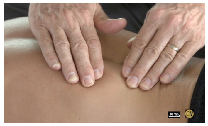
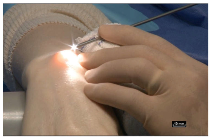
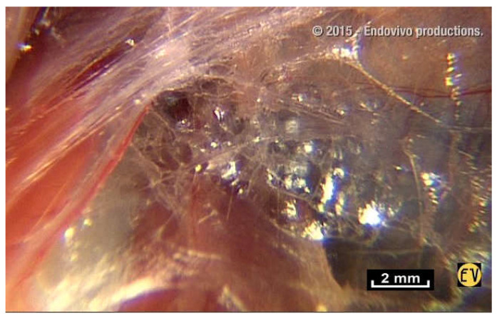
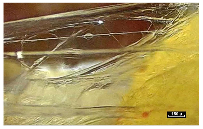
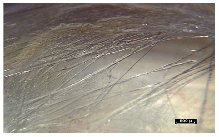
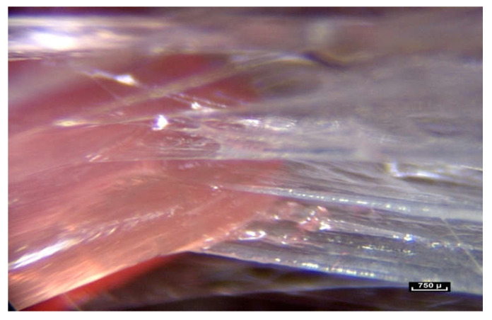
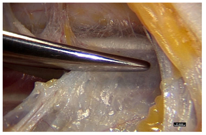
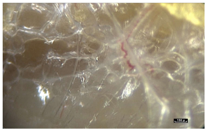
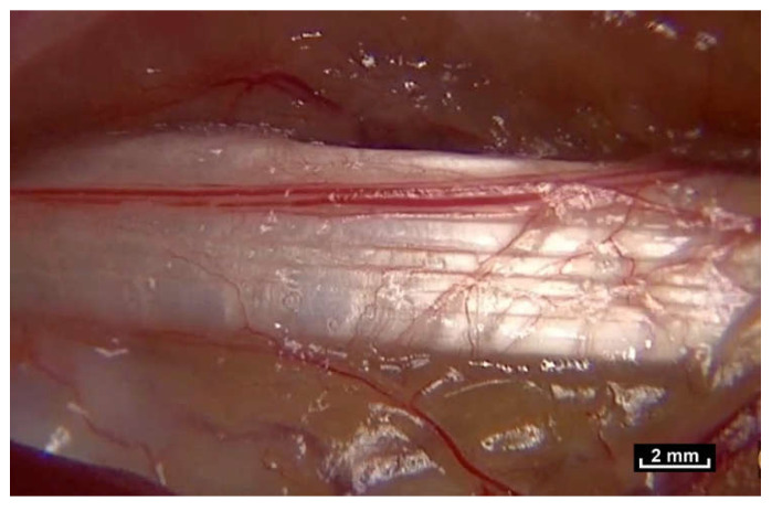
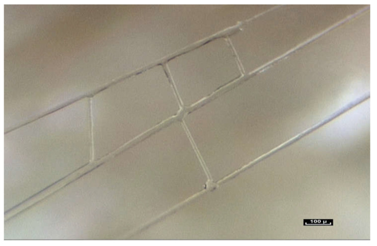
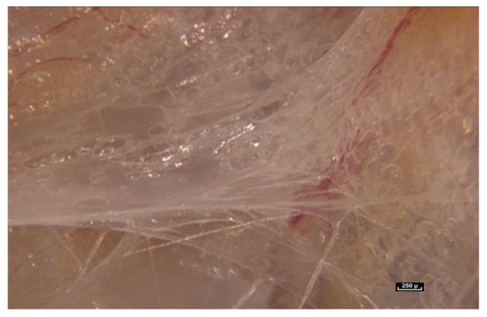
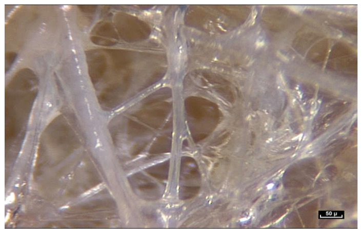
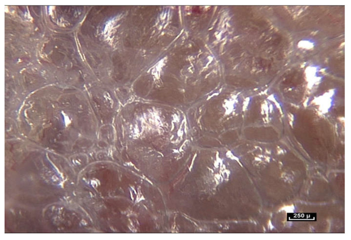
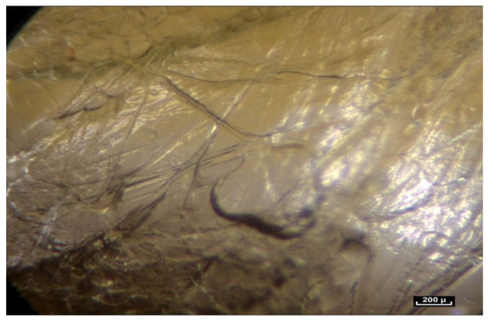
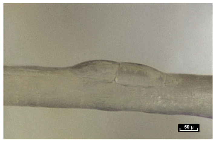
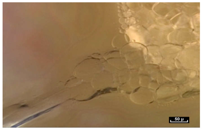
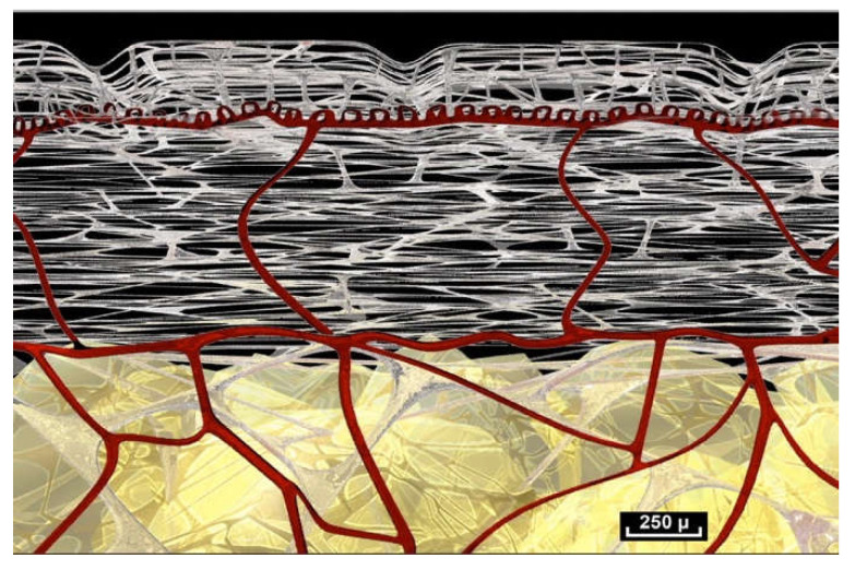
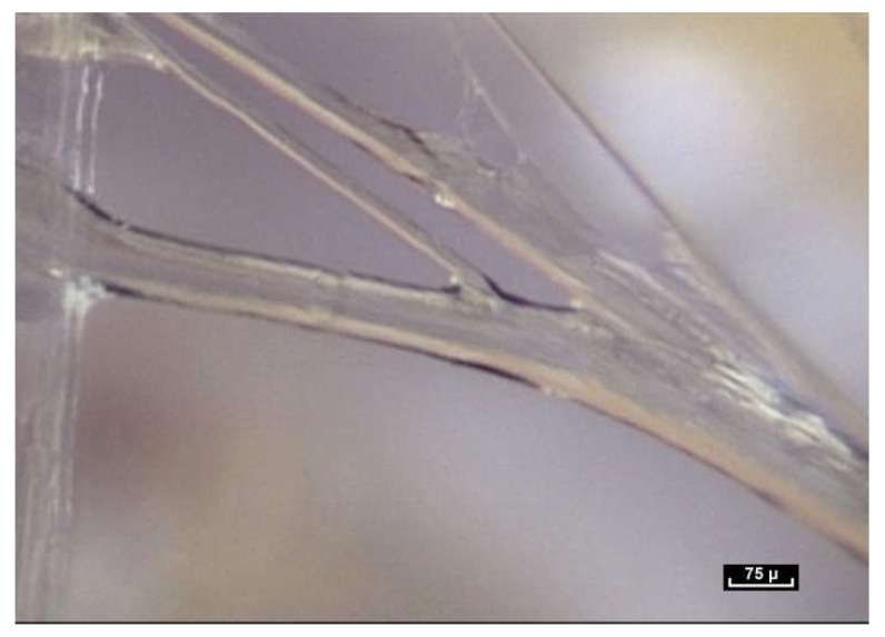
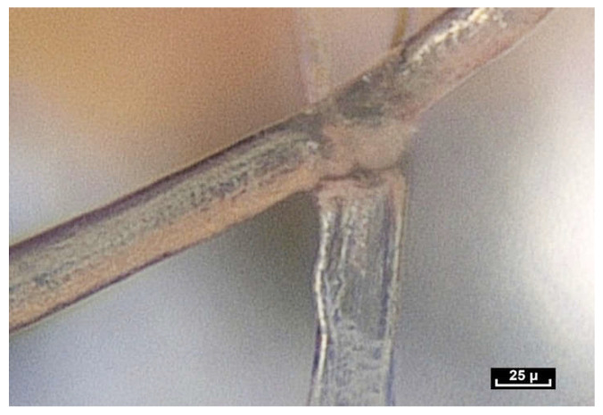
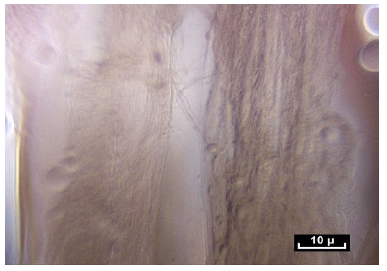
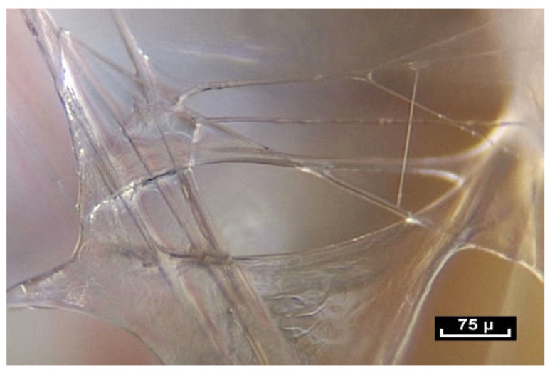
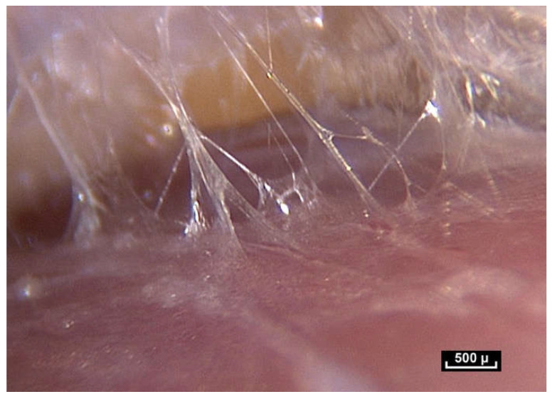
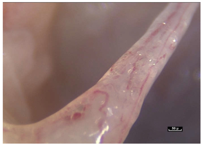
Intra-tissue endoscopy, providing real-time images at all scales, from macroscopic to microscopic, from inside living tissue during surgical procedures, has revealed the existence of a body-wide fibrillar architecture that extends from the surface of the skin to the cell. Different types of cells are housed within this fibrillar architecture and gather together to carry out specific functions. This challenges the commonly accepted notion of the organization of living matter that associates separate organs with connective tissue packaging. We are thus confronted with the global nature of the living human body and its vital processes. This paper sets out to describe the architecture of this fibrillar network which could be assimilated with the fascial tissue and which attributes a more constitutive role to connective tissue. It also demonstrates how movements within this fibrillar network can occur with minimal local distortion while maintaining tissue continuity. The authors propose that the gliding of tissues can be explained by the existence of a highly adaptable fibrillar network that enables the gliding of distinct anatomical structures such as tendons and muscles, without any dynamic influence on the surrounding tissues. The authors propose a new model of tissue movement based on the observation of a ubiquitous dynamic polyhedric fibrillar network with an apparently dispersed and complex pattern of organization, that forms fluid-filled microvolumes, and is found everywhere in the human body. Furthermore, this fibrillar network appears to act as a force absorption system, in addition to providing a framework or scaffolding for cells throughout the body. Observation during intra-tissue endoscopy suggests that this fundamental architectural organization extends into the extracellular matrix that is the natural environment of all cells in the living body, regardless of their size, location or specific function.

Keywords: cell microenvironment; connective tissue; fascia; fibrillar network; force absorption system; intra-tissue endoscopy; microvolumes.


Conflict of interest statement

The authors declare no conflict of interest.

Figures

Figure 1
Figure 1
(Video S1) After massage, the skin always returns to the same place without any modification—there is a real tissue memory.
Figure 2
Figure 2
(Video S2) Use of the endoscope during surgery. Viewing the video in real time on a screen in the operating theatre during surgical endoscopy.
Figure 3
Figure 3
(Video S3) There are no empty spaces in the body. All available space is occupied. A world of fibers. Fibers are everywhere, in every nook and cranny. There is no apparent order. ×20.
Figure 4
Figure 4
(Video S4) (×70 magnification) Fibers penetrate the fatty lobule and are dispersed between the cells. They influence the arrangement of the adipocytes inside the lobules and determine their shapes.
Figure 5
Figure 5
(Video S5) (×10 magnification) Muscular aponeurosis are simply densified areas of the same fibrillar network. However, their structure is in an irregular pattern and is different because of their different functional roles.
Figure 6
Figure 6
(Video S6) (×20 magnification) Perimysial fibers connecting muscle fascicles. The network of fibrils surrounds and penetrates the muscle.
Figure 7
Figure 7
(Video S7) During section of an aponeurosis, the edges retract like an elastic band, evidence of dynamic internal pressure, even at rest. This cannot be observed in a cadaver.
Figure 8
Figure 8
(Video S8) Sharp traction on this fibrillar network causes curious movements to occur due to the bursting of microvolumes at atmospheric pressure, in the operating theatre demonstrating the existence of hydraulic systems under different levels of pressure.
Figure 9
Figure 9
(Video S9) Some vessels do move with the tendon, but others are slower. There is no apparent synchronization or coherence and it can be seen that vessels move at different speeds, with a physical link between the vessels, but this is not a direct link. Once again, movement is irregular and nonlinear. There appears to be some sort of force absorption system with the surrounding tissues.
Figure 10
Figure 10
(Video S10) The fibers display three basic movements: Gliding, then dividing and then lengthening, all within a few tenths of a second. One fiber at first appearing to glide over the other fiber and then dividing into two, and finally another one lengthens. Animated diagram to illustrate this.
Figure 11
Figure 11
(Video S11) When tension is applied the fibers are stretched as the force increases. The adjacent linked element is subjected to tension. The fibers become more aligned in the direction of the applied force. The constraint is gradually diffused, dispersed and absorbed by the fibrillar network due to the capacity of the fibers to distend, divide and glide along each other.
Figure 12
Figure 12
(Video S12) (×100 magnification) The fibers are arranged in a completely disorderly fashion. They may have multiform connections that are very dense and irregular. They divide into smaller-diameter fibrils of a few microns in diameter with extremely variable lengths of 20 to 100 microns and of irregular thickness, giving a disordered and chaotic appearance.
Figure 13
Figure 13
(Video S13) (×40 magnification) As we move the endoscope closer to these areas the light emitted by the endoscope is reflected from the glistening facets of the microvolumes, that resemble a pile of mirrors dumped arbitrarily in a heap. These forms that we see are polyhedral, irregular microvolumes but the physical status of a microvolume is itself unstable. Animated diagram to illustrate.
Figure 14
Figure 14
(Video S14) (×60 magnification). There is no doubt that the position of cells in space is determined by this interwoven latticework of intersecting fibrils. The cells seem to be nestled in this network and to be entirely part of it. This would appear to be evidence that a cell cannot exist outside this fibrillar network. Animated diagram to illustrate.
Figure 15
Figure 15
(Video S15) Along the fibers, cells are found either in pairs, like ladybirds on a blade of grass or in small groups.
Figure 16
Figure 16
(Video S16) The fibers penetrate the cell groups and are therefore interconnected. The fibers seem to be swallowed up by the cell groups via the intercellular membranes, and this fusional relationship between the fiber and the cell is obvious.
Figure 17
Figure 17
(Video S17) Global diagram of the fibrillar intra-tissue organization showing the fibrillar network at different levels.
Figure 18
Figure 18
(Video S18) The migration of a fiber along other fibers. These junctions are mobile. One of the two fibers glides along the other one. In this way, energy is dispersed and absorbed throughout the fibrillar network. As soon as movement begins the fibrils respond by stretching out and lengthening. This ability to lengthen can be clearly seen.
Figure 19
Figure 19
(Video S19) In this case, initially, there are 3 fibers, then 4, then 2, but it is impossible to predict or anticipate the movements. Here, one can see 2 large fibers. One of the two vertical fibers divides into three sub-fibrils, and you might think that one of the three fibers is going to take part in the action. But finally, it is a 4th fiber that is not initially visible, not predictable, that will interact.
Figure 20
Figure 20
(Video S20) Sometimes, fibers are fixed and seem to stabilize the framework.
Figure 21
Figure 21
(Video S21) (×120 magnification) It would seem that these movements of division and gliding could occur in distinct ”separation zones”. This raises the question of morphological determinism, i.e., could these zones be predetermined or not?
Figure 22
Figure 22
(Video S22) No movement of any fiber can be anticipated or predicted, as can be seen in this case. The dynamic potential of the combination of these three mechanical solutions is incalculable. The combined action of these three distinct, yet closely related, types of fibrillar behavior enables the fibrillar network to adapt to the constraint in all three dimensions, while at the same time dispersing and reducing the force of the constraint and preserving the capacity of the structures to return to their resting positions. Animated diagram to illustrate.
Figure 23
Figure 23
(Video S23) This set of trajectories and potentialities means that the mechanical solution is totally unpredictable and non-determined. The conclusion, illustrated by these images, is that a gesture cannot be repeated identically, in terms of its fibrillar execution. This is impossible because the interplay of the fibers will inevitably be different and so we can put forward the hypothesis that the gesture is unique in time but also in space.
Figure 24
Figure 24
(Video S24) This same architecture is used either for mobility or as a cellular habitat. Sometimes you can even see the two combined, as in this case, where a fiber provides shape, mobility, flow of nutrients and cell housing.

Similar articles

Cited by

References

    1. Pratt R.L. Hyaluronan and the Fascial Frontier. Int. J. Mol. Sci. 2021;22:6845. doi: 10.3390/ijms22136845. - DOI - PMC - PubMed
    1. Fede C., Angelini A., Stern R., Macchi V., Porzionato A., Ruggieri P., De Caro R., Stecco C. Quantification of hyaluronan in human fasciae: Variations with function and anatomical site. J. Anat. 2018;233:552–556. doi: 10.1111/joa.12866. - DOI - PMC - PubMed
    1. Fede C., Clair C., Pirri C., Petrelli L., Zhao X., Sun Y., Macchi V., Stecco C. The Human Superficial Fascia: A Narrative Review. Int. J. Mol. Sci. 2025;26:1289. doi: 10.3390/ijms26031289. - DOI - PMC - PubMed
    1. Myers T.W. Anatomy Trains: Myofascial Meridians for Manual Therapists and Movement Professionals. Elsevier; Amsterdam, The Netherlands: 2021.
    1. Characteristics of Connective Tissue. LibreTexts. [(accessed on 18 July 2018)]. Available online: https://med.libretexts.org/@go/page/7357?pdf.

LinkOut - more resources