. 2025 May 15;30(10):2167.
doi: 10.3390/molecules30102167.

AMPEC4: Naja ashei Venom-Derived Peptide as a Stimulator of Fibroblast Migration with Antibacterial Activity

Affiliations

AMPEC4: Naja ashei Venom-Derived Peptide as a Stimulator of Fibroblast Migration with Antibacterial Activity

Ewa Ciszkowicz et al. Molecules. .

Abstract

The treatment of proctological conditions, including hemorrhoids, anal fissures, and perianal abscesses, is often complicated by bacterial infections, particularly those involving multidrug-resistant Escherichia coli. This study presents the synthesis, characterization, and biological evaluation of the newly designed synthetic peptide AMPEC4, inspired by cytotoxin 5 from Naja ashei snake venom. AMPEC4 demonstrated potent antimicrobial properties with MIC values of 100 and 200 µg/mL, effectively inhibiting biofilm formation (up to 84%) and eradicating the pre-formed biofilm by up to 35%. The antibacterial activity of AMPEC4 was further supported by a membrane permeabilization assay, demonstrating its capacity to disrupt bacterial membrane integrity in a dose-dependent manner. Furthermore, AMPEC4 significantly promoted fibroblast migration, a critical step in tissue regeneration, while exhibiting notable biocompatibility, as evidenced by the absence of hemolytic, cytotoxic, and genotoxic effects. By addressing both infection control and tissue regeneration, AMPEC4 represents a promising therapeutic strategy for managing chronic wounds, particularly in the challenging environment of the anorectal region. Its ability to target Escherichia coli reference and clinical strains while accelerating the wound-healing process underscores its potential for future clinical applications.

Keywords: Escherichia coli; Naja ashei; antibiofilm activity; antimicrobial peptide; fibroblasts; proctology; wound healing.


Conflict of interest statement

The authors declare no conflicts of interest.

Figures

Figure 1
Figure 1
Structural representation of identified proteins containing the AMPEC4 domain. (a) The P25517 AMPEC4 domain is presented by magenta, P01441 by green, and the remaining portion of the proteins by gray. (b) The AMPEC4 domain based on the P25517 represented by a cartoon. (c) The AMPEC4 domain structure represented by sticks. (d) The electrostatic surface representation for AMPEC4 domain calculated by APBS Electrostatic PyMOL plugin for P25517 domain. The figure was prepared with PyMol open-source version 3.1.0.
Figure 2
Figure 2
Anti-biofilm activity (a) and ability to eradicate the pre-formed biofilm (b) of AMPEC4 against reference Escherichia coli ATCC 10536 and clinical E. coli 672 strains. The statistical significance between groups treated with different concentrations of AMPEC4 and non-treated control was evaluated using the one-way ANOVA test followed by Dunnett’s multiple comparisons (** p < 0.005, *** p < 0.0005, **** p < 0.0001, no statistical significance compared to untreated control (ns)).
Figure 3
Figure 3
Escherichia coli ATCC 10536 membrane penetration of PI (a) and rhdAMPEC4 (b) after incubation with AMPEC4-rhodamine-labeled peptide in ½ MIC and MIC concentrations; a—no significant differences between 5, 10, 15, 30 and 45 min of incubation of untreated control; statistical significance between groups treated with the appropriate AMPEC4 peptide concentration (½ MIC and MIC) and untreated control after the same incubation time: * p < 0.05, ** p < 0.005, *** p < 0.0005, **** p < 0.0001, ns—no statistical significance compared to untreated control.
Figure 4
Figure 4
AMPEC4 promotes the migration of fibroblasts observed after 6 and 18 h of incubation in EMEM with 5% FBS; scale bar—100 µm (a). Statistical significance between groups treated with the appropriate AMPEC4 peptide concentration (½ MIC and MIC) and untreated control after the same incubation time (6 and 18 h): * p < 0.05, ** p < 0.005, no statistical significance (ns) (b).
Figure 4
Figure 4
AMPEC4 promotes the migration of fibroblasts observed after 6 and 18 h of incubation in EMEM with 5% FBS; scale bar—100 µm (a). Statistical significance between groups treated with the appropriate AMPEC4 peptide concentration (½ MIC and MIC) and untreated control after the same incubation time (6 and 18 h): * p < 0.05, ** p < 0.005, no statistical significance (ns) (b).
Figure 5
Figure 5
Viability of BJ (a) and Caco-2 (b) after 48 h treatment with AMPEC4 (50, 100 and 200 µg/mL); no statistical significance (ns) was observed between groups treated with different concentrations and between assays; the dashed line indicates the viability of the untreated peptide control.
Figure 6
Figure 6
Effect of different concentrations of AMPEC4 (3.13–1600 µg/mL) after 1 h incubation with sRBCs in 37 °C (a) and percentage of hemolysis of sRBC vs. peptide concentration (b).
Figure 7
Figure 7
Biocompatibility of AMPEC4 on BJ cells after treatment with 100 µg/mL peptide and UV for 1 h. One-way ANOVA followed by Dunnett’s multiple comparisons test was performed using GraphPad Prism version 8.1 for Windows, GraphPad Software, Boston, MA, USA, www.graphpad.com (**** p < 0.0001, no statistical significance (ns) between groups).
Figure 8
Figure 8
Graphical representation of cavity prediction and docking simulation results. Panels (ac) present results for P36894 bone morphogenetic protein receptor type-1A, (df) P06756 integrin alpha-V, and (gi) O76093 fibroblast growth factor 18. The secondary structure of the AlphaFold models (access date: 18 March 2025) is colored according to the pLDDT score (red (low confidence)–yellow–blue (high confidence)) [39]. Mesh represents detected cavities colored according to the calculated Coulomb potential and is generated by the CavitOmiX (v. 1.0, https://innophore.com/) plugin. AMPEC4 in purple and cartoon represents top-scoring docking pose and in green and ribbon represents the alternative top 3 binding poses. The figure was prepared with PyMol open-source version 3.1.0.
Figure 9
Figure 9
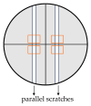
Diagram showing the preparation of scratches for performing the scratch test in a representative well of a 24-well plate: the area filled with BJ cells showing adhesion to the surface (gray areas), marking the observation site (perpendicular black lines), parallel scratches made by 200 µL pipette tips (white rectangles), and precise areas of observation of BJ cell migration (orange rectangles).

References

    1. Oliveira A.L., Viegas M.F., da Silva S.L., Soares A.M., Ramos M.J., Fernandes P.A. The Chemistry of Snake Venom and Its Medicinal Potential. Nat. Rev. Chem. 2022;6:451–469. doi: 10.1038/s41570-022-00393-7. - DOI - PMC - PubMed
    1. Munawar A., Ali S.A., Akrem A., Betzel C. Snake Venom Peptides: Tools of Biodiscovery. Toxins. 2018;10:474. doi: 10.3390/toxins10110474. - DOI - PMC - PubMed
    1. Ageitos L., Torres M.D.T., de la Fuente-Nunez C. Biologically Active Peptides from Venoms: Applications in Antibiotic Resistance, Cancer, and Beyond. Int. J. Mol. Sci. 2022;23:15437. doi: 10.3390/ijms232315437. - DOI - PMC - PubMed
    1. Almeida J.R., Mendes B., Lancellotti M., Franchi G.C., Passos Ó., Ramos M.J., Fernandes P.A., Alves C., Vale N., Gomes P., et al. Lessons from a Single Amino Acid Substitution: Anticancer and Antibacterial Properties of Two Phospholipase A2-Derived Peptides. Curr. Issues Mol. Biol. 2022;44:46–62. doi: 10.3390/cimb44010004. - DOI - PMC - PubMed
    1. Lee A.C.L., Harris J.L., Khanna K.K., Hong J.H. A Comprehensive Review on Current Advances in Peptide Drug Development and Design. Int. J. Mol. Sci. 2019;20:2383. doi: 10.3390/ijms20102383. - DOI - PMC - PubMed

MeSH terms

LinkOut - more resources