. 2025 Apr 23;18(5):612.
doi: 10.3390/ph18050612.

Epigallocatechin Gallate in Camellia sinensis Ameliorates Skin Aging by Reducing Mitochondrial ROS Production

Affiliations

Epigallocatechin Gallate in Camellia sinensis Ameliorates Skin Aging by Reducing Mitochondrial ROS Production

Ji Ho Park et al. Pharmaceuticals (Basel). .

Abstract

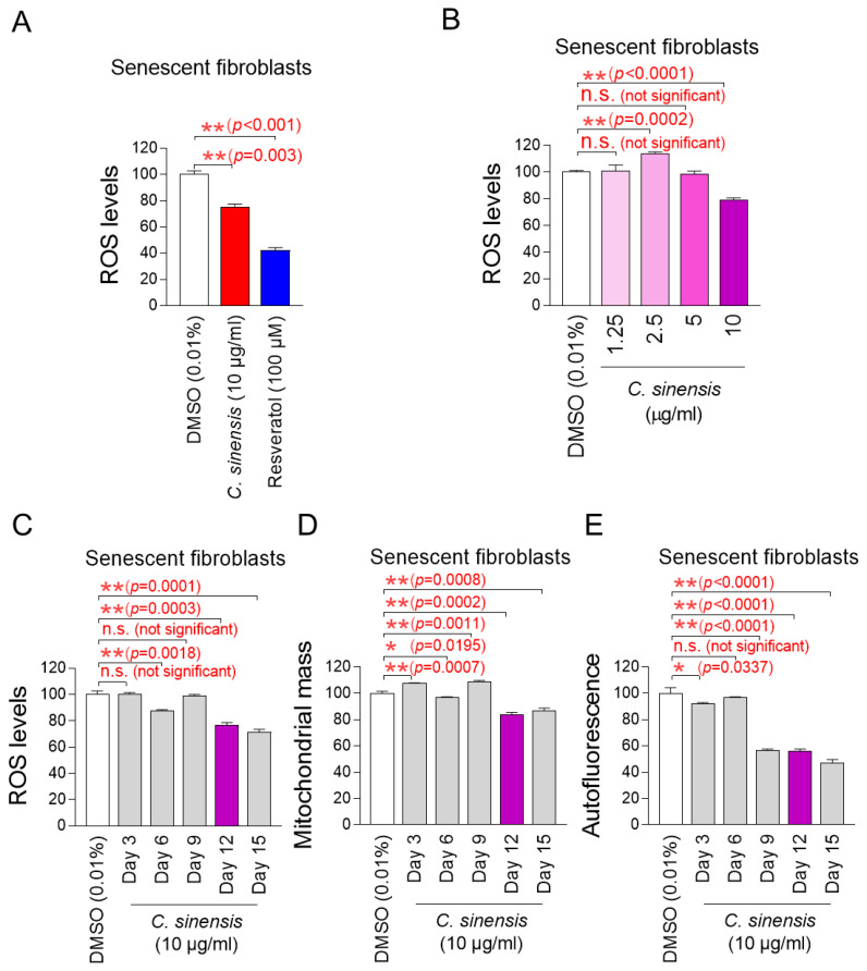
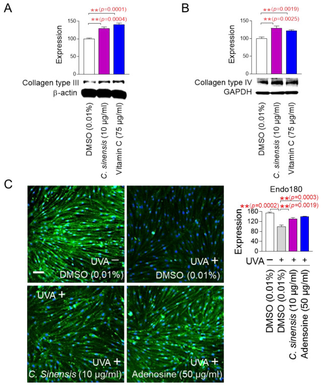
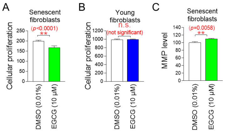
Background: Reactive oxygen species (ROS) generated by mitochondrial dysfunction damage cellular organelles and contribute to skin aging. Therefore, strategies to reduce mitochondrial ROS production are considered important for alleviating skin aging, but no effective methods have been identified. Methods: In this study, we evaluated substances utilized as cosmetic ingredients and discovered Camellia sinensis (C. sinensis) as a substance that reduces mitochondrial ROS levels. Results:C. sinensis extracts were found to act as senolytics that selectively kill senescent fibroblasts containing dysfunctional mitochondria. In addition, C. sinensis extracts facilitated efficient electron transport in the mitochondrial electron transport chain (ETC) by increasing the efficiency of oxidative phosphorylation (OXPHOS), thereby reducing mitochondrial ROS production, a byproduct of the inefficient ETC. This novel mechanism of C. sinensis extracts led to the restoration of skin aging and the skin barrier. Furthermore, epigallocatechin gallate (EGCG) was identified as an active ingredient that plays a key role in C. sinensis extract-mediated skin aging recovery. Indeed, similar to C. sinensis extracts, EGCG reduced ROS and improved skin aging in an artificial skin model. Conclusions: Our data uncovered a novel mechanism by which C. sinensis extract reverses skin aging by reducing mitochondrial ROS production via selective senescent cell death/increased OXPHOS efficiency. Our results suggest that C. sinensis extract or EGCG may be used as a therapeutic agent to reverse skin aging in clinical and cosmetic applications.

Keywords: Camellia sinensis; reactive oxygen species (ROS); senescence rejuvenation; skin aging recovery.


Conflict of interest statement

Authors Eun Young Jeong, Ye Hyang Kim, So Yoon Cha, Ha Yeon Kim, Yeon Kyung Nam, Jin Seong Park, So Yeon Kim and Song Seok Shin was employed by the company Hyundai Bioland Co., Ltd. The remaining authors declare that the research was conducted in the absence of any commercial or financial relationships that could be construed as a potential conflict of interest. The funders had no role in the collection, analyses, or interpretation of data; in the writing of the manuscript; or in the decision to publish the paper.

Figures

Figure 1
Figure 1
Effect of C. sinensis extract on reducing mitochondrial ROS levels. (A) Senescent fibroblasts were treated with dimethyl sulfoxide (DMSO) (0.01%), Camellia sinensis (C. sinensis) extract (10 μg/mL), or resveratrol (100 μM) for 12 days. Use of dihydrorhodamine 123 (DHR123) for fluorescence-activated cell sorting (FACS) analysis. ** p < 0.01, Student’s t-test. Mean ± S.D., n = 3. (B) Senescent fibroblasts were treated with DMSO (0.01%) or different concentrations of C. sinensis extract (1.25, 2.5, 5, and 10 µg/mL) for 12 days. Use of dihydrorhodamine 123 (DHR123) for FACS analysis. n.s. (not significant), ** p < 0.01, Student’s t-test. Mean ± S.D., n = 3. (C) Senescent fibroblasts were treated with DMSO (0.01%) or Camellia sinensis (C. sinensis) extract (10 μg/mL) for 3, 6, 9, 12, and 15 days. Use of dihydrorhodamine 123 (DHR123) for FACS analysis. n.s. (not significant), ** p < 0.01, Student’s t-test. Mean ± S.D., n = 3. (D) Senescent fibroblasts were treated with DMSO (0.01%) or Camellia sinensis (C. sinensis) extract (10 μg/mL) for 3, 6, 9, 12, and 15 days. Use of MitoTracker™ Deep Red FM Dye for FACS analysis. n.s. (not significant), * p < 0.05, ** p < 0.01, Student’s t-test. Mean ± S.D., n = 3. (E) Senescent fibroblasts were treated with DMSO (0.01%) or Camellia sinensis (C. sinensis) extract (10 μg/mL) for 3, 6, 9, 12, and 15 days. Autofluorescence was measured using FACS. n.s. (not significant), * p < 0.05, ** p < 0.01, Student’s t-test. Mean ± S.D., n = 3.
Figure 2
Figure 2
C. sinensis extract selectively kills senescent fibroblasts by inducing apoptosis. (A) Senescent fibroblasts were treated with dimethyl sulfoxide (DMSO) (0.01%) or Camellia sinensis (C. sinensis) extract (10 μg/mL) for 12 days. Then, cellular proliferation was evaluated. ** p < 0.01, Student’s t-test. Mean ± S.D., n = 6. (B) Young fibroblasts were treated with DMSO (0.01%) or C. sinensis extract (10 μg/mL) for 12 days. Then, cellular proliferation was evaluated. n.s. (not significant), Student’s t-test. Mean ± S.D., n = 6. (C) Senescent fibroblasts were treated with DMSO (0.01%) or C. sinensis extract (10 µg/mL) for 12 days. Then, flow cytometric analysis (FACS) of apoptosis was evaluated. The green square represents apoptotic cell populations. ** p < 0.01, Student’s t-test. Mean ± S.D., n = 3.
Figure 3
Figure 3
C. sinensis extract decreases mitochondrial ROS production by enhancing OXPHOS efficiency. (A) Senescent fibroblasts were treated with dimethyl sulfoxide (DMSO) (0.01%) or Camellia sinensis (C. sinensis) extract (10 μg/mL) for 12 days. Oxygen consumption rate (OCR; mpol/min) was measured (black line: DMSO–treated senescent fibroblasts, purple line: C. sinensis extract-treated senescent fibroblasts). ** p < 0.01, two-way ANOVA followed by Bonferroni’s post hoc test. Means ± S.D., n = 3. (B) Senescent fibroblasts were treated with DMSO (0.01%) or C. sinensis extract (10 µg/mL) for 12 days and their ATP production was measured. ** p < 0.01, Student’s t-test. Means ± S.D., n = 3. (C) Senescent fibroblasts were treated with DMSO (0.01%) or C. sinensis extract (10 µg/mL) for 12 days and mitochondrial membrane potential (MMP) was measured. ** p < 0.01, Student’s t-test. Mean ± S.D., n = 3.
Figure 4
Figure 4
Senescence-associated phenotypes are ameliorated by C. sinensis extract. (A) Senescent fibroblasts were treated with dimethyl sulfoxide (DMSO) (0.01%) or Camellia sinensis (C. sinensis) extract (10 μg/mL) for 12 days. Autofluorescence was measured using FACS. ** p < 0.01, Student’s t-test. Mean ± S.D., n = 3. Autofluorescence (green) was also observed using a fluorescence microscope. Scale bar: 10 μm. (B,C) After 12 days of treatment with DMSO (0.01%) or C. sinensis extract (10 µg/mL) in senescent fibroblasts, expression levels of p21 or IL-1β were evaluated. n.s. (not significant), ** p < 0.01, Student’s t-test. Mean ± S.D., n = 3.
Figure 5
Figure 5
C. sinensis extract improves skin aging through collagen synthesis and remodeling. (A,B) After 12 days of treatment with dimethyl sulfoxide (DMSO) (0.01%), Camellia sinensis (C. sinensis) extract (10 µg/mL), or vitamin C (75 µg/mL) in senescent fibroblasts, expression levels of collagen type III and IV were evaluated. ** p < 0.01, Student’s t-test. Mean ± S.D., n = 3. (C) To inhibit the expression of endo 180, senescent fibroblasts were irradiated with 25 J/cm3 ultraviolet A (UVA). Then, senescent fibroblasts were treated with DMSO (0.01%), C. sinensis extract (10 µg/mL), or adenosine (50 μg/mL) for 1 day. ** p < 0.01, student t-test. Mean ± S.D., n = 3. Scale bar: 50 μm. Full-size images of immunofluorescence are shown in Supplementary Figure S1.
Figure 6
Figure 6
C. sinensis extract improves skin aging through enhancing cell-induced collagen contractility. Senescent fibroblasts embedded in collagen gels were treated with dimethyl sulfoxide (DMSO) (0.01%), Camellia sinensis (C. sinensis) extract (10 µg/mL), or vitamin C (50 μg/mL) for 1 day. The cell-induced contractility of collagen is evaluated by measuring the diameter of the collagen disc. ** p < 0.01, student t-test. Mean ± S.D., n = 3.
Figure 7
Figure 7
C. sinensis extract restores the skin barrier function. (A) To inhibit the expression of calpain 1, human epidermal keratinocyte (HEKn) cells were stimulated with 200 ng/mL interleukin-17A (IL-17A). Then, HEKn cells were treated with DMSO (0.01%), C. sinensis extract (10 µg/mL), or ceramide NP (500 μg/mL) for 1 day. ** p < 0.01, Student’s t-test. Mean ± S.D., n = 3. (B) To inhibit the expression of laminin 5, A431 cells were irradiated with 50 mJ/cm3 ultraviolet B (UVB). Then, A431 cells were treated with dimethyl sulfoxide (DMSO) (0.01%), Camellia sinensis (C. sinensis) extract (10 µg/mL), or vitamin C (75 μg/mL) for 1 day. ** p < 0.01, Student t-test. Mean ± S.D., n = 3. (C) To inhibit the expression of collagen type XVII, A431 cells were irradiated with 50 mJ/cm3 UVB. Then, A431 cells were treated with DMSO (0.01%), C. sinensis extract (10 µg/mL), or vitamin C (75 μg/mL) for 1 day. ** p < 0.01, student t-test. Mean ± S.D., n = 3.
Figure 8
Figure 8
Identification of epigallocatechin gallate (EGCG) as the active ingredient in C. sinensis extract. (A,B) Senescent fibroblasts were treated with dimethyl sulfoxide (DMSO) (0.01%), epicatechin (0.01, 0.1, 1, and 10 µM), or Camellia sinensis (C. sinensis) extract (10 µg/mL) for 12 days. Mitochondrial ROS levels using dihydrorhodamine 123 (DHR123) and autofluorescence were evaluated. n.s. (not significant), * p < 0.05, ** p < 0.01, Student’s t-test. Mean ± S.D., n = 3. (C,D) Senescent fibroblasts were treated with DMSO (0.01%), epicatechin gallate (0.01, 0.1, 1, and 10 µM), or C. sinensis extract (10 µg/mL) for 12 days. Mitochondrial ROS levels using DHR123 and autofluorescence were evaluated. n.s. (not significant), ** p < 0.01, Student’s t-test. Mean ± S.D., n = 3. (E,F) Senescent fibroblasts were treated with DMSO (0.01%), epigallocatechin gallate (0.01, 0.1, 1, and 10 µM), or C. sinensis extract (10 µg/mL) for 12 days served as a positive control. Mitochondrial ROS levels using DHR123 and autofluorescence were evaluated. n.s. (not significant), * p < 0.05, ** p < 0.01, Student’s t–test. Mean ± S.D., n = 3.
Figure 9
Figure 9
EGCG, the active ingredient in C. sinensis extract, exhibits similar effects to C. sinensis extract. (A) Senescent fibroblasts were treated with dimethyl sulfoxide (DMSO) (0.01%) or epigallocatechin gallate (EGCG) (10 µM) for 9 days. Then, cellular proliferation was evaluated. ** p < 0.01, Student’s t-test. Mean ± S.D., n = 6. (B) Young fibroblasts were treated with DMSO (0.01%) or EGCG (10 µM) for 9 days. Then, cellular proliferation was evaluated n.s. (not significant), Student’s t-test. Mean ± S.D., n = 6. (C) Senescent fibroblasts were treated with DMSO (0.01%) or EGCG (10 µM) for 9 days. Then, mitochondrial membrane potential (MMP) was measured. ** p < 0.01, Student’s t-test. Mean ± S.D., n = 3.
Figure 10
Figure 10
C. sinensis extracts and EGCG reverse skin aging in artificial skin models. (A,B) To induce skin aging, artificial skin models were irradiated with 20 mJ/cm3 ultraviolet A (UVA) once a day for three times. Then, artificial skin models were treated with dimethyl sulfoxide (DMSO) (0.01%), Camellia sinensis (C. sinensis) extract (10 µg/mL), epigallocatechin gallate (EGCG) (10 µM), or resveratrol (75 μM) for 1 day. To detect ROS in artificial skin models, dihydroethidium (DHE) staining was performed. To distinguish collagen fibers from other tissues, Masson’s trichrome (MT) staining was performed. Scale bar: 10 μm. * p < 0.05, ** p < 0.01, Student’s t-test. Mean ± S.D., n = 3.

References

    1. Baumann L. Skin ageing and its treatment. J. Pathol. 2007;211:241–251. doi: 10.1002/path.2098. - DOI - PubMed
    1. Payeras M.R., Cherubini K., Figueiredo M.A., Salum F.G. Oral lichen planus: Focus on etiopathogenesis. Arch. Oral Biol. 2013;58:1057–1069. doi: 10.1016/j.archoralbio.2013.04.004. - DOI - PubMed
    1. Haltaufderhyde K., Ozdeslik R.N., Wicks N.L., Najera J.A., Oancea E. Opsin Expression in Human Epidermal Skin. Photochem. Photobiol. 2015;91:117–123. doi: 10.1111/php.12354. - DOI - PMC - PubMed
    1. Zorina A., Zorin V., Kudlay D., Kopnin P. Age-Related Changes in the Fibroblastic Differon of the Dermis: Role in Skin Aging. Int. J. Mol. Sci. 2022;23:6135. doi: 10.3390/ijms23116135. - DOI - PMC - PubMed
    1. Sreedhar A., Aguilera-Aguirre L., Singh K.K. Mitochondria in skin health, aging, and disease. Cell Death Dis. 2020;11:444. doi: 10.1038/s41419-020-2649-z. - DOI - PMC - PubMed

LinkOut - more resources