

Alveolar Echinococcosis in 11-Month-Old Dog-Clinical Case
- PMID: 40430770
- PMCID: PMC12114745
- DOI: 10.3390/pathogens14050450
Alveolar Echinococcosis in 11-Month-Old Dog-Clinical Case
Abstract
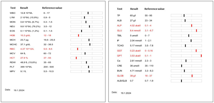
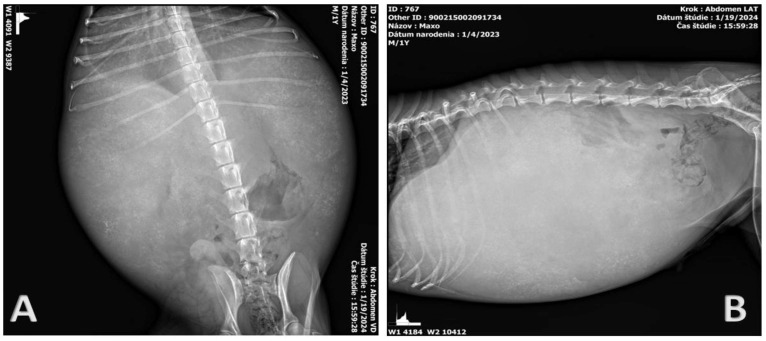
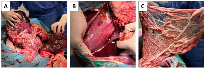
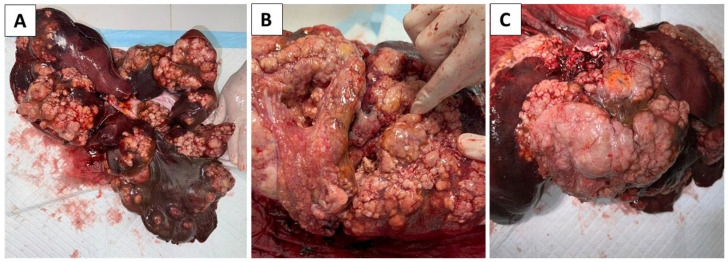
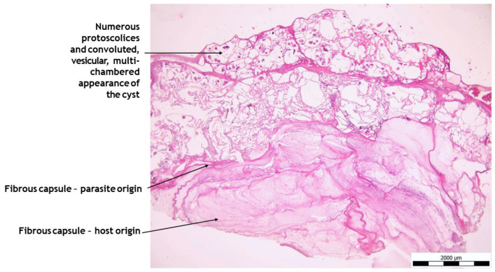
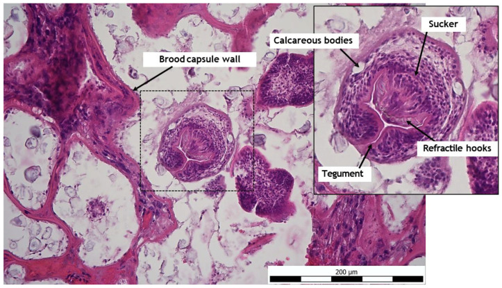
In the present work, we describe the clinical-pathological case of an 11-month-old Border Collie dog, which was presented by its owner to a private veterinary clinic for the purpose of determining the diagnosis and subsequent therapy. The owner reports anamnestic data of abdominal enlargement, persistent apathy, fatigue, and vomiting. A complete examination of the patient was performed, consisting of clinical, hematological, and biochemical blood tests, X-ray, and USG examinations. Based on the findings, a probatory laparotomy was indicated, during which a large multi-lobular cystic irregular mass was detected, affecting the entire liver parenchyma, including macroscopic metastatic foci of the omentum and diaphragm. Due to the inoperable finding, the patient was humanely euthanized during the surgical procedure. Subsequently, an autopsy was performed with the collection of samples for histopathological and PCR examination of the tissue. Serological examination was also performed. The results confirmed a rare generalized form of alveococcosis (Echinococcus multilocularis) in the dog as an intermediate host.
Keywords: Echinococcus multilocularis; alveolar echinococcosis; parasitic zoonoses.
Conflict of interest statement
Michaela Gentil was employed by Laboklin GmbH & Co. KG. The remaining authors declare that the research was conducted in the absence of any commercial or financial relationships that could be construed as a potential conflict of interest.
Figures
Similar articles
-
[Lethal alveolar echinococcosis in a dog: clinical symptoms and pathology].Berl Munch Tierarztl Wochenschr. 2013 Sep-Oct;126(9-10):408-14. Berl Munch Tierarztl Wochenschr. 2013. PMID: 24199383 German.
-
[A case of alveolar hydatid disease in a dog: domestic animals as rare incidental intermediate hosts for Echinococcus multilocularis].Schweiz Arch Tierheilkd. 2007 Mar;149(3):123-7. doi: 10.1024/0036-7281.149.3.123. Schweiz Arch Tierheilkd. 2007. PMID: 17410970 German.
-
Alveolar echinococcosis in a dog; analysis of clinical and histological findings and molecular identification of Echinococcus multilocularis.Acta Parasitol. 2018 Sep 25;63(3):486-494. doi: 10.1515/ap-2018-0058. Acta Parasitol. 2018. PMID: 29975636
-
Alveolar echinococcosis-spreading disease challenging clinicians: a case report and literature review.World J Gastroenterol. 2013 Jul 14;19(26):4257-61. doi: 10.3748/wjg.v19.i26.4257. World J Gastroenterol. 2013. PMID: 23864792 Free PMC article. Review.
-
Cestodes. Echinococcus.Gastroenterol Clin North Am. 1996 Sep;25(3):655-89. doi: 10.1016/s0889-8553(05)70268-5. Gastroenterol Clin North Am. 1996. PMID: 8863045 Review.
References
-
- Echinococcus Infections in Dogs: Prevalence, Symptoms and Diagnosis. LABOKLIN Europe. 2024. [(accessed on 27 March 2024)]. Available online: https://laboklin.com/ch-it/echinococcus-infections-in-dogs-prevalence-sy...
Publication types
MeSH terms
Supplementary concepts
LinkOut - more resources
Full Text Sources
