. 2025 May 6;14(5):455.
doi: 10.3390/pathogens14050455.

Integrated Taxonomic Approaches to Gastrointestinal and Urinary Capillariid Nematodes from Wild and Domestic Mammals

Affiliations

Integrated Taxonomic Approaches to Gastrointestinal and Urinary Capillariid Nematodes from Wild and Domestic Mammals

Masae Tamaru et al. Pathogens. .

Abstract

Fine nematodes of the family Capillariidae parasitize various organs and tissues in fish, amphibians, reptiles, avians, and mammals. Currently classified into more than 20 genera, these nematodes are primarily distinguished based on the caudal structures of male worms. Morphological and molecular analyses were conducted on 15 mammal-parasitic species belonging to the genera Aonchotheca (A. putorii, A. suzukii n. sp., A. suis n. comb. (syn. Capillaria suis), A. riukiuensis, and A. bilobata), Pearsonema (P. neoplica n. sp., P. feliscati, P. iharai n. sp., and P. toriii n. sp.), Liniscus (L. himizu), Calodium (C. hepaticum), Echinocoleus (E. yokoyamae n. sp.), and Eucoleus (E. kaneshiroi n. sp., E. aerophilus, and Eucoleus sp.), using specimens from various wild and domestic animals in Japan and brown rats in Indonesia. As demonstrated in this study, nearly complete SSU rDNA sequencing is a powerful tool for differentiating closely related species and clarifying the phylogenetic relationships among morphologically similar capillariid worms. Additionally, most capillariid worms detected in dogs and cats are suspected to be shared with their respective wildlife reservoir mammals. Therefore, molecular characterization, combined with the microscopic observation of these parasites in wildlife mammals, provides a robust framework for accurate species identification, reliable classification, and epidemiological assessment.

Keywords: Aonchotheca; Calodium; Capillariidae; Echinocoleus; Eucoleus; Liniscus; Pearsonema; SSU rDNA; Trichosomoides crassicauda; morphology; taxonomy.


Conflict of interest statement

The authors declare no conflicts of interest.

Figures

Figure 1
Figure 1
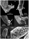
Morphology of Aonchotheca putorii. (a) Caudal end of morphotype “Type A” male worm with lateral alae (LA) and membranous bursa (B) supported with elongated digitiform projections (arrow). Spicule (Sp). (b) Caudal end of morphotype “Type B” male worm with LA and membranous B supported with stumpy digitiform projections (arrow). (c,d) Vulval appendages of female worms at three regions (anterior and posterior swellings of the vulva, and a vulval plate from the anterior edge of the vulval opening (arrows). End of stichosome (S). (e) Posterior end of a female worm with a terminal anus (arrow). (f) Intrauterine egg with striated eggshell surface texture. Photographs (ae) at the same magnification and a scale bar is shown in (b).
Figure 2
Figure 2
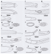
Line drawings of Aonchotheca putorii (ad) and A. suzukii n. sp. (eg). (a) Caudal end of morphotype “Type A” male worm. (b) Caudal end of morphotype “Type B” male worm. (c) Vulval appendages of a female worm at three regions (anterior and posterior swellings of the vulva, and a vulval plate from the anterior edge of the vulval opening), and intrauterine egg with striated eggshell surface texture. (d) Posterior end of a female worm with a terminal anus. (e) Caudal end of A. suzukii n. sp. male worm. (f) Vulval appendages of A. suzukii n. sp. female worm at two regions (small subcuticular elevation anterior to the vulval opening and cuticular ballooned swelling around it), and an intrauterine egg with striated eggshell surface texture. (g) Posterior end of A. suzukii n. sp. female worm with a subterminal anus.
Figure 3
Figure 3
Morphology of Aonchotheca suzukii n. sp. ((ac), specimens from feral alien raccoons, (df), specimens from wild boars). (a,d) Caudal end of male worm with lateral alae (LA) and membranous bursa (B) supported with short digitiform projections. Spicule (Sp), and extruded spicular sheath (SS) showing transverse striations. (b,e) Vulval appendages of female worm; small subcuticular elevation at the anterior to the vulval opening (arrow) and cuticular ballooned swelling around it. End of stichosome (S). (c,f) Posterior end of female worm with subterminal anus (arrow). Photographs (af) at the same magnification, and scale bar is shown in (a,d).
Figure 4
Figure 4
Morphology of Aonchotheca suis n. comb. (ac) and A. riukiuensis (df) from the stomach of wild boars. (a) Caudal end of male worm with conspicuous lateral alae (LA) and membranous bursa (B) supported with two pairs of ventrolateral projections, simple digitiform ones (arrowheads) and hammer-shaped ones (arrow). (b) Caudal end of male worm lacking lateral alae but with membranous bursa (B) supported with two pairs of ventrolateral projections, simple digitiform ones (arrowhead) and hammer-shaped ones (arrow). (c) Evidently and finely roughened cuticular surface around the vulval opening (arrow). End of stichosome (S). (d,e) Caudal end of male worms with rudimentary lateral alae (LA) and membranous bursa (B) supported with two pairs of ventrolateral projections, simple digitiform ones (arrowhead) and thick hammer-shaped ones (arrow). (f) Bell-shaped vulval appendage at the vulval opening (arrow). End of stichosome (S). Photographs (ac) at the same magnification, and scale is shown in (c). Similarly, photographs (df) at the same magnification, and scale bar is shown in (d).
Figure 5
Figure 5
Morphology of Pearsonema neoplica n. sp. (ac) Caudal end of male worm with caudal triangular membranous bursa (B) connected to dorsolateral lobes (arrowheads) and supported with digitiform projections (P). (df) Caudal end of male worm lacking caudal triangular membranous bursa. Dorsolateral lobes (arrowheads) and digitiform projections (P). (g) Vulva (V) without appendages of female worm. (h,i) Posterior end of female worms with terminal anus (arrow). (jm) Bioperculated barrel-shaped eggs, with reticulated eggshell surface texture. Sagittal section (j,l), and reticulated eggshell surface (k,m). Scale bar is shown in each photograph.
Figure 6
Figure 6
Scanning electron microscopic view of Pearsonema neoplica n. sp. (a,b) Caudal end of male worm with caudal triangular membranous bursa (B) connected to dorsolateral lobes (arrowheads) and supported with digitiform projections (arrows). Extruded spicular sheath (SS) from cloaca. (c,d) Caudal end of male worm lacking caudal triangular membranous bursa. Dorsolateral lobes (arrowheads) and digitiform projections (arrows). Extruded spicular sheath (SS) from cloaca. (e,f) Bioperculated barrel-shaped eggs, with reticulated eggshell surface texture. Scale bar is shown in each photograph.
Figure 7
Figure 7
Line drawings of Pearsonema neoplica n. sp. (ae), P. feliscati (fk), P iharai n. sp. (lo), and P. toriii n. sp. (p,q). Caudal end of male worms (a,b,f,g,l,p,q), vulval region of female worms (h,m,r), posterior end of female worms (c,i,n,s), sagittal section of eggs (d,j), eggshell surface (e,k), and jointed view of sagittal egg section and eggshell surface (o,t).
Figure 8
Figure 8
Morphology of Pearsonema feliscati. (a,b) Ventral and lateral views of caudal end of male worms, lacking apparent membranous bursa. Dorsolateral lobes (arrowheads) and a nodular projection (arrow). (c) Vulva (arrow) without appendages of female worm. (dg) Bioperculated barrel-shaped egg, with punctuated eggshell surface texture, collected from urine of a feral alien raccoon (d,e), or a domestic cat (f,g). (h) Scanning electron microscopic view of male caudal end with dorsolateral lobes (arrowheads) in ventral view. (i) Scanning electron microscopic view of P. feliscati egg surface. Scale bar is shown in each photograph.
Figure 9
Figure 9
Morphology of Pearsonema iharai n. sp. (a) Ventral view of caudal end of male worm, lacking apparent membranous bursa. Circular cuticular swelling (arrowheads) before caudal end with dorsolateral lobes (arrows). Protruded spicule (Sp). (b) Vulva (arrow) without appendages of female worm. (c) Caudal end of female worm with a terminal anus (arrow). (d) Intrauterine bioperculated lemon-shaped egg, with vermiculated eggshell surface texture. (eg) Scanning electron microscopic view of male caudal end with dorsolateral lobes (arrows) in ventral view. Circular cuticular swelling (arrowheads) prior to the caudal end. (h) Scanning electron microscopic view of P. iharai n. sp. egg with vermiculated eggshell surface. Scale bar is shown in each photograph.
Figure 10
Figure 10
Morphology of Pearsonema toriii n. sp. (a) Dorsal view of caudal end of male worm, lacking apparent membranous bursa. Lateral cuticular swelling (arrowheads) before caudal end with conspicuous spheroid dorsolateral lobes (arrows). Protruded spicular sheath (SS) and spicule (Sp). (b) Lateral view of caudal end of male worm. Dorsolateral lobes (arrows). (c) Vulva (arrow) without appendages of female worm. End of stichosome (S). (d) Caudal end of female worm with subterminal anus (arrow). (e,f) Bioperculated barrel-shaped egg, with vermiculated eggshell surface texture. (g) Scanning electron microscopic view of male caudal end with spheroid dorsolateral lobes (arrows). (h) Scanning electron microscopic view of transversely wrinkled spicular sheath. (i) Scanning electron microscopic view of P. toriii n. sp. egg with vermiculated eggshell surface. Scale bar is shown in each photograph.
Figure 11
Figure 11
Morphology of Echinocoleus yokoyamae n. sp. (ac) specimens from badgers; (df) specimens from wild boars. (a,d) Ventral view of caudal end of male worm, terminated in two lobes, extending to sickle-shaped thick projections (arrows on right side), which support two-lobed membranous pseudobursa (PB). In addition, a pair of digitiform ventrolateral projections at precloacal level (arrows on left side) border anterior edge of membranous bursal lobes. (b,e) Vulva (arrow) with cylindrical vulvar appendage. (c,f) Caudal end of female worm with terminal anus (arrow). All photograph at the same magnification, and scale is shown in (c,f).
Figure 12
Figure 12
Line drawings of Echinocoleus yokoyamae n. sp. (ac), and Eucoleus kaneshiroi n. sp. (df). (a,d) Ventral view of caudal end of male worms. (b,e) Vulval region of female worms, and intrauterine eggs. (c,f) Caudal end of female worm with terminal anus.
Figure 13
Figure 13
Morphology of Eucoleus kaneshiroi n. sp. (a) Ventral view of caudal end of male worm, terminated in two small lobes (arrows), supporting clear semilunar-shape membranous pseudobursa (PB). A pair of papillae, close to each other, on each caudal lobe. (b) Vulva (arrow) without vulvar appendage. (c) Caudal end of female worm with terminal anus (arrow). (d) Intrauterine bioperculated lemon-shaped egg, with minutely pitted eggshell surface. Photographs (ac) at the same magnification, and scale is shown in (c).
Figure 14
Figure 14
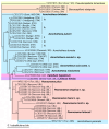
ML phylogenetic tree based on the SSU rDNA sequence. The GenBank accession number is followed by the host and isolation organ in parentheses and the species name. ABO, abomasum; CEM, cecum; ESP, esophagus; LIN, large intestine; LIV, liver; RES, respiratory system; SIN, small intestine; STM, stomach; TRA, trachea; and UBL, urinary bladder. Capillariid species reported in this study are indicated in bold letters. Sequences of isolates belonging to the same genus are placed on a single background color: Aonchotheca, light blue; Baruscapillaria, light orange; Calodium, pink; Capillaria, light violet; Echinocoleus, light gree; Eucoleus, emerald green, Liniscus, violet; Pearsonema, orange; and Pseudocapillaria, light yellow.
Figure 15
Figure 15
ML phylogenetic tree based on the SSU rDNA sequence, focusing on a clade of Aonchotheca/Pearsonema/Calodium/Liniscus. The labeling of each sequence, organ abbreviation, and species name are expressed as explained in the legend for Figure 14.
Figure 16
Figure 16
Locations of interspecific and intraspecific nucleotide changes (substitutions and indels) of 88 isolates (classified into 28 species of 9 genera) plotted on a putative SSU rDNA secondary structure of A. putorii (DDBJ/EMBL/GenBank accession no. LC052349). Eighty-eight sequences (isolates) are classified as Aonchotheca (34 sequences of 7 species), Baruscapillaria (5 sequences of B. obsignata), Capillaria (15 sequences of 5 species), Echinocoleus (2 sequences of E. yokoyamae n. sp.), Eucoleus (13 sequences of 8 species), Pseudocapillaria (one sequence of P._tomentosa), Calodium (one sequence of C. hepaticum), Pearsonema (16 sequence of 5 species), and Liniscus (one sequence of L. himizu). Base sites of intergeneric and interspecific nucleotide variation are expressed in red letters on a yellow background color, and those of intraspecific nucleotide variation are with blue letters. Bases expressed in black letters are highly conserved nucleotides with all capillariid isolates analyzed here.

Similar articles

References

    1. Freitas T.J.F., Lent H. Estudo sobre os Capillariinae parasitos de mammiferos: (Nematoda: Trichuroidea) Mem. Inst. Oswaldo Cruz. 1936;31:85–160. doi: 10.1590/S0074-02761936000100006. - DOI
    1. López-Neyra C.R. Los Capillarinae. Mem. Real Acad. Cienc. Exactas Fis. Nat. Madrid. Ser. Giencias Nat. 1947;12:1–248.
    1. Skrjabin K.I., Shikhobalova N.P., Orlov I.V. Essentials of Nematology. Trichocephalidae and Capillariidae of Animals and Man and Diseases Caused by Them. Volume VI Academy of Sciences of the USSR, Moscow, USSR; Moscow, Russia: 1957. pp. 1–599. Jerusalem, Israel Program for Scientific Translations.
    1. Butterworth E.W., Beverley-Burton M. The taxonomy of Capillaria spp. (Nematoda: Trichuridea) in carnivorous mammals from Ontario, Canada. Syst. Parasitol. 1980;1:211–236. doi: 10.1007/BF00009847. - DOI
    1. Moravec F. Proposal of a new systematic arrangement of nematodes of the family Capillariidae. Folia Parasitol. 1982;29:119–132. - PubMed