. 2025 Apr 24;17(5):557.
doi: 10.3390/pharmaceutics17050557.

The Molecular Chaperone TCP1 Affects Carcinogenicity and Is a Potential Therapeutic Target for Acute Myeloid Leukemia

Affiliations

The Molecular Chaperone TCP1 Affects Carcinogenicity and Is a Potential Therapeutic Target for Acute Myeloid Leukemia

Yong Wu et al. Pharmaceutics. .

Abstract

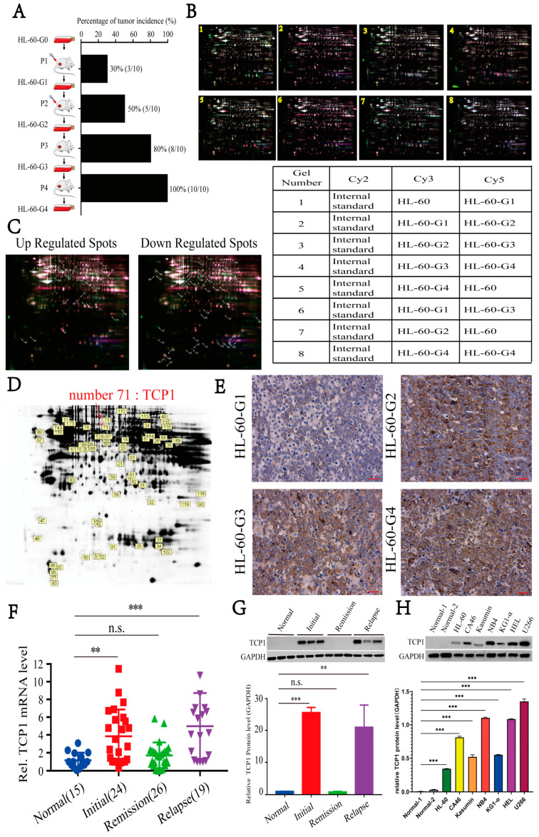
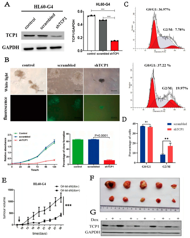
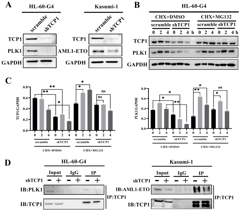
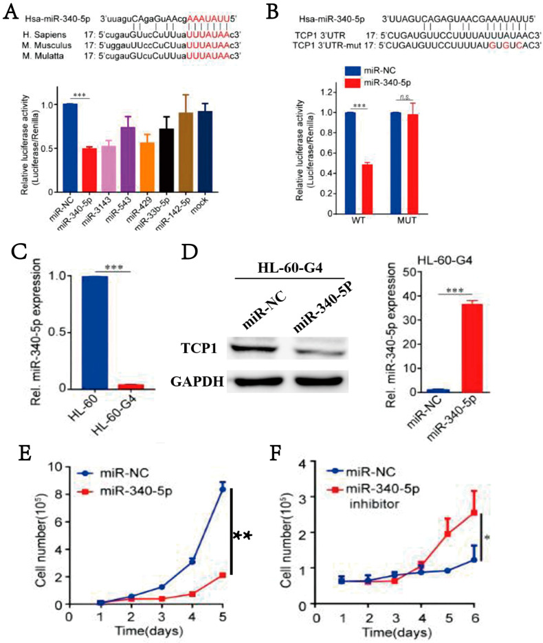
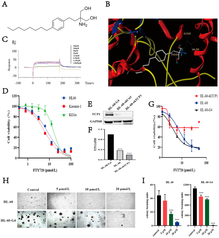
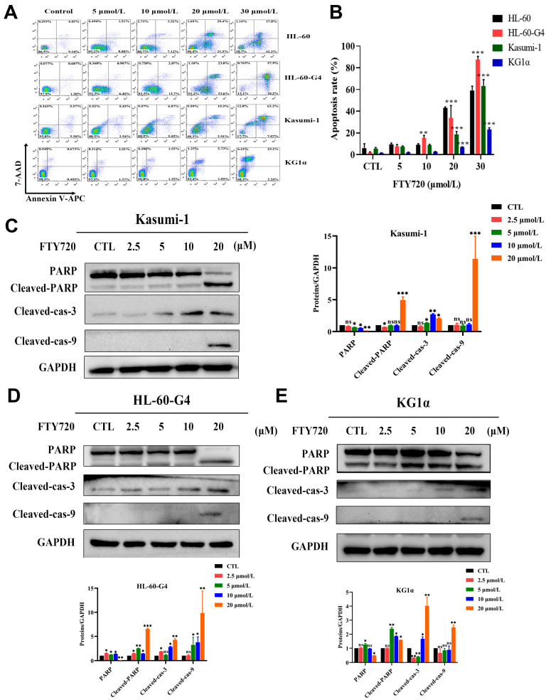
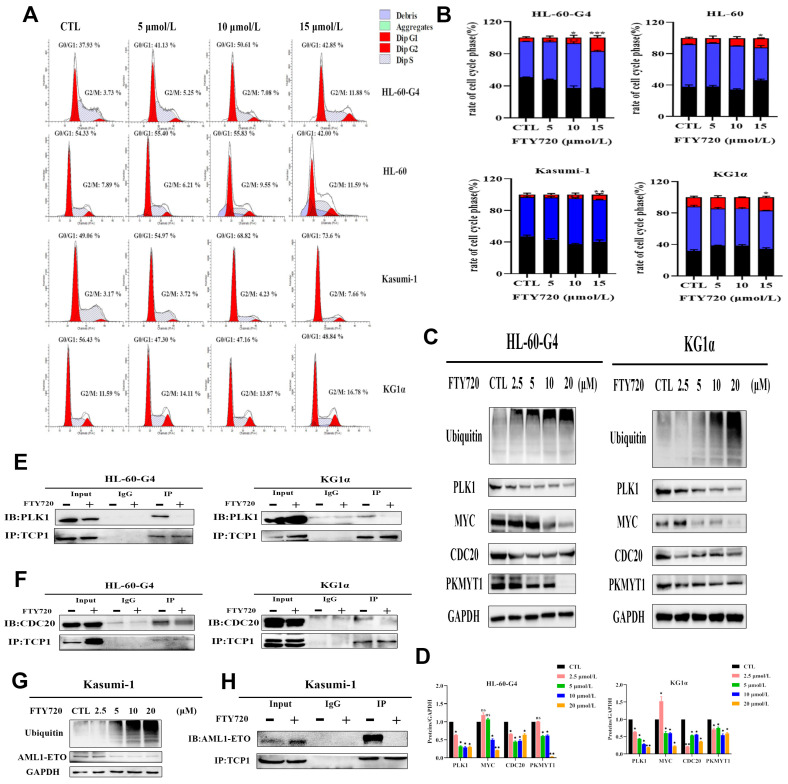
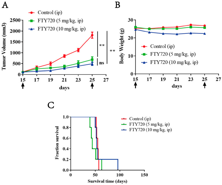
Background/Objectives: Acute myeloid leukemia (AML) is an aggressive malignancy marked by high relapse rates and molecular heterogeneity, necessitating the identification of novel therapeutic targets. T-complex protein 1 (TCP1), a chaperonin implicated in protein folding, remains underexplored in AML pathogenesis. This study investigates the functional role of TCP1 in AML progression and evaluates its therapeutic potential. Methods: Using successive generations of xenografted tumor models, we systematically assessed the correlation between TCP1 expression and AML tumorigenicity. Functional consequences of TCP1 silence were evaluated through in vitro proliferation assays and in vivo tumor growth monitoring. Two distinct inhibitory strategies were employed: miR-340-5p-mediated transcriptional silencing and FTY720-induced disruption of TCP1 chaperone activity. Mechanistic insights were derived from ubiquitin-proteasome pathway analysis, cell cycle profiling, and apoptosis assays. Results: High TCP1 expression correlated strongly with enhanced AML tumorigenicity. Knockdown of TCP1 significantly inhibited AML cell growth and induced degradation of AML1-ETO and PLK1 proteins through the ubiquitin-proteasome pathway. miR-340-5p effectively silenced TCP1 expression, exhibiting an inverse correlation with TCP1 levels. FTY720 disrupted TCP1's chaperone function, leading to cell cycle arrest, apoptosis, and reduced xenograft tumor growth in murine models. Conclusion: Our findings establish TCP1 as a promising therapeutic target for AML. Both miR-340-5p and FTY720 demonstrate potent anti-leukemic effects by suppressing TCP1 activity, highlighting their potential as novel strategies to inhibit AML proliferation and improve therapeutic outcomes.

Keywords: FTY720; TCP1; acute myeloid leukemia; cell cycle arrest; chaperone function; miR-340-5p.


Conflict of interest statement

The authors declare that they have no known competing financial interests or personal relationships that could have appeared to influence the work reported in this paper.

Figures

Figure 1
Figure 1
TCP1 is positively related to the tumorigenesis and development of AML. (A) In the successive generations, the tumor formation rate increased in the HL-60 cell line. (B) Mass spectrometry combined with two-dimensional in-gel electrophoresis revealed that TCP1 is a highly oncogenic gene. (C) Differentially expressed protein spots marked with master numbers displayed in 2D-DIGE images. The arrows indicate the 33 significantly upregulated protein spots (marked with master numbers) and 54 downregulated protein spots (marked with master numbers) displayed in 2D-DIGE images. (D) Proteomic analysis of HL-60 cells using 2D-DIGE. The arrows indicate protein spots that differed significantly between the HL-60 cells and the control cells. Relative spot intensity was calculated on the basis of the spot volume. Only the spots indicated with arrows were used for identification. (E) IHC staining examined TCP1 expression in tumor tissues of different generations. The TCP1 expression in tissue samples of different stage of AML patients was detected by RT-qPCR (F) and Western blotting (G). (H) Western blotting was used to examine the expression of TCP1 in different leukemia cell lines. Data are presented as the means ± SDs from three independent experiments. ** p < 0.01, *** p < 0.001, ns: no significance.
Figure 2
Figure 2
The effect of TCP1 on AML cell development and proliferation in vitro and in vivo. (A) The efficiency of shTCP1 was measured in HL-60-G4 cells by Western blotting. (B) A clonogenic cell survival assay was performed to evaluate the growth of HL-60-G4-shTCP1 cells in vitro. (C,D) Representative cell cycle distribution of HL-60-G4 cells after knocking down TCP1 with lentivirus transfection. (E) Detection of xenograft tumor formation in nude mice using Tet-on technology. (F) Representative image of Dox- and Dox+ groups of tumors at the end of the experiment. (G) Western blotting was used to analyze the protein level of TCP1 in tumor tissues in each group. Data are presented as the means ± SD from three independent experiments. ** p < 0.01, *** p < 0.001, ns: no significance.
Figure 3
Figure 3
TCP1 regulated the stability of PLK1 and AML1-ETO. (A) Western blotting analysis of TCP1, PLK1, and AML1-ETO expression in HL-60-G4-shTCP1 and Kasumi-1-shTCP1 cells. (B) Cells were incubated with 10 μM CHX alone or combined with 10 μM MG132, and WB assays were used to determine the levels for TCP1 and PLK1. (C) The image shown in Figure 3B was quantified using ImageJ software (Version 1.8.0). (D) Co-IP assays were performed to observe the pull-down levels of PLK1 and AML1-ETO in the shTCP1 group and scramble group. Data are presented as the means ± SD from three independent experiments. * p < 0.05, ** p < 0.01, ns: no significance.
Figure 4
Figure 4
MiR-340-5P inhibits AML cell proliferation by silencing TCP1. (A) Dual-luciferase reporter system analysis of targeted transcriptional regulation of TCP1 by miRNAs. (B) HEK293 cells were co-transfected with miR-340-5p mimics or control and reporter plasmid or the mutant 3′-UTR of TCP1, and the luciferase activity was measured after 48 h. (C) MiR-340-5p levels in AML cell lines were tested by RT-qPCR. (D) HL-60-G4 cells overexpressing (O/E) the vector control or miR-340-5p. Detection of TCP1 expression in HL-60-G4 cells overexpressing miR-340-5p by Western blot. (E) The cellular proliferation of HL-60-G4O/E cells was determined by cell counting. (F) HL-60-G4O/E cells were treated with 100 nmol L−1 of an hsa-miR-340-5p inhibitor for 24 h, and cellular proliferation was examined. Data are presented as the means ± SDs from three independent experiments. * p < 0.05, ** p < 0.01, *** p < 0.001, ns: no significance.
Figure 5
Figure 5
FTY720 has an affinity for TCP1 and can inhibit AML cell proliferation. (A) The chemical structure of FTY720. (B) TCP1 subunit protein and FTY720 molecular docking virtual screening. (C) Surface plasmon resonance detected binding between FTY720 and TCP1. (D) The effect of FTY720 on the viability of AML cells. (E) TCP1 protein levels in the three cell lines were examined by Western blotting and (F) normalized to the corresponding density of GAPDH. (G) The sensitivity of HL-60 cells to FTY720 increased as the TCP1 protein level increased. (H) HL-60-G4 cells formed many more colony units than HL-60 cells, and their colony numbers decreased as FTY720 increased. (I) The quantification and plot were created using ImageJ software (Version 1.8.0) for the image shown in Figure 5H. Data are presented as the means ± SDs from three independent experiments. *** p < 0.001.
Figure 6
Figure 6
Induction of apoptosis by FTY720 in AML cells. (A) Representative flow cytometry images. (B) Quantitative analysis of the percentage of apoptotic cells after treatment with FTY720 for 24 h. (CE) Western blotting analysis of caspase family-related proteins in AML cells exposed to different concentrations of FTY720. Data are presented as the means ± SDs from three independent experiments. * p < 0.05, ** p < 0.01, *** p < 0.001, ns: no significance.
Figure 7
Figure 7
FTY720 induces cell cycle arrest and decreases protein levels by inhibiting TCP1 chaperone function in AML cells. (A) Representative cell cycle distribution images. (B) Column chart of the cell cycle ratio of HL-60-G4, HL-60, Kasumi-1, and KG1α cells. (C) Changes in intracellular protein levels in HL-60-G4 and KG1α cells incubated with FTY720 for 24 h. (D) The image shown in Figure 8C was quantified using ImageJ software (Version 1.8.0). (E) The interaction between the PLK1 protein and TCP1 protein in HL-60-G4 and KG1α cells exposed to FTY720 for 24 h was assayed by Co-IP. (F) The interaction between the CDC20 protein and TCP1 protein in HL-60-G4 and KG1α cells exposed to FTY720 for 24 h was assayed by Co-IP. (G) Changes in intracellular protein levels in Kasumi-1 cells incubated with FTY720 for 24 h were detected by Western blotting. (H) TCP1 interacts with AML1-ETO in Kasumi-1 cells, but FTY720 disrupts the interaction. Means ± SDs of three independent experiments are shown. * p < 0.05, ** p < 0.01, *** p < 0.001, ns: no significance.
Figure 8
Figure 8
Anti-tumor effect of FTY720 on xenograft AML tumors. (A) The tumor volume in each group, n = 5. (B) Weight of tumor-bearing mice during the treatment period. (C) Survival curves of mice treated with FTY720. ** p < 0.01, ns: no significance.

Similar articles

References

    1. Vadakekolathu J., Rutella S. Escape from T-cell–targeting immunotherapies in acute myeloid leukemia. Blood. 2024;143:2689–2700. doi: 10.1182/blood.2023019961. - DOI - PMC - PubMed
    1. Shimony S., Stahl M., Stone R.M. Acute myeloid leukemia: 2023 update on diagnosis, risk-stratification, and management. Am. J. Hematol. 2023;98:502–526. doi: 10.1002/ajh.26822. - DOI - PubMed
    1. Bhansali R.S., Pratz K.W., Lai C. Recent advances in targeted therapies in acute myeloid leukemia. J. Hematol. Oncol. 2023;16:29. doi: 10.1186/s13045-023-01424-6. - DOI - PMC - PubMed
    1. Chen X., Chen X., Huang Y., Lin J., Wu Y., Chen Y. TCP1 increases drug resistance in acute myeloid leukemia by suppressing autophagy via activating AKT/mTOR signaling. Cell Death Dis. 2021;12:1058. doi: 10.1038/s41419-021-04336-w. - DOI - PMC - PubMed
    1. Piano V., Alex A., Stege P., Maffini S., Stoppiello G.A., Veld P.J.H.I., Vetter I.R., Musacchio A. CDC20 assists its catalytic incorporation in the mitotic checkpoint complex. Science. 2021;371:67–71. doi: 10.1126/science.abc1152. - DOI - PubMed

LinkOut - more resources