Review
. 2025 May 2;17(5):603.
doi: 10.3390/pharmaceutics17050603.

Nano-Drug Delivery Systems for Bone Metastases: Targeting the Tumor-Bone Microenvironment

Affiliations
Review

Nano-Drug Delivery Systems for Bone Metastases: Targeting the Tumor-Bone Microenvironment

Mohamad Bakir et al. Pharmaceutics. .

Abstract

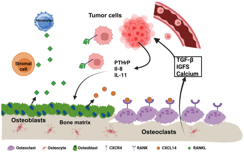
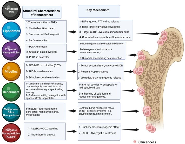
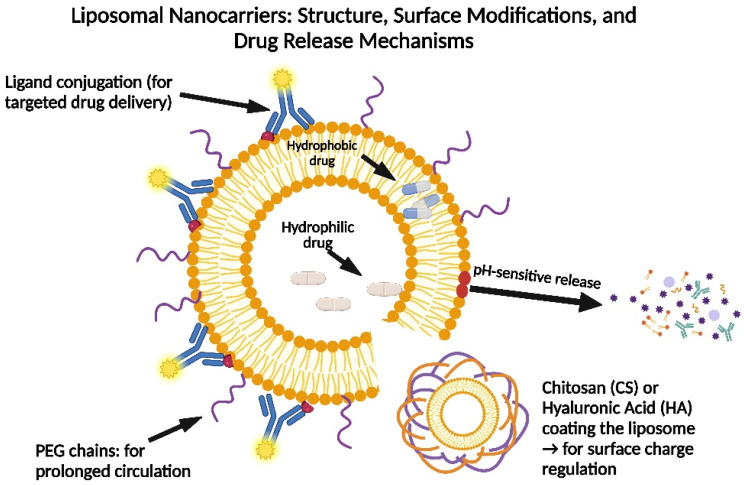
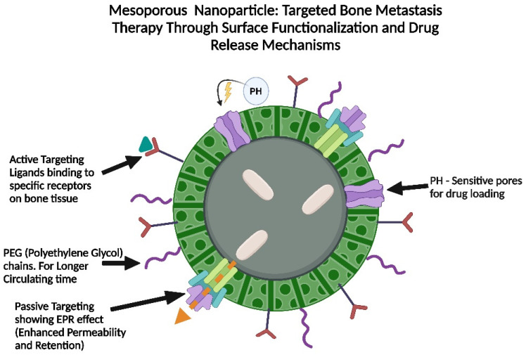
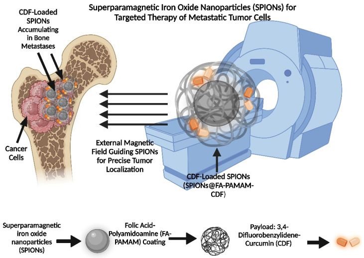
Bone metastases are a prevalent and debilitating consequence of various cancers, including breast and prostate carcinomas, which significantly compromise patient quality of life due to pain, fractures, and other skeletal-related events (SREs). This review examines the pathophysiology of bone metastases, emphasizing the role of the bone microenvironment in tumor progression through mechanisms such as osteotropism and the dysregulated bone remodeling cycle. The primary focus is on the emerging nano-drug delivery systems (DDS) designed to target the bone microenvironment and improve the therapeutic index of anticancer agents. Current treatments, mainly comprising bisphosphonates and radiotherapy, provide palliative benefits but often have limited efficacy and significant side effects. Innovative strategies, such as bisphosphonate-conjugated nanoparticles and targeted therapies that utilize the unique bone marrow niche, are explored for their potential to enhance drug accumulation at metastatic sites while minimizing systemic toxicity. These approaches include the use of liposomes, polymeric nanoparticles, and inorganic nanoparticles, which can be functionalized to exploit the biological barriers within the bone microenvironment. This review also discusses the challenges and future directions for nano-DDS in clinical settings, emphasizing the need for multidisciplinary research to effectively integrate these technologies into standard care protocols.

Keywords: bone metastases; bone-targeting nanoparticles; nano-therapeutics for bone metastasis; nanocarrier systems; nanoparticles in oncology; targeted drug delivery.


Conflict of interest statement

The authors declare no conflicts of interest.

Figures

Figure 1
Figure 1
Bidirectional crosstalk that amplifies osteolytic bone metastasis. CXCL14, released by osteoblasts and stromal cells (orange), chemo-attracts circulating CXCR4+ tumor cells to bone. Lodged cancer cells secrete PTHrP, IL-8, and IL-11, prompting nearby osteoblasts (green) to produce RANKL (green diamonds). RANKL activates RANK on osteoclasts (purple), accelerating bone resorption. The resulting release of TGF-β, IGFs, and calcium from the matrix fuels tumor growth and survival, sustaining the “vicious cycle” of destruction. Osteocytes (pink) are shown in situ for anatomical context.
Figure 2
Figure 2
Engineered nanocarrier platforms and their therapeutic logic for treating bone-located malignancies. Six major nanocarrier classes are depicted with their design features and therapeutic mechanisms at the bone–tumor interface. Liposomes functionalized with gold nanorods (GNRs) enable NIR photothermal therapy (PTT)-triggered drug release, while glutamate or glucose modifications support bone- or tumor-targeting via hydroxyapatite or GLUT1 binding. Polymeric nanoparticles, such as PLGA- or chitosan-based systems, deliver osteogenic, antibacterial, and immunomodulatory cues via sustained release from bone scaffolds. Amphiphilic micelles (e.g., PEG-b-PCLm- and TPGS-based ones) concentrate doxorubicin (DOX), overcome P-gp-mediated multidrug resistance (MDR), and enable stimuli-responsive drug liberation. Dendrimers encapsulate hydrophobic drugs and prolong circulation through PEGylation or peptide conjugation. Mesoporous silica nanoparticles employ tunable pore structures and redox- or pH-responsive gatekeepers for on-demand release. Gold nanoparticles (Au@PDA) combine chemotherapy with immunogenic cell death and photothermal effects via localized surface plasmon resonance (LSPR). Arrows indicate how each system targets or is retained within bone metastases (pink), disrupting tumors and promoting bone repair. Figure created with BioRender.com. Abbreviations: PLGA, poly(lactic-co-glycolic acid); PCL, poly(ε-caprolactone); PEG, polyethylene glycol; TPGS, d-α-tocopheryl polyethylene glycol succinate; DOX, doxorubicin; LSPR, localized surface plasmon resonance.
Figure 3
Figure 3
Structure and surface modifications of liposomal nanocarriers for bone metastasis therapy. This diagram illustrates the key components of liposomal nanocarriers, including hydrophilic and hydrophobic drugs, PEG chains for prolonged circulation, and surface modifications such as chitosan or hyaluronic acid for charge regulation. The figure also highlights ligand conjugation for targeted drug delivery and pH-sensitive release mechanisms.
Figure 4
Figure 4
Design of mesoporous nanoparticles for targeted bone metastasis therapy. The figure shows a mesoporous nanoparticle featuring pH-sensitive pores for drug loading, PEG chains for enhanced circulation, and surface ligands for active targeting of bone tissue. It also details the EPR effect for passive targeting.
Figure 5
Figure 5
Superparamagnetic iron oxide nanoparticles (SPIONs) for targeted therapy of metastatic tumor cells. The diagram displays the structure of CDF-loaded SPIONs, featuring an external magnetic field application for tumor localization, and details components like the superparamagnetic iron oxide core, folic-acid-polyamidoamine coating, and 3,4-difluorobenzylidene-curcumin payload.

Similar articles

References

    1. Zhang J., Cai D., Hong S. Prevalence and Prognosis of Bone Metastases in Common Solid Cancers at Initial Diagnosis: A Population-Based Study. BMJ Open. 2023;13:e069908. doi: 10.1136/bmjopen-2022-069908. - DOI - PMC - PubMed
    1. Knapp B.J., Cittolin-Santos G.F., Flanagan M.E., Grandhi N., Gao F., Samson P.P., Govindan R., Morgensztern D. Incidence and Risk Factors for Bone Metastases at Presentation in Solid Tumors. Front. Oncol. 2024;14:1392667. doi: 10.3389/fonc.2024.1392667. - DOI - PMC - PubMed
    1. Waning D.L., Guise T.A. Molecular Mechanisms of Bone Metastasis and Associated Muscle Weakness. Clin. Cancer Res. 2014;20:3071–3077. doi: 10.1158/1078-0432.CCR-13-1590. - DOI - PMC - PubMed
    1. Gomis R.R., Gawrzak S. Tumor Cell Dormancy. Mol. Oncol. 2017;11:62–78. doi: 10.1016/j.molonc.2016.09.009. - DOI - PMC - PubMed
    1. Kollet O., Dar A., Shivtiel S., Kalinkovich A., Lapid K., Sztainberg Y., Tesio M., Samstein R.M., Goichberg P., Spiegel A., et al. Osteoclasts Degrade Endosteal Components and Promote Mobilization of Hematopoietic Progenitor Cells. Nat. Med. 2006;12:657–664. doi: 10.1038/nm1417. - DOI - PubMed

LinkOut - more resources