. 2025 May 5;17(5):614.
doi: 10.3390/pharmaceutics17050614.

Neutrophil-Camouflaged Stealth Liposomes for Photothermal-Induced Tumor Immunotherapy Through Intratumoral Bacterial Activation

Affiliations

Neutrophil-Camouflaged Stealth Liposomes for Photothermal-Induced Tumor Immunotherapy Through Intratumoral Bacterial Activation

Xinxin Chen et al. Pharmaceutics. .

Abstract

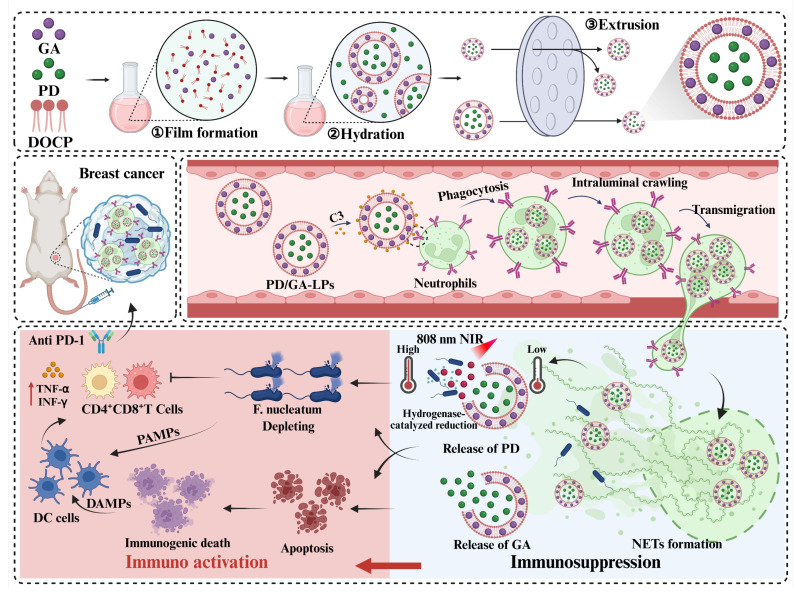
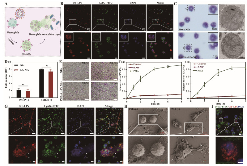
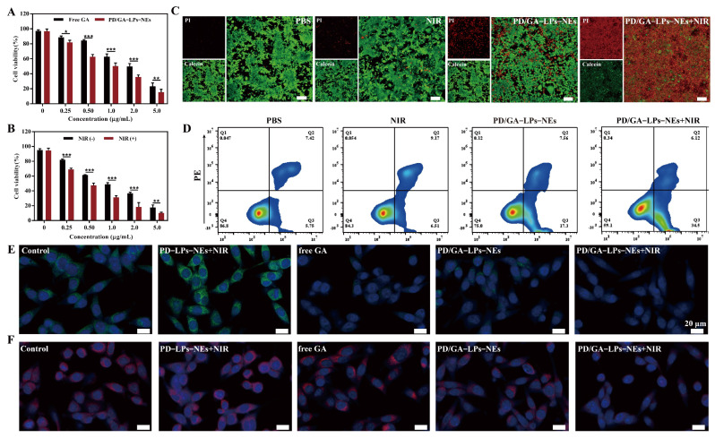
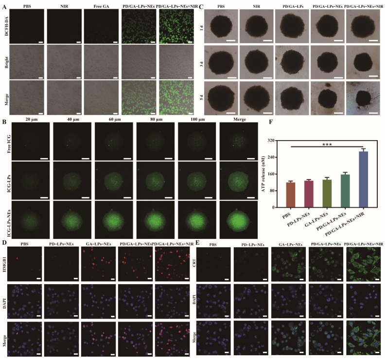
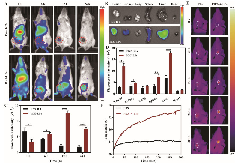
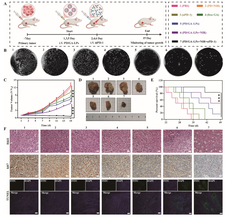
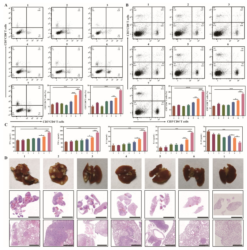
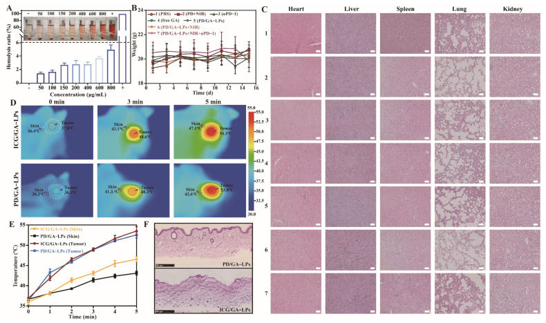
Objective: F. nucleatum, a tumor-resident bacterium colonizing breast cancer (BC), results in an immunosuppressive microenvironment and facilitates tumor growth and metastasis. This study aimed to develop a neutrophil-based liposome delivery system designed for dual-targeted elimination of tumor cells and F. nucleatum, while simultaneously upregulating pathogen-associated molecular patterns and damage-associated molecular patterns to potentiate tumor immunotherapy. Methods: The liposomes (PD/GA-LPs) loaded with the perylene diimide complex (PD) and gambogic acid (GA) were fabricated via the extrusion method. Subsequently, comprehensive evaluations including physicochemical characteristics, antibacterial activity, antitumor effect, and immunomodulatory effect evaluation were systematically conducted to validate the feasibility of this delivery system. Results: The resulting PD/GA-LPs exhibited a dynamic size (121.3 nm, zeta potential -44.1 mV) and a high encapsulation efficiency of approximately 78.1% (PD) and 91.8% (GA). In addition, the optimized PD/GA-LPs exhibited excellent photothermal performance and antibacterial efficacy. In vitro cellular experiments revealed that PD/GA-LPs exhibited enhanced internalization by neutrophils, followed by extracellular trap-mediated release, ultimately significantly inhibiting tumor cell proliferation and inducing immunogenic cell death. During in vivo treatment, PD/GA-LPs exhibited targeted tumor accumulation, where F. nucleatum-driven PD reduction activated near-infrared-responsive photothermal ablation. When combined with GA, this delivery system effectively eliminated tumor cells and F. nucleatum, while facilitating the subsequent T-cell infiltration. Conclusions: This strategy amplified the antitumor immune response, thus leading to effective treatment of BC and prevention of metastasis. In summary, this approach, grounded in the distinct microecology of tumor and normal tissues, offers novel insights into the development of precise and potent immunotherapies for BC.

Keywords: breast cancer; intratumor bacteria; neutrophil; photoimmunotherapy; tumor immune microenvironment.


Conflict of interest statement

The authors declare no conflicts of interest. All authors have read and agreed to the published version of the manuscript.

Figures

Scheme 1
Scheme 1
The schematic diagram of the mechanism of the PD/GA-LPs triggered by intratumoral bacteria to enhance anti-tumor immunity.
Figure 1
Figure 1
Preparation and characterization of PD/GA-LPs. (A) Schematic illustration of PD/GA-LP preparation. (B) The representative photograph and TEM images of PD/GA–LPs (Scale bar: 200 μm, 500 μm). (C) Hydrodynamic size distributions of PD/GA-LPs. (D) UV–vis spectra of free GA, free PD, and PD/GA-LPs. (E) The DL and EE of PD/GA-LPs (n = 3). (F) The cumulative GA and PD release from PD/GA-LPs (n = 3). (G) UV–vis absorption spectra of PD/GA-LPs before and after incubation of F. nucleatum. (H) Infrared thermographic maps of PD/GA-LPs in the presence of F. nucleatum under NIR irradiation (808 nm, 2 W/cm2) (n = 3). (I) Temperature profile of PD/GA–LPs with 808 nm laser irradiation for different durations. (J) Colony-forming unit (CFU) ratio of F. nucleatum with various formulations under 808 nm NIR irradiation for 10 min (n = 3). (K) Colony plate images of F. nucleatum treated with various formulations (ns indicates p > 0.05; ** p < 0.01).
Figure 2
Figure 2
Evaluation of the biological function of PD/GA–LPs–NEs. (A) Schematic illustration of PD/GA–LPs–NEs formation and release in vitro. (B) CLSM images of NEs after co-incubation with DiI–LPs for 4 h (Scale bar: 50 μm, 10 μm). (C) Morphological images of NEs and PD/GA–LPsc–NEs stained with Diff-Quick, SA–β–Gal, and DAPI. (D) Cell numbers of NEs and PD/GA–LPs–NEs after incubation with 10 nM fMLP for 30 min (n = 3). (E) The chemotaxis of NEs before and after loading with PD/GA–LPs (Scale bar: 50 μm). (F) The amount of PD and GA released from PD/GA–LPs–NEs with varied treatments (n = 3). (G) CLSM images of DiI-LPs releasing from DiI–LPs–NEs (Scale bar: 50 μm). (H) SEM images of NETs before and after PMA incubation (Scale bar: 10 μm, 5 μm). (I) CLSM images of DiI–LP uptake by 4T1 cells (Scale bar: 10 μm, 5 μm) (ns indicates p > 0.05).
Figure 3
Figure 3
In vitro anticancer effects of PD/GA-LPs against 4T1 cells. (A) Cell viability of 4T1 cells incubated with various formulations for 24 h (n = 4). (B) Cell viability of 4T1 cells after PD/GA-LPs-NEs treatment for 24 h with or without laser irradiation (n = 4). (C) Live-dead staining of 4T1 treated with PBS and different formulations for 4 h (Scale bar: 100 μm). (D) Apoptosis analysis via an Annexin V-FITC/PI assay of 4T1 cells incubated with PBS and different formulations for 4 h (n = 3). (E) CLSM images of the expression of the HSP90 protein in 4T1 cells after various treatments (Scale bar: 20 μm). (F) CLSM images of the expression of the HSP70 protein in 4T1 cells after various treatments (Scale bar: 20 μm). (* p < 0.05, ** p < 0.01, *** p < 0.001).
Figure 4
Figure 4
The inhibitory effect of PD/GA–LPs on tumor spheroid proliferation and the PTT–induced ICD in vitro. (A) ROS production in 4T1 cells treated with PBS, free GA, PD/GA–LPs-NEs with or without irradiation. (B) CLSM images of the in vitro penetration of ICG-LPs-NEs into the 3D tumor spheroids (Scale bar: 100 μm). (C) Cell viability of 3D tumor spheroids after incubation with different formulations for 5 d (Scale bar: 200 μm). (D) CLSM images of exposed HMGB1 in 4T1 cells with various formulations as indicated (Scale bar: 20 μm). (E) CLSM images of exposed CRT in 4T1 cells with various formulations as indicated (Scale bar: 20 μm). (F) ATP secretion in 4T1 cells after treatment with various formulations (n = 3) (*** p < 0.001).
Figure 5
Figure 5
In vivo biodistribution and photothermal profile of PD/GA–LPs in 4T1 tumor-bearing mice. (A) In vivo fluorescence images of 4T1 tumor-bearing mice captured at the indicated time points after intravenous administration of free ICG and ICG–LPs. (B) The quantification of the relative fluorescence signals of free ICG and ICG-LPs in the tumor site at different time points. (C) Ex vivo fluorescence images of major organs excised from mice intravenously injected with free ICG and ICG–LPs at 24 h of postinjection (n = 3). (D) Quantification analysis of relative fluorescence intensity in the organs at 24 h after intravenous injection (n = 3). (E) Infrared thermographic images of tumors of mice injected with PBS and PD/GA–LPs with laser irradiation at 24 h after injection. (F) Time-dependent tumor temperature increase of 4T1 tumor-bearing mice irradiated by the 808 nm laser at 24 h after i.v. injection of PBS and PD/GA–LPs (n = 3) (* p < 0.05, ** p < 0.01, *** p < 0.001).
Figure 6
Figure 6
In vivo antitumor evaluation. (A) Schematic illustration of the procedure of administration in vivo. (B) The growth profiles of 4T1 tumors in mice that received different treatments. (C) Images of tumors harvested from 4T1-bearing mice on day 15 after various treatments. (D) Survival curves of 4T1 tumor-bearing mice after various treatments. (E) Colony plate images of tumors harvested from mice receiving different treatments on their 15th day. (F) H&E staining, Ki-67, and TUNEL immunohistochemical images of the tumor harvested from 4T1-bearing mice after different treatments (Scale bar: 100 μm) (*** p < 0.001).
Figure 7
Figure 7
Immune activation and antimetastatic effect of the PD/GA-LPs in vivo. (A) Flow cytometry analysis and corresponding quantitative analysis of cytotoxic T lymphocytes gated at CD3+CD4+ CD8+ T cells from primary tumors (n = 3). (B) Flow cytometry analysis and corresponding quantitative analysis of cytotoxic T lymphocytes gated at CD3+CD4+ CD8+ cells from spleens (n = 3). (C) IFN-γ, TNF-α, IL-2, IL-6, and IL-10 cytokine levels in serum isolated from treated mice (n = 3). (D) Representative photographs and H&E staining of lungs in each group (Scale bar: 5 μm, 1 μm) (* p < 0.05, ** p < 0.01, *** p < 0.001, *** p < 0.0001).
Figure 8
Figure 8
The biosafety evaluation of PD/GA-LPs. (A) Hemolysis ratios and hemolysis images of PD/GA-LPs (n = 3). (B) H&E staining of vital organ sections after various treatments on 4T1 tumor-bearing. (C) Body weight of 4T1 tumor-bearing mice after being treated with various formulations ale bar: 100 μm). (D) Infrared thermal images and the temperature evolution of the tumor and skin tissue of mice treated with ICG/GA-LPs and PD/GA-LPs at 808 nm laser irradiation. (E) Time-dependent tumor temperature increase of 4T1 tumor-bearing mice irradiated by the 808 nm laser (n = 3). (F) H&E section of the skin of 4T1 tumor-bearing mice irradiated by the 808 nm laser (Scale bar: 500 μm).

Similar articles

References

    1. Asleh K., Riaz N., Nielsen T.O. Heterogeneity of triple negative breast cancer: Current advances in subtyping and treatment implications. J. Exp. Clin. Cancer Res. 2022;41:265. doi: 10.1186/s13046-022-02476-1. - DOI - PMC - PubMed
    1. Siegel R.L., Miller K.D., Wagle N.S., Jemal A. Cancer statistics, 2023. CA Cancer J. Clin. 2023;73:17–48. doi: 10.3322/caac.21763. - DOI - PubMed
    1. Keenan T.E., Tolaney S.M. Role of immunotherapy in triple-negative breast cancer. J. Natl. Compr. Cancer Netw. 2020;18:479–489. doi: 10.6004/jnccn.2020.7554. - DOI - PubMed
    1. Dvir K., Giordano S., Leone J.P. Immunotherapy in breast cancer. Int. J. Mol. Sci. 2024;25:7517. doi: 10.3390/ijms25147517. - DOI - PMC - PubMed
    1. Pivot X. Pembrolizumab in the treatment of breast cancer. N. Engl. J. Med. 2022;387:273–274. doi: 10.1056/NEJMe2207532. - DOI - PubMed

LinkOut - more resources