Review
. 2025 Apr 24;13(5):969.
doi: 10.3390/microorganisms13050969.

Some Insights into the Inventiveness of Dinoflagellates: Coming Back to the Cell Biology of These Protists

Affiliations
Review

Some Insights into the Inventiveness of Dinoflagellates: Coming Back to the Cell Biology of These Protists

Marie-Odile Soyer-Gobillard. Microorganisms. .

Abstract

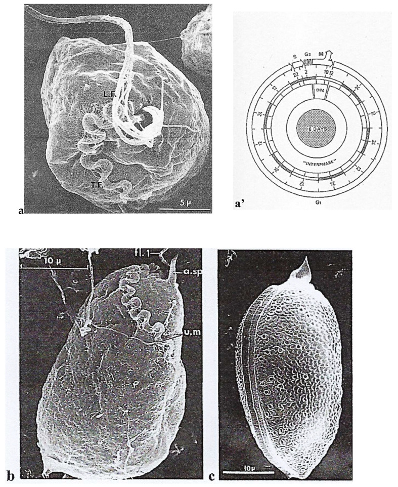
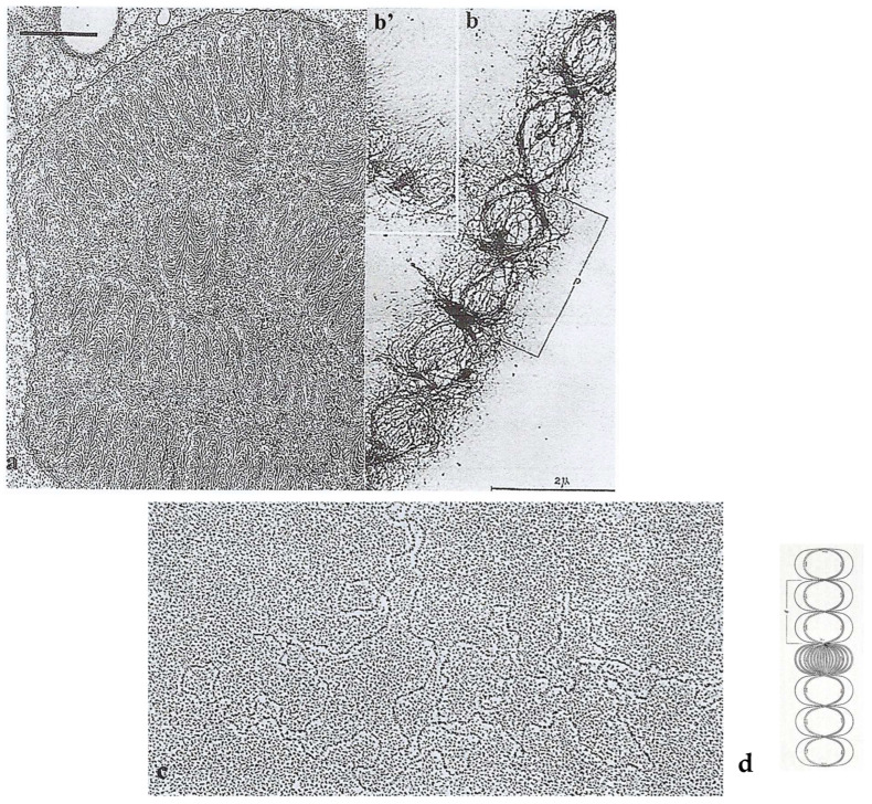
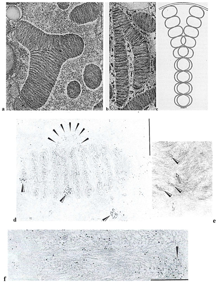
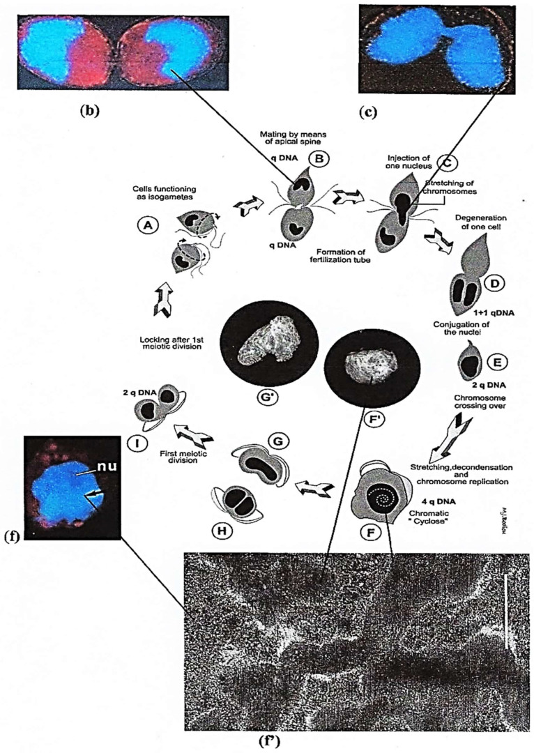
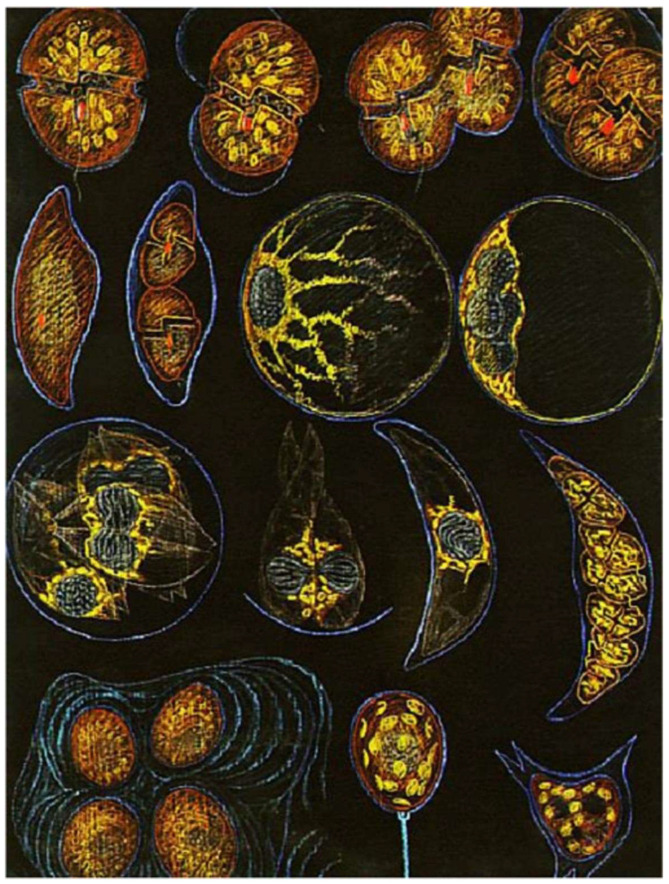
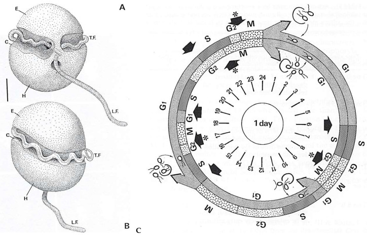
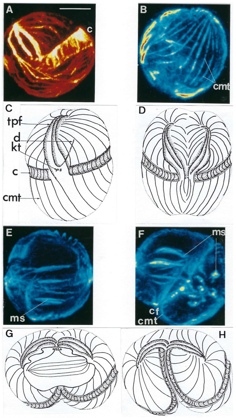
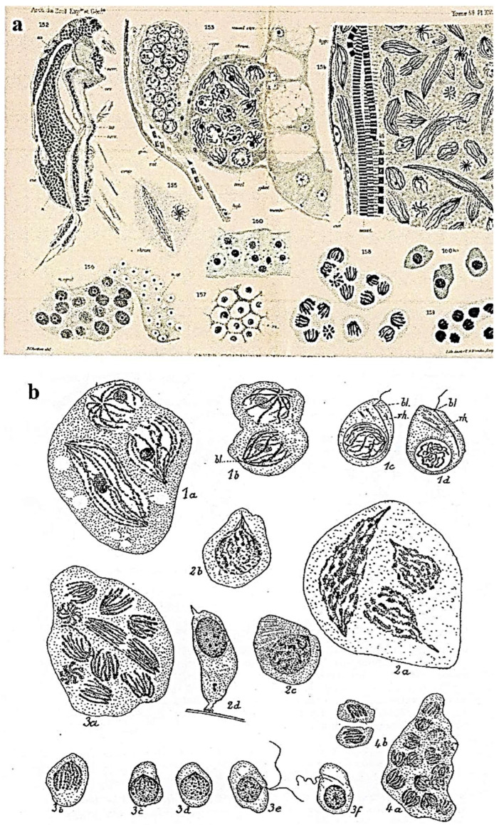
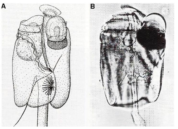
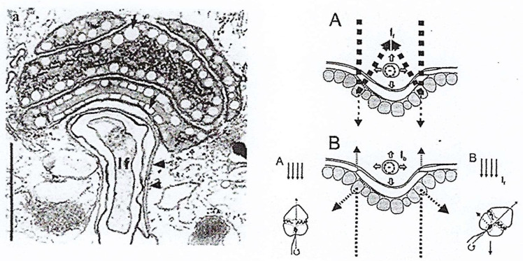
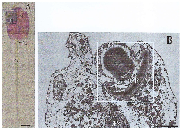
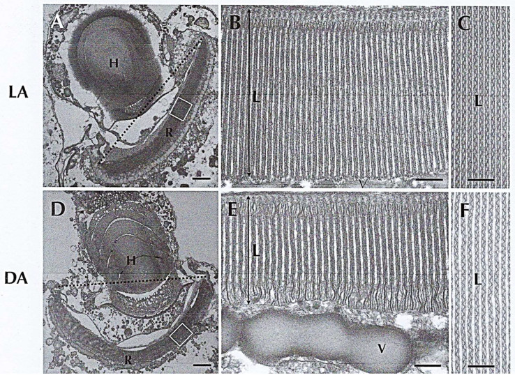
In this review dedicated to the great protistologist Edouard Chatton (1883-1947), I wanted to highlight the originality and remarkable diversity of some dinoflagellate protists through the lens of cell biology. Their fossilized traces date back to more than 538 million years (Phanerozoic eon). However, they may be much older because acritarchs from the (Meso) Proterozoic era (1500 million years ago) could be their most primitive ancestors. Here, I described several representative examples of the various lifestyles of free-living (the autotrophic thecate Prorocentrum micans Ehrenberg and the heterotrophic athecate Noctiluca scintillans McCartney and other "pseudo-noctilucidae", as well as the thecate Crypthecodinium cohnii Biecheler) and of parasitic dinoflagellates (the mixotroph Syndinium Chatton). Then, I compared the different dinoflagellate mitotic systems and reported observations on the eyespot (ocelloid), an organelle that is present in the binucleated Glenodinium foliaceum Stein and in some Warnowiidae dinoflagellates and can be considered an evolutionary marker. The diversity and innovations observed in mitosis, meiosis, reproduction, sexuality, cell cycle, locomotion, and nutrition allow us to affirm that dinoflagellates are among the most innovative unicells in the Kingdom Protista.

Keywords: cell biology; dinoflagellates; evolution; innovative features.


Conflict of interest statement

The author declares no conflicts of interest.

Figures

Figure 1
Figure 1
Scanning electron microscope images of the autotrophic dinoflagellate P. micans Ehr. (a) Apical view showing the peripheral polysaccharidic envelope (epitheca) conserved after soft centrifugation. Two longitudinal flagella (L.F.) of this pre-dividing cell run more or less parallel to each other; the transverse(oblique) flagellum (T.F.) is also visible. Scale bar = 5 µm. (a’) P. micans cell cycle and duration of the different phases: S < 4 h; G2 short (<4 h), G2 + M = 8 h; S + G2 + M ≤ 12 h; G1 = 120 h. From Bhaud and Soyer-Gobillard [19]. Courtesy of Elsevier. (b) One undulating, transverse (oblique) flagellum lined with an undulating membrane (u.m) arises from the same opening as the longitudinal flagella and adheres to the outer layer. Scale bar = 10 µm (a.sp., apical spine). (a,b) Reproduced from Soyer-Gobillard et al. [20]. Courtesy of Elsevier. (c) After stronger centrifugation, two halves with numerous pores, separated by a central suture, are visible, with the apical spine visible at the top. Scale bar = 10 µm. From M.-O. Soyer-Gobillard et al. [21]. Courtesy of Vie Milieu Life & Environment.
Figure 2
Figure 2
(a) TEM observation of an ultrathin section of the nucleus of a non-dividing P. micans Ehrenberg cell after specific fixation (Karnovsky-Soyer technique) showing regular arch-shaped chromosomes. Scale bar = 1 µm. From Soyer-(Gobillard) M.-O. [17]. Courtesy of Wiley. (b) P. micans chromosome spread in water showing the regular unwound periodic organization of the nucleofilaments supporting the hypothesis that P. micans chromosome is composed of numerous circular chromatids as shown on the schematic drawing of (d) p: pitch (around 2 µm). (b’) Extrachromosomal filaments. (c) Circular chromatid after spreading and carbon-platin shadowing of the molecules from spread extracts of another free-living dinoflagellate, Crypthecodinium cohnii Biecheler. In this species, chromosomes are ten times shorter than in P. micans Ehr. The length of de DNA molecule is about 120 µm [24]. Unpublished image (M.-O. Soyer-Gobillard). (d) Model of circular chromatids illustrating the dinoflagellate P. micans chromosomes structure as shown after stretching on water. p: pitch (b,b’,d): From Haapala, O.-K. and Soyer-Gobillard M.-O. [22]. Courtesy of Springer Nature.
Figure 3
Figure 3
(a,b) TEM images of P. micans Ehr. dividing chromosomes: (a) The ultrathin section of dividing chromosomes fixed to the nuclear envelope (arrows) in which the nucleofilament regular organization can be observed. (b) Segregating chromosomes, close to the nuclear envelope (not visible on this picture), supporting the hypothesis that chromosomes are made of many circular chromatids, as shown in the schematic representation of our model (c). From Soyer-(Gobillard) M.-O. [21]. Courtesy of Wiley. (d) Double labeling of chromosome nucleofilaments with anti-B-DNA antibody (black arrowheads, 5 nm gold particles) forming a loop in the chromosome periphery and Z-DNA antibody (white and black arrowheads); observe the clusters of 7 nm gold particles. Bar = 0.5 µm. (e) Immunolocalization of Z-DNA on dividing chromosomes. The white arrow is located in the fission zone. Observe the clusters of 7 nm gold particles (black and white arrow heads). Bar = 0.5 µm. (f) High magnification showing immunolocalization of B-DNA with 5 nm gold particles on the chromosome nucleofilaments visible in the background. Bar = 0.2 µm. From Soyer-Gobillard et al. [18]. Courtesy of Rockefeller University Press.
Figure 4
Figure 4
Sexual reproduction phases of the dinoflagellate P. micans Ehr. (AI) Diagram based on in vivo observations of the nuclei after exposure of cultured P. micans cells to low temperature (4 °C) in the dark for 12 h. Vegetative cells, which function as isogametes (A) and contain n = qDNA, become paired through their apical spines (B). Then, the donor cell injects its nucleus with stretched chromosomes into the receiver cell (C). (b,c,f,f’) In vivo DAPI-stained nuclei of P. micans Ehr cells (blue color): (b) corresponds to B, (c) to (C), (f) to (F). After the conjugation of the two nuclei (D) in the zygote containing n = 2qDNA (E), the chromosomes cross over. Chromosomes become completely unwound and stretched (f’). In the nucleus containing n = 4qDNA (F,F’), chromatin begins to spin around on the right (chromatic cyclosis), giving a round shape to the nucleus (nu) as shown in (f). (f’) TEM image of the disorganized chromosome structure during chromatic cyclosis corresponding to microscope optical pic-tures (f,F,F’). Scale bar = 1 µm. Only one meiotic division occurs (G,G′,H), leading to n = 2qDNA-containing cells (I). From Soyer-Gobillard, M.-O. et al. [29]. By copyright permission from Vie Milieu Life and Environment.
Figure 5
Figure 5
Intranuclear microcables in the nucleoplasm observed after P. micans cell vitrification (−269 °C) followed by cryosubstitution according to the methods described in [17]. (a) At this meiosis stage, during the chromatic cyclosis, chromosomes (Ch.) are unwound (× 37,000), and (a’,b), nucleoplasmic microfilaments are supercoiled (black arrowhead) (× 110,000). (c) The 450A diameter structure represents the longitudinal section of the greatest measured width of four supercoiled microfilaments organized in microcables. From Soyer-(Gobillard), M.-O. [32]. Courtesy of Elsevier. (b,c) Unpublished images of M-O. S-G.
Figure 6
Figure 6
The heterotrophic dinoflagellate N. scintillans McCartney. (a) Adult trophozoite (diploid) observed in vivo with its tentacle X 65. Photo J. Lecomte, Arago Laboratory. (b) Uniflagellated spores (haploid) about to be released. n. nucleus, fl. flagellum. X 1075. (c) Overview of N. scintillans McCartney about to release its spores. X 65. (ac) From M.-O. Soyer-(Gobillard [39]. Courtesy of Springer Nature. (d) Schematic representation of a vegetative cell (a, left), its binary fission (b, middle), and sporocytes (c, d, right). (e) Dividing cells in vivo. (d,e) https://www.istockphoto.com/fr/vectoriel/image-de-zoologie-de-biologie-antique-noctiluca-miliaris-gm1454192599-489914637 (accessed on 17 April 2025).
Figure 7
Figure 7
(ad) The ultrathin section of the nuclear envelope of the trophozoite of Noctiluca scintillans McCartney and the differentiation of its whole internal surface into hundreds of ampullae. (a) A partial view of a N. scintillans trophozoite nucleus (Nu.) showing the nuclear ampullae filled with nuclear pores, the organization of which constitutes the whole nuclear membrane (Zf. Fibrous zone). Scale bar = 2 µm. (b) Higher magnification image showing nuclear ampullae (a) filled with pores (p) that communicate with the cytoplasm through an opening set with a collar (c) and revealing a granular substance (gr) of nucleolar origin (i.e., ribosomes). In the trophozoite, the chromatin is fully decondensed (mf: microfibrils). Scale bar = 1 µm. (c) A schematic representation of a nuclear ampulla with its collar and the organization of the nuclear pores. A, bulb; a, ring; c, collar, Cy, cytoplasm; d, diaphragm; m.i., inner membrane; p, pore. (d) Fusion of the nuclear membranes at the level of the ampullae (a) (stage: 2–4 nuclei). Reproduced from M.-O. Soyer-(Gobillard). [40]. II. Rôle des ampoules nucléaires et de certains constituants cytoplasmiques dans la mécanique mitotique [41]. Courtesy of Wiley, Society of Protistologists.
Figure 8
Figure 8
N. scintillan McCartney during sporulation. At the first stage of division, the trophozoite chromatin is never organized into chromosomes and the nucleofilament mass will separate into two parts, while giant mitochondria (M) (a,b) enter the channels bordered by the nuclear envelope (en.). As divisions progress, the chromatin compacts, revealing an organization in a series of arches (c) with axial formations (arrows) that initiate the future separation between the chromosome masses. From Soyer-(Gobillard) M.-O. [42]. Courtesy of Springer Nature.
Figure 9
Figure 9
N. scintillans McCartney sporulating nucleus (stage: 8 × 2 nuclei) in which the chromatin is being organized into chromosomes with arch-shaped nucleofilaments. Reproduced from M.-O. [42]. Courtesy of Springer Nature.
Figure 10
Figure 10
Phase contrast microscopy images of a N. scintillans McCartney trophozoite. (A) Relaxed tentacle (T). (B) Contracted tentacle with its tip close to the cytostome (Cy). Tracts of myonemes (arrows) are anchored between the cytostome and the supporting rod (SR). Scale bar = 200 µm. Photographs by J. Lecomte [43]. Courtesy of Springer Nature.
Figure 11
Figure 11
Fine structure of the N. scintillans McCartney tentacle. (a) TEM image of an ultrathin transverse section of the tentacle showing the striated myonemes inserted in the epiplasm (E) lined with microtubules. (b) Schematic drawing of a transverse section of the N. scintillans tentacle showing a knot (Kn) of myonemes on the axis, the plasma membrane (PM), vacuoles (V), mitochondria (mi), microtubular rows, and the peripheral alveolar space (AS). (b’,c) Double striation (S, s, arrows) of striated myofibers organized in myonemes and forming a node in the tentacle axis. c: bar = 0.5 µm. (D) Higher magnification of a transverse section of the tentacle showing the insertion of myonemes fixed on the epiplasm (E) between several (5) rows of microtubules linked together (arrows). (ac) Reproduced from Soyer-(Gobillard) M.-O. [39]. Courtesy of Springer Nature. (b’,D) Reproduced from Métivier, Ch. and Soyer-Gobillard, M.-O. [43]. Courtesy of Springer Nature.
Figure 12
Figure 12
The fine structure of the cytostome (oral apparatus) in N. scintillans McCartney. (a) A schematic representation of a N. scintillans: the cytostome (Cy) is connected to a fixed rod structure or sulcus (S) by a curtain of many myofilaments (mf). N, nucleus, c, cytoplasmic span. (b) A transverse section showing the fine structure of the cytostome (Cy) bordered by a thickened lip (arrow). (c) Anchoring of striated myonemes (arrows) to the cytostome border. Scale bar = 0.5 µm. (d) A striated myoneme (My) composed of many myofibrils. Scale bar = 1 µm. (e) A schematic representation of the connection between the anchoring sulcus (S) and the cytostome (Cy) by long ribbons of myonemes (My) mp: plasmic membrane. (ae): Reproduced from Soyer-(Gobillard), M.-O. [39]. Courtesy of Springer Nature.
Figure 13
Figure 13
Several free-living bioluminescent (athecate and thecate) dinoflagellates and the life cycle of the genus Pyrocystis Murray ex Haeckel, 1890. The species Pyrocystis pseudonoctiluca Wyville-Thomson, 1876, is in the second row on the right; Pyrocystis lunula Schütt 1896 is in the third row, on the right. On the first row, on the right, is represented Lingulodinium spp. J.D. Dodge, 1989, (Gonyaulax polyedra F. Stein, 1883), a thecate bioluminescent dinoflagellate. The blue color of the cell cover simulates their bioluminescence at night. In these species, this phenomenon, as well as its chemistry and molecular control, has been described [35,36]. Reproduced from Soyer-(Gobillard), M.-O. and Schrével, J. [3]. Copyright of page 115 is courtesy of “Bibliothèque du Laboratoire Arago-Sorbonne Université”, bequest Lwoff.
Figure 14
Figure 14
(A,B) A schematic representation of a Crypthecodinium cohnii Biecheler cell. Upper part: Ventral view; lower part: dorsal view. Also indicated are the episome (E), hyposome (H), longitudinal flagellum (LF), transverse flagellum (TF) and cingulum (C). Bar = 5 µm. Reproduced from Perret, E. et al. [48]. Courtesy of Wiley (The Company of Biologists Limited). (C) A diagram of two complete successive C. cohnii cell cycles (16 h), represented by two circles inside each other. At the end of each, four daughter cells are released. The transition points G1-S (‘start’ point) and G2-M are represented by arrows and by arrows plus an asterisk, respectively. Reproduced from Bhaud, Y. et al. [47]. Courtesy of Wiley (The Company of Biologists Limited).
Figure 15
Figure 15
(AH) The organization of the cortex microtubules and the microtubular spindle during mitosis as observed by confocal microscopy. Images (A,B,E,F) are reconstructions of each 16 confocal laser scanning sections of C. cohnii cells after labeling with anti-β-tubulin antibody. Images (C,D,G,H) are schematic drawings interpreting the confocal images. Bar = 10 µm. (tpf: three-pronged fork, d: desmose, ms: mitotic spindle, cf: cleavage furrow, c: cingulum, cmt: cortical microtubular rows, kt: kinetosomes. Reproduced from Soyer-Gobillard M.-O. et al. [49]. Courtesy of Elsevier.
Figure 16
Figure 16
(a) Syndinium turbo Chatton, coelomic parasite of pelagic copepods. At the top, a plasmodium with nuclei containing five chromosomes. At the bottom, on the right, sporocytes with their five chromosomes. In Thèse de CHATTON, E. [1]. (b) Plasmodia with dividing nuclei and dinospores (bottom) rh: rhizoplast, bl: blepharoplast (organelles of the flagellar structure). 1. Syndinium rostratum; 2. S. corycoei; 3. S. turbo; 4. S. microsporum. E. Chatton del. From Titres et Travaux Scientifiques by Chatton [2]. E. Chatton del.
Figure 17
Figure 17
TEM images of Syndinium sp. (a) One dividing cell showing one of the five V-shaped chromosomes linked by microtubules of the mitotic spindle to one of the two centrioles that compose the centrosome (white arrows). Scale bar = 0.5 µm From Ris, H. and Kubai, D.F. [58]. Courtesy of Rockefeller University Press. (b) Sporocyte of Syndinium sp. just before its emission from the coelomic cavity of the Copepod. No plastid is visible. The chromosomes are fragmented in the nucleus and an external flagellum is visible in cross section (arrow). Scale bar = 0.5 µm. From Soyer-(Gobillard) M.-O. [59]. Courtesy of Vie Millieu Life & Environment.
Figure 18
Figure 18
A schematic representation of a dinoflagellate (except Syndinium, Oxyrrhis and, probably, Noctiluca) mitotic apparatus in anaphase with the list of remarkable centrosome-associated proteins. Microtubular mitotic spindles lie throughout the nucleus, pass into the archoplasmic spheres (Golgi apparatus), and are linked to the two pairs of kinetosomes or flagellar bases. AS, archoplasmic sphere (containing Golgi bodies); C, centrosome (without centrioles); ECC, extranuclear cytoplasmic channel; MMS, microtubular mitotic spindle; MD, microtubular desmose; Kt, kinetosomes; N, nucleus; NE, nuclear envelope (permanent); Ch, chromosomes. Reproduced from Soyer-Gobillard, M.-O. et al. [49]. Courtesy of Elsevier.
Figure 19
Figure 19
A schematic representation of nuclear division of Syndinium sp. that contains five V-shape chromosomes. (a) Interphase. (b) Early division: centrosome (two centrioles), kinetochores and chromosomes have duplicated. (c) An early stage of chromosome segregation. The central spindle between separating centrioles. (d) A late stage of chromosome segregation. The central spindle in cytoplasmic channel throughout the nucleus. (e) Division of nucleus. (f) An early daughter nucleus with persisting channel and microtubules. Reproduced from Ris, H. and Kubai, D.F. [58]. Courtesy of Rockefeller University Press.
Figure 20
Figure 20
The evolution of the mitotic apparatus in alveolates according to the phylogeny by Bachvaroff et al. [7] and reproduced from Moon, E., et al. [60]. Courtesy of Elsevier.
Figure 21
Figure 21
Ocelloid of the dinoflagellate Erythropsis pavillardi Hertwig. (A) A schematic drawing of E. pavillardi in which the eyespot is represented on the right of the cell anterior part. (B) Light microscopy image showing the hyalosome, which plays the role of the lens (upper part), and the pigment, which plays the role of the retina. From Greuet C. [61].
Figure 22
Figure 22
TEM images of the eyespot of the binucleated dinoflagellate G. foliaceum in the transverse section (a) localized in the posterior part of the cell. The images show the layers of refracting globules that form the pigment cup, composed of carotenoid-rich lipids through which light passes (A) and/or is reflected (B), which determines the cell orientation, according to Kreimer’s hypothesis. Dotted arrows indicate the direction of the light reflected or passing through the cell. The longitudinal flagellum (lf) is visible in the hollow of the posterior sulcus. Bar = 0.5 µm. Reproduced from Kreimer G. (with permission). [69]. Courtesy of Elsevier.
Figure 23
Figure 23
Erythropsidinium spp. dinoflagellate equipped with a piston (PS) involved in its locomotion in water. (A) Light microscopy view of Erythropsidinium spp. with the ocelloid in the left anterior part of the cell (square). PS: piston; R: retina-like body; H: hyalosome; Fl: flagellum. Scale bar = 20 µm. (B) TEM view of the ocelloid. The hyalosome (H), (crystalline body) plays the role of a lens; observe at its base the lamellated retina-like body (R). Scale bar = 10 µm. From Hayakawa, S. et al. [72]. Courtesy of PLoS ONE.
Figure 24
Figure 24
(AF). Detail of ocelloid of Erythropsidinium spp. and the variations of its morphology in different conditions of light. Light state (LA: AC) and dark state (DA: DF). (B,E): longitudinal sections of retinal body; (C,F). cross sections of retinal body are shown. L. lamellae; V. vesicular layer. A,D. Bar = 2 µm; B,C,E,F, Bar= 0.2 µm. From Hayakawa, S. et al. [72]. Courtesy of PLoS ONE.

Similar articles

References

    1. Chatton E. Les Péridiniens Parasites: Morphologie, Reproduction, Ethologie. Librairie, H. Le Soudier; Paris, France: 1920. pp. 1–475.
    1. Chatton E. Titres et Travaux Scientifiques (1906–1937) Imp-Editor Sottano; Sète, France: 1938. pp. 1–405.
    1. Soyer-Gobillard M.-O., Schrével J. The Discoveries and Artistic Talents of Edouard Chatton and André Lwoff, Famous Biologists. 1st ed. Cambridge Scholars Publishing; Cambridge, UK: 2021. pp. 1–228.
    1. Chatton E. Pansporella perplexa. Ann. Sci. Nat. Zool. 1925;8:1–85.
    1. Taylor F.J.R. Phylum Dinoflagellata. In: Margulis L., Corliss J., Melkonian M., Chapman D.J., McKhan H.I., editors. Handbook of Protoctista. 1st ed. Jones and Bartlett Publishers; Boston, MA, USA: 1990. pp. 419–437.

LinkOut - more resources