

Fecal Microbiota Transplantation Using Donor Stool Obtained from Exercised Mice Suppresses Colonic Tumor Development Induced by Azoxymethane in High-Fat Diet-Induced Obese Mice
- PMID: 40431182
- PMCID: PMC12114393
- DOI: 10.3390/microorganisms13051009
Fecal Microbiota Transplantation Using Donor Stool Obtained from Exercised Mice Suppresses Colonic Tumor Development Induced by Azoxymethane in High-Fat Diet-Induced Obese Mice
Abstract
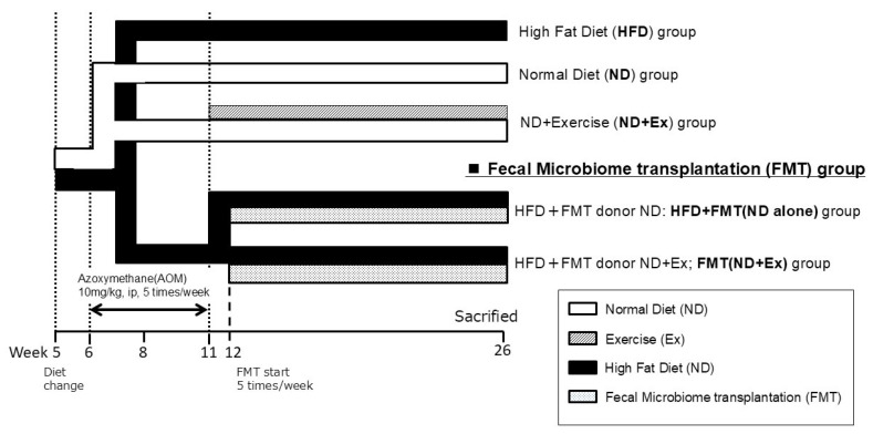
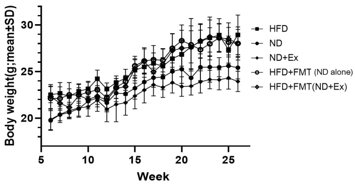
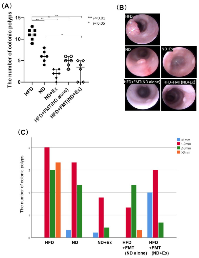
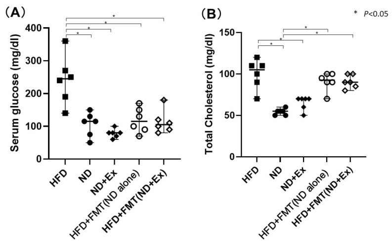
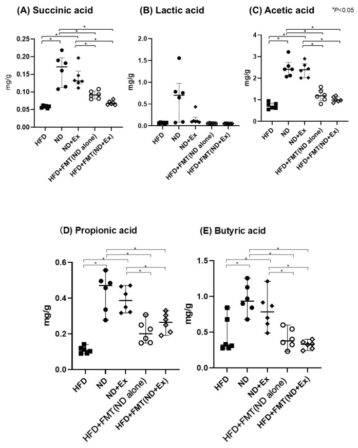
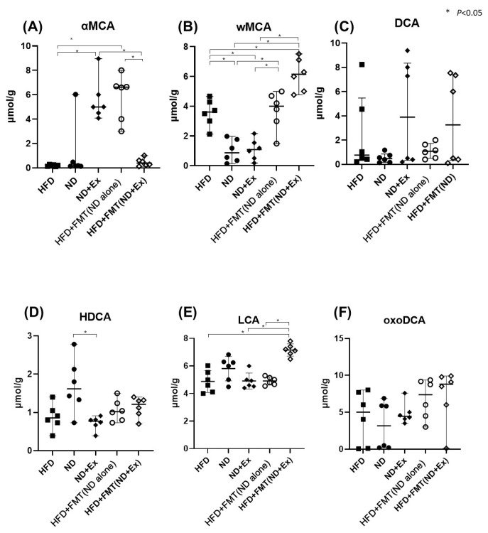
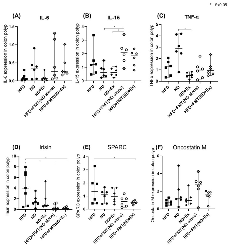
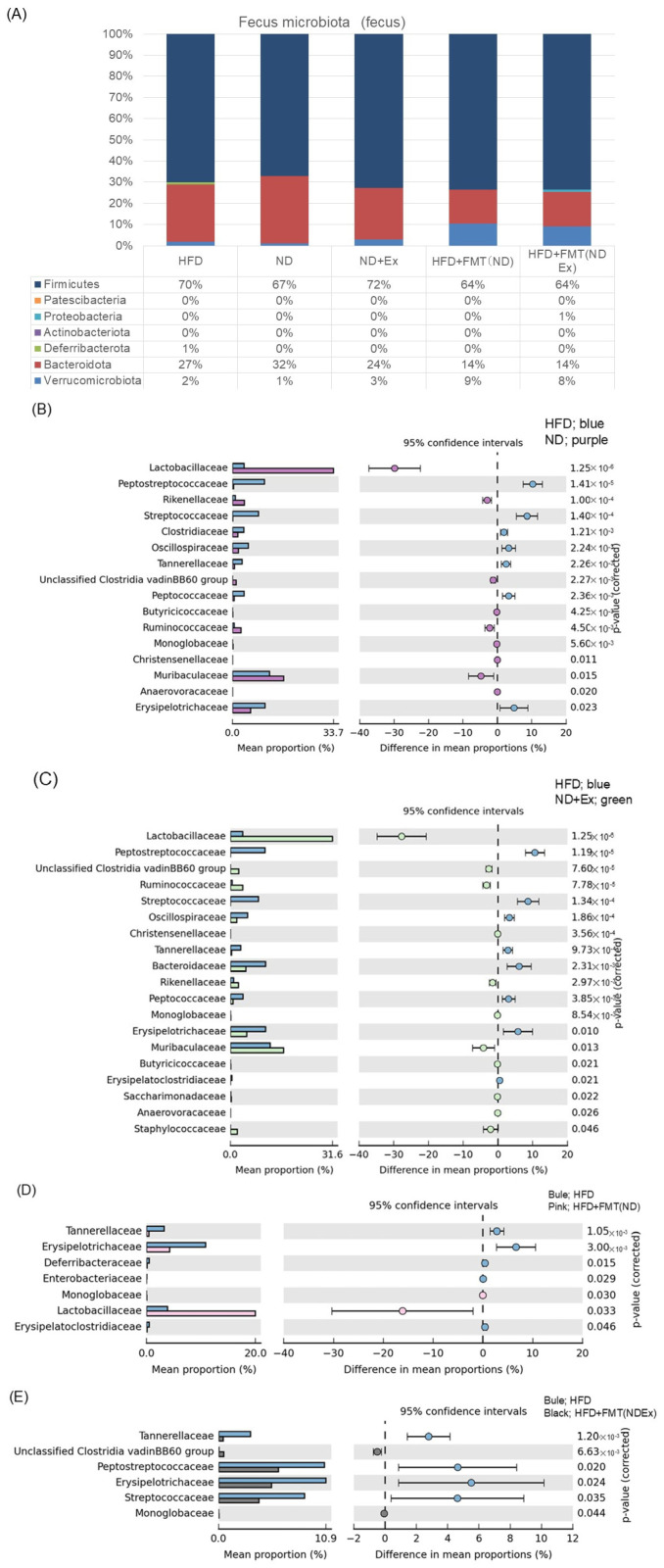
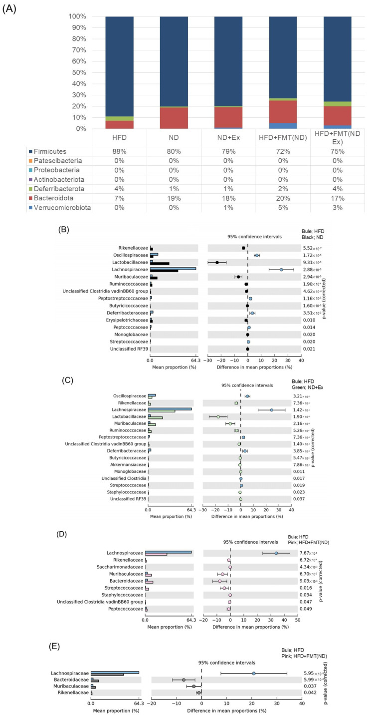
The gut microbiota plays an important role in the development of colorectal tumors. However, the underlying mechanisms remain unclear. In this study, we examined the effects of fecal microbiota transplantation (FMT) on azoxymethane (AOM)-induced colorectal tumors in obese mice. We divided the study subjects into the following five groups: high-fat diet (HFD), normal diet (ND), ND+exercise (Ex), HFD+FMT from ND-alone donor (HFD+FMT(ND alone)), and HFD+FMT from ND+Ex donor (HFD+FMT(ND+Ex)). The Ex group performed treadmill exercise for 15 weeks. Thereafter, fecal and colonic mucus samples were extracted for microbiome analysis. The deoxyribonucleic acid sample was collected from the feces and colonic mucosa, and V3-V4 amplicon sequencing analysis of the 16S rRNA gene was performed using MiSeq. The number of polyps was significantly lower in the ND (6.0 ± 1.6) and ND+Ex (1.8 ± 1.3) groups than in the HFD group (11.4 ± 1.5). The ND+Ex group had significantly fewer polyps than the ND group. The HFD+FMT(ND alone) (5.2 ± 0.8) and HFD+FMT(ND+Ex) (2.8 ± 2.6) groups also had significantly fewer polyps than the HFD group. The IL-15 mRNA levels in the colonic tissues were significantly higher in the HFD+FMT(ND alone) group than in the ND group. Fecal ω-muricholic acid concentrations were significantly higher in the HFD+FMT(ND alone) group than in the ND group and in the HFD+FMT(ND+Ex) group than in the ND+Ex group. The ND, ND+Ex, HFD+FMT(ND alone), and HFD+FMT(ND+Ex) groups had a significantly higher abundance of Lacyobacillaceae than the HFD group. In the FMT group, Erysipelotrichaceae and Tannerellaceae were significantly less abundant. Compared with the HFD group, the ND, ND+Ex, HFD+FMT(ND alone), and HFD+FMT(ND+Ex) groups had a significantly higher abundance of Muribaculaceae and a significantly higher abundance of Lactobacillaceae and Rikenellaceae in common among the ND and ND+Ex groups. The common and significantly less common species were Bacteroidaceae in the FMT group and Lactobacillaceae and Rikenellaceae in the ND alone and ND+Ex groups. Bacteroidaceae and Lachnospiraceae were significantly less common in the FMT group. We found that FMT inhibited AOM-induced colorectal tumorigenesis in obese mice. Furthermore, the fecal concentrations of short-chain fatty acids, bile acids, microbiota, and mucosa-associated microbiota differed between the FMT and diet/EX groups, suggesting that the inhibitory effect of FMT on colorectal tumorigenesis may be due to mechanisms different from those of ND alone and ND+Ex.
Keywords: colorectal cancer; exercise; fecal microbiota transplantation (FMT); high-fat diet; obesity.
Conflict of interest statement
The authors declare no conflicts of interest.
Figures
Similar articles
-
Exercise Affects Mucosa-Associated Microbiota and Colonic Tumor Formation Induced by Azoxymethane in High-Fat-Diet-Induced Obese Mice.Microorganisms. 2024 May 9;12(5):957. doi: 10.3390/microorganisms12050957. Microorganisms. 2024. PMID: 38792787 Free PMC article.
-
Gut microbial and metabolomics profiles reveal the potential mechanism of fecal microbiota transplantation in modulating the progression of colitis-associated colorectal cancer in mice.J Transl Med. 2024 Nov 15;22(1):1028. doi: 10.1186/s12967-024-05786-4. J Transl Med. 2024. PMID: 39548468 Free PMC article.
-
Hepatobiliary and pancreatic: Multi-donor fecal microbiota transplantation attenuated high-fat diet-induced hepatic steatosis in mice by remodeling the gut microbiota.J Gastroenterol Hepatol. 2023 Dec;38(12):2195-2205. doi: 10.1111/jgh.16359. Epub 2023 Oct 3. J Gastroenterol Hepatol. 2023. PMID: 37787118
-
High-fat diet promotes gestational diabetes mellitus through modulating gut microbiota and bile acid metabolism.Front Microbiol. 2025 Jan 28;15:1480446. doi: 10.3389/fmicb.2024.1480446. eCollection 2024. Front Microbiol. 2025. PMID: 39935515 Free PMC article.
-
Lingguizhugan decoction attenuates diet-induced obesity and hepatosteatosis via gut microbiota.World J Gastroenterol. 2019 Jul 21;25(27):3590-3606. doi: 10.3748/wjg.v25.i27.3590. World J Gastroenterol. 2019. PMID: 31367159 Free PMC article.
References
Grants and funding
LinkOut - more resources
Full Text Sources
