. 2025 May 8;13(5):1091.
doi: 10.3390/microorganisms13051091.

Reducing Functional Domain of Histatin 5 Improves Antifungal Activity and Prevents Proteolytic Degradation

Affiliations

Reducing Functional Domain of Histatin 5 Improves Antifungal Activity and Prevents Proteolytic Degradation

Carolina R Zambom et al. Microorganisms. .

Abstract

Histatin 5 (Hst5) is an antifungal peptide (AFP) naturally produced by parotid glands with strong activity against Candida albicans. One of its mechanisms of action is the generation of reactive oxygen species (ROS) inside the C. albicans cells. Despite being an important peptide for the human innate immune response, its activity is reduced or inactivated by proteolytic degradation caused by salivary enzymes. To overcome this barrier, we used solid phase peptide synthesis (SPPS) to modify the Hst5 amino acid sequence improving its antifungal action and minimizing its degradation. We synthesized five peptides, three of which were based on the Hst5 functional domain. We determined that the smallest peptides (8WH5, 7WH5 and 6WH5) demonstrated the greatest antifungal action against C. albicans, including one fluconazole-resistant strain. Besides that, cationic-PAGE and HPLC assays showed that the degradation in saliva was slower for the smaller peptides than for 0WHst5 and WP113. Furthermore, 8WH5, 7WH5 and 6WH5 were found in the samples even after 8 h in whole saliva, while 0WHst5 and WP113 completely disappear after 1.5 h. Finally, we found that the smaller peptides were less fragmented than the 0WHst5 and WP113, so they were the smallest fragments of Hst5 to preserve its antifungal action with reduced degradation in whole saliva. Thus, they can be considered promising molecules for the treatment of C. albicans in the oral cavity.

Keywords: P-113; SPPS; antifungal peptides; degradation; histatin 5; peptides; proteolysis; saliva.


Conflict of interest statement

The authors declare no conflicts of interest.

Figures

Figure 1
Figure 1
Inhibition of C. albicans ATCC 90028 by 0WHst5, WP113, 8WH5, 7WH5 and 6WH5. The experiment was performed in triplicate. * Peptides were statistically compared between treatments of the same concentration. Equal letters indicate statistically equal means while different letters indicate statistically different means.
Figure 2
Figure 2
Inhibition of C. albicans ATCC 18801 by 0WHst5, WP113, 8WH5, 7WH5 and 6WH5. The experiment was performed in triplicate. * Peptides were statistically compared between treatments of the same concentration. Equal letters indicate statistically equal means while different letters indicate statistically different means.
Figure 3
Figure 3
Inhibition of C. albicans ATCC 10231 by 0WHst5, WP113, 8WH5, 7WH5 and 6WH5. The experiment was performed in triplicate. * Peptides were statistically compared between treatments of the same concentration. Equal letters indicate statistically equal means while different letters indicate statistically different means.
Figure 4
Figure 4
Cell viability of C. albicans ATCC 90028 cells after 2 h incubation at 37 °C with dilution series of peptides 0WHst5, WP113, W8H5, 7WH5 and 6WH5. The dilutions were then plated in Sabouraud Dextrose Agar (SDA) media and the logarithm values of CFU mL−1 were calculated for the determination of cell viability.
Figure 5
Figure 5
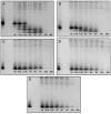
Peptide degradation profile in human saliva (WSS): (A) 0WHst5; (B) WP113; (C) 8WH5; (D) 7WH5; (E) 6WH5. An aliquot of 100 µL was collected after each incubation period and evaluated by cationic-PAGE. The first column on the left (column 1) corresponds to the standard band for all peptides tested; columns 2 to 9 show the different incubation periods (t = 0, 0.5, 1.5, 4, 6, 8, 24 and 48 h).
Figure 6
Figure 6
Peptide degradation over time for 0WHst5, WP113, 8WH5, 7WH5 and 6WH5. Data were obtained after the cationic-PAGE and the amount of peptide in each aliquot was determined using the software Image Lab 6.1 (Bio-Rad Inc.). Error bars correspond to the standard deviations on duplicate samples.
Figure 7
Figure 7
Proteolytic cleavage sites in the peptides target to salivary proteases. The stars indicate all primary cleavage sites and the circle indicates preferred primary cleavage sites.
Figure 8
Figure 8
0WHst5 and WP113 degradation profile in human saliva. Aliquots of 100 µL were boiled and analyzed by HPLC after 0 h, 1.5 h and 8 h of incubation. Black arrows indicate intact 0WHst5 and WP113, retention time in 15.35 and 15.02 min, respectively.
Figure 9
Figure 9
8WH5, 7WH5 and 6WH5 degradation profile in human saliva. Aliquots of 100 µL were boiled and analyzed by HPLC after 0 h, 1.5 h and 8 h of incubation. Black arrows indicate intact 8WH5, 7WH5 and 6WH5, retention time at 14.00, 13.99 and 14.61 min, respectively.

References

    1. Zasloff M. Antimicrobial Peptides of Multicellular Organisms. Nature. 2002;415:389–395. doi: 10.1038/415389a. - DOI - PubMed
    1. Lazzaro B.P., Zasloff M., Rolff J. Antimicrobial Peptides: Application Informed by Evolution. Science. 2020;368:eaau5480. doi: 10.1126/science.aau5480. - DOI - PMC - PubMed
    1. Seo M.D., Won H.S., Kim J.H., Mishig-Ochir T., Lee B.J. Antimicrobial Peptides for Therapeutic Applications: A Review. Molecules. 2012;17:12276–12286. doi: 10.3390/molecules171012276. - DOI - PMC - PubMed
    1. Mwangi J., Hao X., Lai R., Zhang Z.Y. Antimicrobial Peptides: New Hope in the War against Multidrug Resistance. Zool Res. 2019;40:488–505. doi: 10.24272/j.issn.2095-8137.2019.062. - DOI - PMC - PubMed
    1. Bechinger B., Gorr S.U. Antimicrobial Peptides: Mechanisms of Action and Resistance. J. Dent. Res. 2017;96:254–260. doi: 10.1177/0022034516679973. - DOI - PMC - PubMed

LinkOut - more resources