. 2025 May 12;17(10):1643.
doi: 10.3390/nu17101643.

Protective Effects of a Standardized Water Extract from the Stem of Ipomoea batatas L. Against High-Fat Diet-Induced Obesity

Affiliations

Protective Effects of a Standardized Water Extract from the Stem of Ipomoea batatas L. Against High-Fat Diet-Induced Obesity

Chae-Won Lee et al. Nutrients. .

Abstract

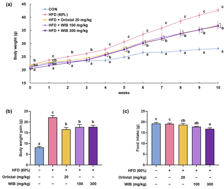
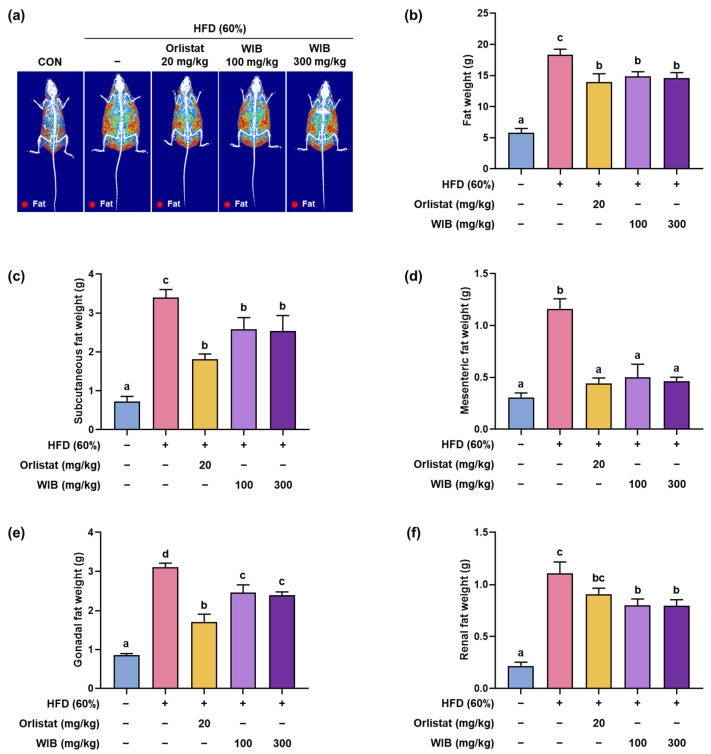
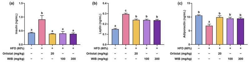
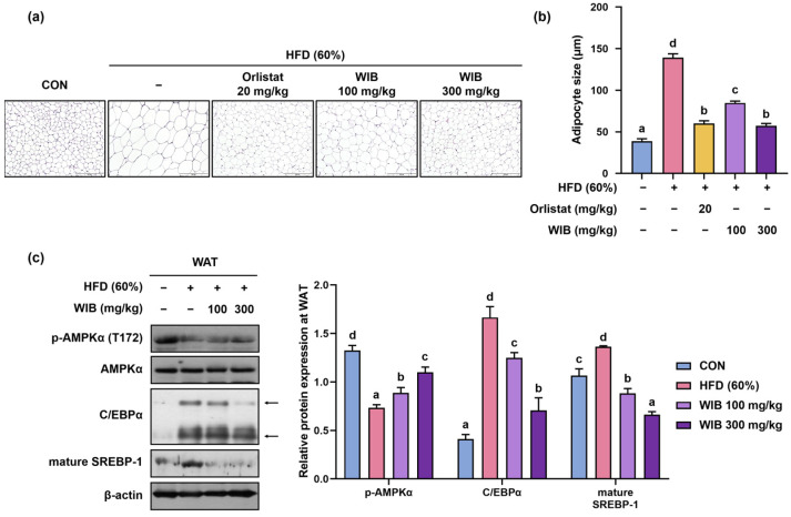
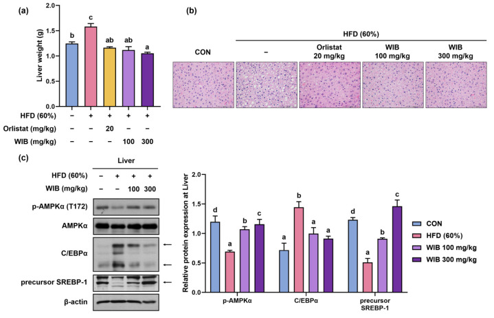
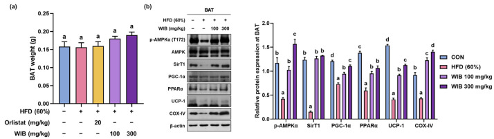
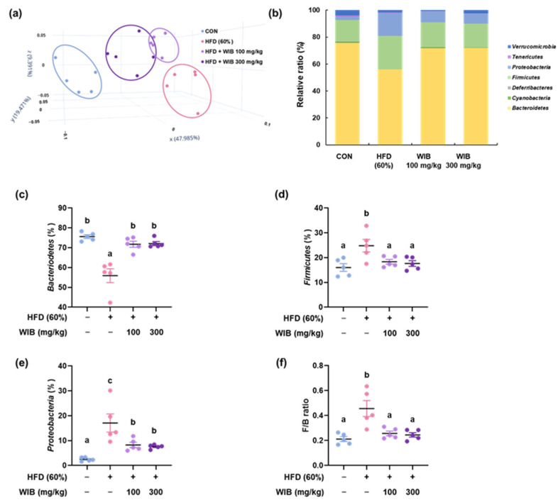
Background/Objectives: Obesity is a major health concern that can lead to various chronic diseases. Little is known about the anti-obesity effect of a standardized hot water extract from the stems of Ipomoea batatas (WIB). This study aimed to evaluate the therapeutic potential of WIB as a natural alternative to conventional anti-obesity treatments by assessing its effects on body weight, fat accumulation, and key metabolic biomarkers in a high-fat diet-induced obesity model. Methods: A high-fat diet (HFD) induced obesity in C57BL/6 mice. The mice were then treated orally with either orlistat (positive control) or WIB. Changes in body weight, food intake, and fat weight were measured, along with blood lipid profiles and adipokines. Western blot analyses were conducted to determine protein levels in each tissue. H&E staining in white adipose tissue and liver, and the gut microbiota composition were analyzed. Results: WIB treatment significantly reduced body weight and fat mass compared to the HFD group and demonstrated comparable effects to orlistat. WIB improved blood lipid profiles and adipokine levels. H&E staining revealed reduced fat accumulation in the white adipose tissue and liver. Also in those tissues, WIB restored expression levels of sterol regulatory element-binding protein-1 (SREBP-1) and CCAAT/enhancer-binding protein α (C/EBPα) and increased AMP-activated protein kinase (AMPK) phosphorylation. In brown adipose tissue, WIB enhanced AMPK phosphorylation and upregulated thermogenic-related proteins, including peroxisome proliferator-activated receptor-gamma coactivator-1α (PGC-1α), peroxisome proliferator-activated receptor α (PPARα), sirtuin 1 (SIRT1), uncoupling protein-1 (UCP-1), and cytochrome C oxidase subunit 4 (COX-IV). Analysis of gut microbiota revealed that WIB normalized β-diversity and reversed HFD-induced phyla imbalances (notably in Bacteroidetes, Firmicutes, and Proteobacteria). Conclusions: By reducing adiposity under the conditions tested in a murine model, improving metabolic markers, and favorably modulating gut microbiota, WIB demonstrates potential in mitigating obesity-related risks. These findings suggest that WIB may serve as a promising natural substance for the management of obesity. Further studies are warranted to confirm its efficacy and explore the potential underlying mechanisms in overweight or obese humans as a health supplement to help manage or prevent obesity.

Keywords: AMP-activated protein kinase; Ipomoea batatas L.; anti-obesity; brown adipose tissue; lipogenesis; liver; microbiota; thermogenesis; white-adipose tissue.


Conflict of interest statement

Authors Ye Seul Yoon and Mi-ju Kim were employed by the Dalim Biotech Co., Ltd. Authors Young-Seo Yoon and Kyung-Sook Chung were employed by the BELABEL BIO. Inc. The remaining authors declare that the research was conducted in the absence of any commercial or financial relationships that could be construed as a potential conflict of interest. The authors declare that this study received funding from DALIM BIOTECH Corporation. The funder was not involved in the study design, collection, analysis, interpretation of data, the writing of this article or the decision to submit it for publication.

Figures

Figure 1
Figure 1
The UPLC-PDA chromatogram and peak-specific UV spectrum of identified WIB compounds; (1) L-tryptophan, (2) neochlorogenic acid, (3) caffeic acid, (4) chlorogenic acid, (5) cryptochlorogenic acid, (6) quercetin 3-O-β-D-sophoroside, (7) hyperoside, (8) isoquercetin, (9) isochlorogenic aicd B, (10) isochlorogenic acid A, (11) isochlorogenic acid C.
Figure 2
Figure 2
Repressive effects of WIB on body weight in HFD-induced obese mice. HFD-induced mice were administered the indicated doses of WIB (100 and 300 mg/kg) daily during the experiment. (a) Body weights, (b) total body weight gain for 10 weeks, and (c) food intake in 1 week (n = 8). Values are represented as the mean ± standard error of the mean (SEM). Values with different letters are significantly different, p < 0.05 (a < b < c).
Figure 3
Figure 3
Effects of WIB on body composition and fat mass in HFD-induced obese mice. (a) Radiography images of fat tissues. (b) The fat weight in each group was measured through DEXA analysis. Fat was obtained from each tissue after sacrificing the mice, and their weights were measured. (cf) Subcutaneous, mesenteric, gonadal, and renal fat weights. Values are represented as the mean ± SEM (DEXA analysis; n = 3, fat in tissue weight; n = 5). Values with different letters are significantly different, p < 0.05 (a < b < c < d).
Figure 4
Figure 4
Effects of WIB on obesity-related hormones in HFD-induced obese mice. (ac) Insulin, leptin, and adiponectin levels. Values are represented as the mean ± SEM (n = 5). Values with different letters are significantly different, p < 0.05 (a < b < c).
Figure 5
Figure 5
Effects of WIB on adipocyte size and lipogenesis in subcutaneous fat tissues of HFD-induced obese mice. (a) Histological analysis of subcutaneous fat tissue via H&E staining at ×20 magnification. (b) Representative diameters of adipocytes in the tissue. (c) Levels of p-AMPKα and lipogenic transcription factors were determined by Western blot analysis. Arrowed bands are the main band of that protein. Quantitative values for the Western blot of p-AMPKα to AMPKα, CEBP/α, and SREBP-1 are shown. β-Actin was used as an internal control. Values are represented as the mean ± SEM (adipocyte size; n = 5, density; n = 3). Values with different letters are significantly different, p < 0.05 (a < b < c < d).
Figure 6
Figure 6
Effects of WIB on lipid accumulation in the liver tissues of HFD-induced obese mice. (a) Comparison of liver weights of each group. (b) The images of H&E-stained liver tissues at ×40 magnification. (c) Expression levels of p-AMPKα and lipogenic transcription factors were determined by Western blot analysis. Arrowed bands are the main band of that protein. Quantitative values for the Western blot of p-AMPKα to AMPKα, CEBP/α, and SREBP-1 are shown. β-Actin was used as an internal control. Values are represented as the mean ± SEM (liver weight; n = 5, density; n = 3). Values with different letters are significantly different, p < 0.05 (a < b < c < d).
Figure 7
Figure 7
Effect of WIB on thermogenesis-related factors in the BAT of HFD-induced obese mice. (a) Weight of BAT in each group. (b) Expression levels of p-AMPKα, AMPKα, and thermogenesis-related proteins were determined by Western blot analysis. Quantitative values for Western blot are shown in graphs. β-Actin was used as an internal control. Values are represented as the mean ± SEM (BAT weight; n = 5, density; n = 3). Values with different letters are significantly different, p < 0.05 (a < b < c < d).
Figure 8
Figure 8
Regulatory effects of WIB on microbiota composition and colonization. (a) β-Diversity between groups was analyzed using a PCoA plot. (b) Phylum-level composition ratios in each group. (cf) Relative ratios of Bacteroidetes, Firmicutes, and Proteobacteria and F/B ratio. Values are represented as the mean ± SEM (n = 5). Values with different letters are significantly different, p < 0.05 (a < b < c).

Similar articles

References

    1. Prattichizzo F., Ceriello A., Shah V.N. Obesity, NAFLD/NASH, and Diabetes. Diabetes Technol. Ther. 2024;26:S231–S240. doi: 10.1089/dia.2024.2516. - DOI - PubMed
    1. Blackstone R.P., Blackstone R.P. Obesity-related diseases and syndromes: Insulin resistance, type 2 diabetes mellitus, non-alcoholic fatty liver disease, cardiovascular disease, and metabolic syndrome. Obes. Med. Pract. Essent. Guide. 2016:83–108.
    1. Longo M., Zatterale F., Naderi J., Parrillo L., Formisano P., Raciti G.A., Beguinot F., Miele C. Adipose Tissue Dysfunction as Determinant of Obesity-Associated Metabolic Complications. Int. J. Mol. Sci. 2019;20:2358. doi: 10.3390/ijms20092358. - DOI - PMC - PubMed
    1. Cheng L., Wang J., Dai H., Duan Y., An Y., Shi L., Lv Y., Li H., Wang C., Ma Q., et al. Brown and beige adipose tissue: A novel therapeutic strategy for obesity and type 2 diabetes mellitus. Adipocyte. 2021;10:48–65. doi: 10.1080/21623945.2020.1870060. - DOI - PMC - PubMed
    1. Petito G., Cioffi F., Magnacca N., de Lange P., Senese R., Lanni A. Adipose Tissue Remodeling in Obesity: An Overview of the Actions of Thyroid Hormones and Their Derivatives. Pharmaceuticals. 2023;16:572. doi: 10.3390/ph16040572. - DOI - PMC - PubMed

LinkOut - more resources