. 2025 Apr 22;17(5):592.
doi: 10.3390/v17050592.

Lassa Virus Infection of Primary Human Airway Epithelial Cells

Affiliations

Lassa Virus Infection of Primary Human Airway Epithelial Cells

Helena Müller-Kräuter et al. Viruses. .

Abstract

Lassa mammarenavirus (LASV), a member of the family Arenaviridae, is a highly pathogenic virus capable of causing severe systemic infections in humans. The primary host reservoir is the Natal multimammate mouse (Mastomys natalensis), with human infections typically occurring through mucosal exposure to virus-containing aerosols from rodent excretions. To better understand the molecular mechanisms underlying LASV replication in the respiratory tract, we utilized differentiated primary human airway epithelial cells (HAECs) grown under air-liquid interface conditions, closely mimicking the bronchial epithelium in vivo. Our findings demonstrate that HAECs are permissive to LASV infection and support productive virus replication. While LASV entry into polarized HAECs occurred through both apical and basolateral surfaces, progeny virus particles were predominantly released from the apical surface, consistent with an intrinsic apical localization of the envelope glycoprotein GP. This suggests that apical virus shedding from infected bronchial epithelia may facilitate LASV transmission via airway secretions. Notably, limited basolateral release at later stages of infection was associated with LASV-induced rearrangement of the actin cytoskeleton, resulting in compromised epithelial barrier integrity. Finally, we demonstrate that LASV-infected HAECs exhibited a pronounced type III interferon response. A detailed understanding of LASV replication and host epithelial responses in the respiratory tract could facilitate the development of targeted future therapeutics.

Keywords: IFN-λ; Lassa mammarenavirus; primary airway epithelial cells; virus–host interactions.


Conflict of interest statement

The authors declare no conflicts of interest. The funders had no role in the design of this study; in the collection, analyses, or interpretation of data; in the writing of the manuscript, or in the decision to publish the results.

Figures

Figure 1
Figure 1
Formation of a polarized phenotype in HAECs. (a,b) HAECs and HFF cells were seeded on coverslips. After fixation with 4% PFA and permeabilization, cells were immunostained for pan-cytokeratin (a) or the fibroblast marker TE-7 (b). Nuclei were visualized by DAPI staining. Scale bars, 20 μm. (c) HAECs were cultivated on Transwell inserts under ALI culture conditions. TEER was measured after medium changes every 2–3 days following ALI induction. Mean values and standard errors of the mean (SEM) of three independent experiments are shown. (d) ALI cultures grown for 35 days were fixed with 4% PFA and permeabilized with Triton X-100, followed by immunostaining of tight and adherens junction proteins (ZO-3, red; E-cadherin, green). Nuclei were visualized by DAPI staining. Confocal images are shown. Scale bars, 20 μm.
Figure 2
Figure 2
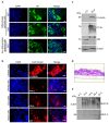
Formation of a differentiated respiratory epithelium. HAECs were seeded on Transwell inserts and cultivated under ALI conditions. (a) Immunostaining of specific cytokeratins (CK7, CK8, and CK17). (b) Immunostaining of markers for ciliated cells (β-tubulin), goblet cells (TFF-3), club cells (CC16), or basal cells (p63). Cells were analyzed between days 28 and 35 post-airlift. Nuclei were visualized using DAPI staining. Scale bars, 20 μm. (c) Western blot detection of ß-tubulin, CC16, and p63 in non-differentiated HAECs (0 d) and HAECs cultured for 50 days under ALI conditions (50 d). (d) HAECs were grown on Transwell inserts, followed by histological sectioning and HE staining. Representative histological sections were examined by light microscopy (40× magnification). (e) ALI cultures were rinsed weekly, and the apical secretion of mucus proteins (MUC16 and MUC4) was monitored by western blot analysis.
Figure 3
Figure 3
Effect of airway mucus on virus entry. MDCK II (a) or VeroE6 (b) cells were seeded in a 96-well plate and cultivated for 24 h. Mucus collected from the apical sides of HAECs was diluted and incubated with 1400 FFU of IAV (a) or 800 PFU of VSVΔG/LASVGP (b). Mixtures of mucus and virus were then added to the cells and incubated for 8 h. Infected cells were detected by immunostaining using anti-IAV NP (a) or anti-VSV antibodies (b). The total numbers of infected cells per well were quantified. The numbers of infected cells in control samples (virus without mucus) were set as 100% (indicated by dashed lines). Error bars represent SEM of three biological replicates.
Figure 4
Figure 4
LASV entry and egress in primary human airway epithelial cells. (a,b) Differentiated HAECs cultured on Transwell inserts under ALI conditions were infected with 2.4 × 10⁴ PFU of LASV via either the apical or basolateral membrane. Infected cells were fixed with 4% PFA either at 48 hpi, and virus-positive cells were detected by immunostaining with a rabbit anti-LASV NP antibody (a), or at 4 dpi, where virus-positive cells were detected using a human anti-LASV GP antibody (b). Actin filaments were visualized with Phalloidin-TRITC staining, and cell nuclei were stained with DAPI. Scale bars, 20 µm (a) and 25 µm (b). (c) HAECs were infected with 8.75 × 10⁴ PFU of LASV via the apical or basolateral route. Virus titers in the apical medium were determined at the indicated time points post-infection using TCID50 analysis. Error bars represent the SEM of three biological replicates.
Figure 5
Figure 5
LASV infection induces actin cytoskeleton rearrangement. (a) Differentiated HAECs were infected with 1.2 × 10⁵ PFU of LASV simultaneously via both the apical and basolateral routes. Virus release was determined by TCID50 analysis at the indicated time points post-infection. Error bars represent the SEM of three biological replicates. (b) HAECs were infected with 2.4 × 10⁴ PFU of LASV via the apical side. Infected cells and mock-infected cells were fixed with 4% PFA at 4 dpi. Virus-positive cells were detected by immunostaining using a human monoclonal anti-LASV GP antibody. Actin filaments were visualized by Phalloidin-TRITC staining, and cell nuclei were stained with DAPI. Enlarged regions from the top-view images and z-scans are indicated by white frames. Scale bars, 25 µm.
Figure 6
Figure 6
IFN induction by LASV infection. Differentiated HAECs were infected with 1.2 × 10⁵ PFU of LASV simultaneously via the apical and basolateral routes. Induction of IFN (a) and ISGs (b) was analyzed by quantitative RT-PCR. Error bars represent the SEM of three biological replicates. (c) Differentiated HAECs were infected with 1.2 × 10⁵ PFU of LASV via the apical or the basolateral route, and IFN-λ induction was determined by quantitative RT-PCR. Error bars represent the SEM of three biological replicates.
Figure 7
Figure 7
IFN induction and IFN-mediated antiviral responses during LASV infection. (a) Differentiated HAECs were stimulated for 16 h with human IFN-λ2, and ISG induction was analyzed by quantitative RT-PCR. (bd) IFN-λ preincubated cells were infected with 1.2 × 10⁵ PFU of LASV simultaneously via the apical and basolateral routes. (b) At 48 hpi, viral replication was assessed by quantitative RT-PCR using viral RNA isolated from cell lysates. (c) Virus titers in the apical medium were determined by TCID50 analysis. Error bars represent the SEM of three biological replicates. Statistical significance was calculated using an unpaired t-test (* p = 0.032; *** p < 0.01). (d) Infected cells were fixed with 4% PFA at 48 hpi. Virus-positive cells were detected by immunostaining with anti-LASV NP or anti-LASV GP antibodies. Cell boundaries were visualized by actin or E-cadherin staining, and nuclei were stained with DAPI. Scale bars, 25 μm.

Similar articles

References

    1. Basinski A.J., Fichet-Calvet E., Sjodin A.R., Varrelman T.J., Remien C.H., Layman N.C., Bird B.H., Wolking D.J., Monagin C., Ghersi B.M., et al. Bridging the gap: Using reservoir ecology and human serosurveys to estimate Lassa virus spillover in West Africa. PLoS Comput. Biol. 2021;17:e1008811. doi: 10.1371/journal.pcbi.1008811. - DOI - PMC - PubMed
    1. McCormick J.B. Epidemiology and control of Lassa fever. Curr. Top Microbiol. Immunol. 1987;134:69–78. - PubMed
    1. McCormick J.B., Webb P.A., Krebs J.W., Johnson K.M., Smith E.S. A prospective study of the epidemiology and ecology of Lassa fever. J. Infect. Dis. 1987;155:437–444. doi: 10.1093/infdis/155.3.437. - DOI - PubMed
    1. McCormick J.B., King I.J., Webb P.A., Scribner C.L., Craven R.B., Johnson K.M., Elliott L.H., Belmont-Williams R. Lassa fever. Effective therapy with ribavirin. N. Engl. J. Med. 1986;314:20–26. doi: 10.1056/NEJM198601023140104. - DOI - PubMed
    1. Eberhardt K.A., Mischlinger J., Jordan S., Groger M., Gunther S., Ramharter M. Ribavirin for the treatment of Lassa fever: A systematic review and meta-analysis. Int. J. Infect. Dis. 2019;87:15–20. doi: 10.1016/j.ijid.2019.07.015. - DOI - PubMed

Publication types

LinkOut - more resources