. 2025 Apr 24;17(5):606.
doi: 10.3390/v17050606.

Dysfunctional Senescent Herpes Simplex Virus-Specific CD57+CD8+ T Cells Are Associated with Symptomatic Recurrent Ocular Herpes in Humans

Affiliations

Dysfunctional Senescent Herpes Simplex Virus-Specific CD57+CD8+ T Cells Are Associated with Symptomatic Recurrent Ocular Herpes in Humans

Aziz A Chentoufi et al. Viruses. .

Abstract

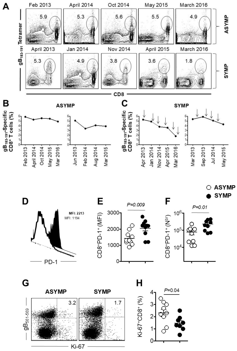
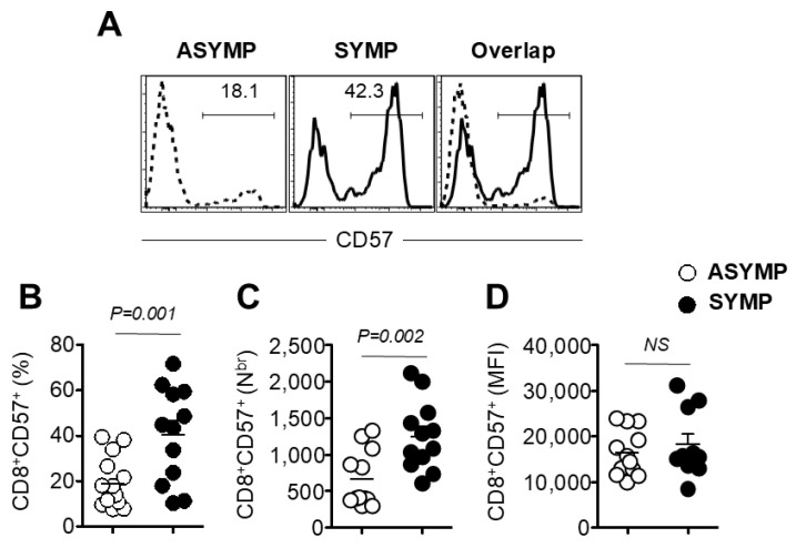
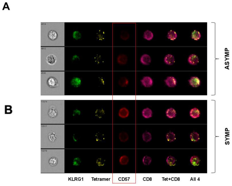
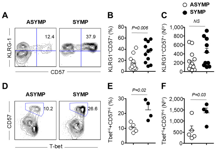
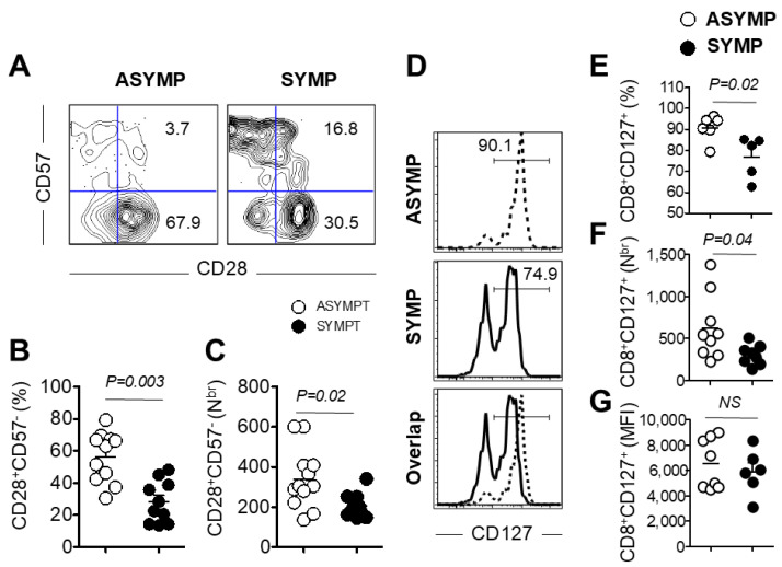
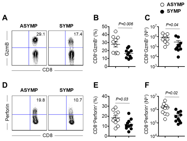
Herpes simplex virus (HSV)-specific CD8+ T cells protect mice from herpes infection and disease. However, the phenotype and function of HSV-specific CD8+ T cells that play a key role in the "natural" protection seen in HSV-1-seropositive healthy asymptomatic (ASYMP) patients (who have never had clinical herpes disease) remain to be determined. We previously reported that symptomatic (SYMP) patients (who have frequent bouts of recurrent ocular herpes disease) had more undifferentiated and dysfunctional HSV-specific CD8+ T cells. In contrast, healthy ASYMP individuals maintained a significantly higher proportion of differentiated polyfunctional CD8+ T cells. Here, we report that HSV-specific CD8+ T cells from 10 SYMP patients, but not HSV-specific CD8+ T cells from 10 ASYMP patients, have phenotypic and functional characteristics of cellular senescence, including: (i) high frequency of senescent (CD57+) and exhausted (PD-1+) CD8+ T cells; (ii) late terminally differentiated (KLRG1+), non-proliferating CD8+ T cells; (iii) HSV-specific CD8+ T cells which decreased in number over time and were not homeostatically maintained, as indicated by a reduction in the number of CD127+CD8+ T cells; (iv) loss of the co-stimulatory molecule CD28 on HSV-specific CD8+ T cells; and (v) decreased production of effector molecules (granzyme B and perforin) by HSV-specific CD8+ T cells. Our findings provide insights into the role of senescence in HSV-specific CD8+ T cells in susceptibility to recurrent herpes and have implications for T-cell-based immunotherapeutic strategies against recurrent herpes in humans.

Keywords: CD57; CD8+ T cells; cornea; ocular herpes; senescent; trigeminal ganglia.


Conflict of interest statement

The authors declare no conflict of interest.

Figures

Figure 1
Figure 1
The decline of gB183–191-specific CD8+ T cells in SYMP individuals but not in ASYMP individuals. gB183–191-specific CD8+ T cells were measured in HLA-A*02:01-positive, HSV-1 seropositive SYMP and ASYMP individuals at different time points. Each time history of recurrent disease was recorded. (A) The frequency of gB183–191-specific CD8+ T cells is shown as FACS dot plots. The top panel shows a dot plot of gB183–191-specific CD8+ T cells at the different time points of ASYMP individuals. The bottom panel shows a dot plot of gB183–191-specific CD8+ T cells at the different time points of SYMP individuals. (B) The line graph shows the kinetic of gB183–191-specific CD8+ T cells at different time points of ASYMP individuals. The frequency of the gB183–191-specific CD8+ T cells was maintained over time. (C) The line graph shows the kinetic of gB183–191-specific CD8+ T cells at the different time points of SYMP individuals. Unlike ASYMP individuals, the frequency of the gB183–191-specific CD8+ T cells declined over time. The arrow on the line graph shows the frequency of recurrent disease in SYMP individuals. (D) The representative histogram of PD-1 expression on gB183–191-specific CD8+ T cells. A filled histogram represents an expression of PD-1 in SYMP individuals, and an open histogram represents ASYMP individuals. (E) Expression level and (F) The absolute number of PD-1 was significantly higher in SYMP individuals compared to ASYMP individuals. (G) Representative dot plot of gB183–191-specific CD8+ T cells stained for proliferation marker Ki-67. (H) Frequency of Ki-67+CD8+ T cells gated on gB183–191 tetramer. Open circles indicate data for ASYMP individuals and filled circles represent data for SYMP individuals. The results are representative of two independent experiments for each individual. The indicated p values, calculated using one-way ANOVA, show the statistical significance of differences between SYMP and ASYMP individuals. The data are representative of two independent experiments, and the bars represent SD between the experiments.
Figure 2
Figure 2
Increased frequency of senescent CD8+ T cells in SYMP individuals compared to ASYMP individuals. PBMC isolated from HLA-A*02:01-positive, HSV-1 seropositive SYMP and ASYMP individuals was stained for senescent marker CD57. (A) Representative histogram of CD57 expression on gB183–191-specific CD8+ T cells. (B) Frequency of CD8+CD57+ on gB183–191-specific CD8+ T cells. (C) The absolute number of CD8+CD57+ on gB183–191-specific CD8+ T cells. (D) Mean Fluorescent Intensity (MFI) of CD57 expression gated on gB183–191-specific CD8+ T cells. Open circles indicate data for ASYMP individuals and filled circles represent data for SYMP individuals.
Figure 3
Figure 3
Increased expression of CD57 on gB183–191-specific CD8+ T cells in SYMP individuals compared to SYMP individuals. PBMC isolated from HLA-A*02:01-positive, HSV-1 seropositive SYMP and ASYMP individuals was stained for senescent marker CD57 along with CD8, gB183–191 tetramer, and KLRG-1. Images of individual PBMC stained with different markers were visualized and captured using Image Stream. Data were analyzed using the software IDEA version 4. Representative images of individual PMBC isolated from HSV-1 seropositive (A) ASYMP patients and (B) SYMP patients. Cells are shown as individually stained with KLRG-1, gB183–191 tetramer, CD57, CD8, gB183–191 tetramer, and CD8, and superimposed image of PBMC stained with KLRG-1, gB183–191 tetramer, CD57, and CD8+ T cells.
Figure 4
Figure 4
HSV-1-specific CD8+ T cells from SYMP individuals were terminally differentiated and could not divide. PBMC isolated from HLA-A*02:01-positive, HSV-1 seropositive SYMP and ASYMP individuals was stained for the terminally differentiated marker KLRG-1 and transcription factor T-bet, along with CD57. Cells were also stained for Ki-67 to see the percentage of proliferating cells. (A) Representative contour plot of CD57 expression on terminally differentiated CD8+ T cells gated on gB183–191 tetramer in PBMC isolated from SYMP and ASYMP individuals. (B) Frequency of KLRG-1+CD57+ on gB183–191-specific CD8+ T cells. (C) The absolute number of KLRG-1+CD57+ on gB183–191-specific CD8+ T cells. (D) A representative contour plot of T-betHi+CD57+ expression gated on gB183–191 tetramer in PBMC isolated from SYMP and ASYMP individuals. (E) Frequency of T-betHi+CD57+ on gB183–191-specific CD8+ T cells. (F) The absolute number of T-betHi+CD57+ on gB183–191-specific CD8+ T cells. Open circles indicate data for ASYMP individuals and filled circles represent data for SYMP individuals. The results are representative of two independent experiments for each individual. The indicated p values, calculated using one-way ANOVA, show the statistical significance of differences between SYMP and ASYMP individuals. The data are representative of two independent experiments, and the bars represent SD between the experiments.
Figure 5
Figure 5
Decreased expression of co-stimulatory molecule CD28 and survival molecule CD127 on PBMC isolated from SYMP individuals. PBMC isolated from HLA-A*02:01-positive, HSV-1 seropositive SYMP and ASYMP individuals was stained for co-stimulatory and survival molecules. (A) Representative contour plot of CD28 and CD57 expression gated on gB183–191 tetramer in PBMC isolated from SYMP and ASYMP individuals. (B) Frequency of CD28+CD57 on gB183–191-specific CD8+ T cells. (C) The absolute number of CD28+CD57 on gB183–191-specific CD8+ T cells. (D) Representative histogram showing the expression level of survival molecule CD127 gated on gB183–191-specific CD8+ T cells. (E). Frequency of CD127 gated on gB183–191-specific CD8+ T cells. (F) The absolute number of CD127 gated on gB183–191-specific CD8+ T cells. (G) Mean Fluorescent Intensity (MFI) of CD127 expression on gB183–191-specific CD8+ T cells. Open circles indicate data for ASYMP individuals and filled circles represent data for SYMP individuals. The results are representative of two independent experiments for each individual. The indicated p values, calculated using one-way ANOVA, show the statistical significance of differences between SYMP and ASYMP individuals. The data are representative of two independent experiments and the bars represent SD between the experiments.
Figure 6
Figure 6
Decreased production of effector molecules Granzyme B and Perforin by HSV-specific CD8+ T cells isolated from HSV-1 seropositive SYMP individuals: PBMC isolated from HLA-A*02:01-positive, HSV-1 seropositive SYMP and ASYMP individuals were stimulated and then stained for effector molecules granzyme B and perforin (A). Representative contour plot of CD8+GzmB+ cells gated on gB183–191-specific CD8+ T cells in PBMC isolated from SYMP and ASYMP individuals. (B) The frequency of CD8+GzmB+ gated on gB183–191-specific CD8+ T cells in SYMP and ASYMP individuals (C). The absolute number of CD8+GzmB+ gated on gB183–191-specific CD8+ T cells in SYMP and ASYMP individuals. (D) The contour plot of CD8+Perforin+ cells gated on gB183–191-specific CD8+ T cells in PBMC isolated from SYMP and ASYMP individuals. (E). Frequency of CD8+Perforin+ gated on gB183–191-specific CD8+ T cells in SYMP and ASYMP individuals. (F). The absolute number of CD8+Perforin+ gated on gB183–191-specific CD8+ T cells in SYMP and ASYMP individuals. Open circles indicate data for ASYMP individuals and filled circles represent data for SYMP individuals. The results are representative of two independent experiments for each individual.
Figure 7
Figure 7
Illustration of the mechanisms of senescent and exhausted HSV-specific CD8+ T cells in SYMP individuals. Symptomatic patients (left) exhibited senescent and exhausted HSV-specific CD8+ T cells. While the majority of the cells exhibited a senescent profile, characterized by high CD57+ PD-1+ and KLGR1+ expression, low CD127+ and CD28+, and reduced cytotoxic function, a small fraction of newly recruited cells exhibited functional profiles. This major dysfunction led to more frequent HSV-1 reactivation and increased viral spread, resulting in greater cell damage and disease severity. In contrast, Asymptomatic patients (right) show high numbers of functional HSV-specific CD8+ T cells expressing CD127+, CD28+, Granzyme B+, and Perforin+, effectively controlling virus reactivation and limiting disease progression.

Similar articles

References

    1. Kuo T., Wang C., Badakhshan T., Chilukuri S., BenMohamed L. The Challenges and Opportunities for the Development of a T-Cell Epitope-Based Herpes Simplex Vaccine. Vaccine. 2014;32:6733–6745. doi: 10.1016/j.vaccine.2014.10.002. - DOI - PMC - PubMed
    1. Samandary S., Kridane-Miledi H., Sandoval J.S., Choudhury Z., Langa-Vives F., Spencer D., Chentoufi A.A., Lemonnier F.A., BenMohamed L. Associations of HLA-A, HLA-B and HLA-C Alleles Frequency with Prevalence of Herpes Simplex Virus Infections and Diseases Across Global Populations: Implication for the Development of an Universal CD8+ T-Cell Epitope-Based Vaccine. Hum. Immunol. 2014;75:715–729. doi: 10.1016/j.humimm.2014.04.016. - DOI - PMC - PubMed
    1. Zhang X., Dervillez X., Chentoufi A.A., Badakhshan T., Bettahi I., BenMohamed L. Targeting the genital tract mucosa with a lipopeptide/recombinant adenovirus prime/boost vaccine induces potent and long-lasting CD8+ T cell immunity against herpes: Importance of MyD88. J. Immunol. 2012;189:4496–4509. doi: 10.4049/jimmunol.1201121. - DOI - PMC - PubMed
    1. Chentoufi A.A., Dasgupta G., Christensen N.D., Hu J., Choudhury Z.S., Azeem A., Jester J.V., Nesburn A.B., Wechsler S.L., BenMohamed L. A novel HLA (HLA-A*0201) transgenic rabbit model for preclinical evaluation of human CD8+ T cell epitope-based vaccines against ocular herpes. J. Immunol. 2010;184:2561–2571. doi: 10.4049/jimmunol.0902322. - DOI - PMC - PubMed
    1. Chentoufi A.A., Zhang X., Lamberth K., Dasgupta G., Bettahi I., Nguyen A., Wu M., Zhu X., Mohebbi A., Buus S., et al. HLA-A*0201-restricted CD8+ cytotoxic T lymphocyte epitopes identified from herpes simplex virus glycoprotein D. J. Immunol. 2008;180:426–437. doi: 10.4049/jimmunol.180.1.426. - DOI - PubMed

Publication types

LinkOut - more resources