Review
. 2025 Apr 24;17(5):611.
doi: 10.3390/v17050611.

Alphaflexiviridae in Focus: Genomic Signatures, Conserved Elements and Viral-Driven Cellular Remodeling

Affiliations
Review

Alphaflexiviridae in Focus: Genomic Signatures, Conserved Elements and Viral-Driven Cellular Remodeling

Jesús R Úbeda et al. Viruses. .

Abstract

The family Alphaflexiviridae comprises plant- and fungus-infecting viruses with single-stranded, positive-sense RNA genomes ranging from 5.4 to 9 kb. Their virions are flexuous and filamentous, measuring 470-800 nm in length and 12-13 nm in diameter. The family includes 72 recognized species, classified into six genera: Allexivirus, Lolavirus, Platypuvirus, Potexvirus (plant-infecting), and Botrexvirus and Sclerodarnavirus (fungus-infecting). The genus Potexvirus is the largest, with 52 species, including Potexvirus ecspotati (potato virus X), an important crop pathogen and plant virology model. The genera are distinguished by genome organization and host range, while species differentiation relies on nucleotide and protein sequence identity thresholds. In this review, we summarize the current knowledge on the genomic structure, conserved genes, and phylogenetic relationships within Alphaflexiviridae, with a particular focus on the replicase and coat protein genes as signature markers. Additionally, we update the model of cellular remodeling driven by the triple gene block proteins, which are essential for virus movement, among other viral functions. Beyond their biological significance, alphaflexiviruses serve as valuable models for studying virus-host dynamics and hold potential applications in plant disease control and biotechnology. This review provides an updated framework for understanding Alphaflexiviridae and their broader impact on plant virology.

Keywords: Allexivirus; Botrexvirus; Lolavirus; Platypuvirus; Potexvirus; Sclerodarnavirus; coat protein; replicase; triple gene block proteins.


Conflict of interest statement

The authors declare no conflicts of interest.

Figures

Figure 3
Figure 3
Organization of replicase motifs of exemplar viruses within each genus and subgenus of the family Alphaflexiviridae. Coordinates of each motif refer to amino acid positions. The amino acid length of each replicase protein is shown on the right. MET: type 1 RNA methyltransferase; HEL: RNA helicase-like; RdRp: RNA-dependent RNA polymerase [30].
Figure 4
Figure 4
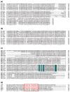
Amino acid alignments of replicase motifs of exemplar viruses within each genus and subgenus of the family Alphaflexiviridae. Amino acid alignments of MET (a), HEL (b), and RdRp (c) motifs. The characteristic core motif S/TGX3TX3NS/TX22GDD is highlighted in (c), where conserved amino acids are in dark green and any amino acid (X) in grey. (d) Amino acid alignment showing the predicted location of the amphipathic alpha-helix downstream of the MET domain. The red square indicates the location of the alpha-helix in Plantago asiatica mosaic virus (Potexvirus marmorplantagonis), and the red letters indicate the positions with predicted helical structure as in [30]. (*) Fully conserved residues; (:) Conservation between groups with strongly similar properties; (.) Conservation between groups with weakly similar properties.
Figure 1
Figure 1
Genome organization of exemplar viruses within each genus and subgenus of the family Alphaflexiviridae. 5′ and 3′ untranslated regions are represented by a black line. Boxes in different colors mark regions encoding known viral proteins; regions encoding hypothetical proteins or proteins with unknown function are marked with white boxes. TGB: triple gene block; CP: coat protein; NABP: nucleic acid binding protein; MP: movement protein. A scale bar of 500 nucleotides is shown.
Figure 2
Figure 2
5′ and 3′ untranslated regions (UTRs) predicted structures of exemplar viruses within each genus and subgenus of the family Alphaflexiviridae. RNA structures include the 5′ and 3′-UTRs plus 60 nts downstream of the translation initiation codon, or upstream of the termination codon, respectively. The structure with the lowest initial Gibbs free energy (DG) was selected using the UNAFold web server. The UTR sequences are highlighted in dark blue. The positions of the putative conserved octanucleotide (ACCNNACC) and the GAAA sequence in the 5′ stem-loop 1 (5′SL1) are highlighted in red. 5′ end is denoted with a triangle and 3′ end with an asterisk.
Figure 5
Figure 5
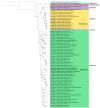
Phylogenetic analysis of the replicase protein of the family Alphaflexiviridae. The multiple sequence alignment of 72 replicase sequences was performed using MUSCLE [41]. The best amino acid substitution method was inferred using MEGA 11 [42]. The maximum likelihood (ML) trees were inferred using RAxML-NG software [43] using the LG method considering the proportion of invariable sites (+I) and the variation of the substitution rate among sites according to a gamma distribution (+G). The best ML tree with bootstrap support values (1000 replicates) is shown. Only bootstrap values higher than 50% are displayed. Carnation latent virus (QJX15400.1) (Carlavirus latensdianthi, family Quinivirinae) was used as an outgroup, and the tree was rooted with this sequence.
Figure 6
Figure 6
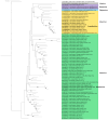
Phylogenetic analysis of full protein sequences of the coat protein (CP) of the family Alphaflexiviridae. The multiple sequence alignment of 72 CP sequences was performed using MUSCLE [41]. The best amino acid substitution method was inferred using MEGA 11 [42]. The maximum likelihood (ML) trees were inferred using RAxML-NG software [43] using the LG method considering the variation of the substitution rate among sites according to a gamma distribution (+G). The best ML tree with bootstrap support values (1000 replicates) is shown. Only bootstrap values higher than 50% are displayed. Carnation latent virus (QJX153999.1) (Carlavirus latensdianthi, family Quinivirinae) was used as an outgroup, and the tree was rooted with this sequence.
Figure 7
Figure 7
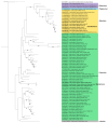
Phylogenetic analysis of the core region of the coat protein (CP) of the family Alphaflexiviridae. Phylogenetic tree was constructed excluding the variable N- and C- terminus of the CP sequences (Supplementary Figure S2). The description of the figure is as in Figure 6. Carnation latent virus (QJX153999.1) (Carlavirus latensdianthi, family Quinivirinae) was used as an outgroup, and the tree was rooted with this sequence.
Figure 8
Figure 8
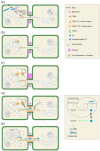
Model of the cellular remodeling coordinated by the triple gene block (TGB) proteins of the family Alphaflexiviridae. (a) Viral entry and initial replication. The virion penetrates into the host cell through a microlesion and undergoes uncoating via phosphorylation of capsid protein (CP) subunits by host factors. The free viral RNA (+vRNA) recruits the host translation machinery, synthesizing the replicase, which transcribes negative-strand vRNA (−vRNA) and subgenomic RNAs (sgRNA) that serve as templates for replication and translation of the viral proteins, respectively. N: nucleus; nu: nucleolus; ER: endoplasmic reticulum; PD: plasmodesmata. (b) Formation of endoplasmic reticulum (ER)-associated granules and TGB2-vesicles. TGB2 and TGB3 move through the ER, forming complexes that recruit the viral replicase and free vRNA, leading to the formation of ER-associated granules that act as early viral replication complexes (VRCs). Additionally, TGB2 induces the formation of trafficking vesicles that recruit TGB3. (c) Peripheral body formation. TGB3 contains localization signals directing it to the distal ER, where it undergoes oligomerization and/or conformational changes forming punctate structures. In the distal ER, TGB2 acquires a reticulon-like conformation, remodeling the ER into highly curved membranes and forming peripheral bodies. These structures function as intermediate VRCs, coupling transcription and translation with movement through the plasmodesmata (PD). TGB1 is recruited to PD by TGB3 and TGB2, increasing the size exclusion limit. Through distinct molecular mechanisms, the host perceives the infection and enhances callose deposition to seal the PD and restrict the viral spread. As a countermeasure, TGB2 interacts with TGB2 interacting proteins (TIPs) to activate β-1,3-glucanase, leading to callose degradation and facilitating viral movement through the PD. (d) RNP formation and infection of the adjacent cell. Within the VRC, CP may undergo S-oxidation, modifying its redox state and suppressing its affinity for vRNA, freeing it for translation and transcription. In contrast, outside the VRC, the reduced CP exhibits affinity for vRNA, promoting the formation of RNPs. Newly synthesized vRNA begins to encapsidate upon exiting the VRC, with TGB1 subunits binding to the 5′ end, forming the movement ribonucleoprotein (RNP). RNPs associate with TGB2-TGB3, which have colonized the desmotubule. The RNP complex colonizes adjacent cells. TGB1 triggers vRNA uncoating, making it accessible to the host translational machinery. This newly infected cell already possesses a set of viral proteins before the arrival of the vRNA. (e) Viral replication pseudo-organelle (VRO) formation. At the final stages of infection, all virus-induced structures concentrate into a VRO through restructuring the endomembrane system. As described by [61], the different VRCs, arranged in a spiral-like formation, associate with reorganized actin filament and contain TGB1 along with TGB2-TGB3 granules. Replication and translation take place in the space between TGB1 and the TGB2-TGB3 granules. This pseudo-organelle harbors large amounts of free vRNA, and nascent virions are located at its periphery.

Similar articles

References

    1. Koonin E.V., Kuhn J.H., Dolja V.V., Krupovic M. Megataxonomy and Global Ecology of the Virosphere. ISME J. 2024;18:wrad042. doi: 10.1093/ismejo/wrad042. - DOI - PMC - PubMed
    1. Scholthof K.-B.G., Adkins S., Czosnek H., Palukaitis P., Jacquot E., Hohn T., Hohn B., Saunders K., Candresse T., Ahlquist P., et al. Top 10 Plant Viruses in Molecular Plant Pathology. Mol. Plant Pathol. 2011;12:938–954. doi: 10.1111/j.1364-3703.2011.00752.x. - DOI - PMC - PubMed
    1. Verchot J. Potato Virus X: A Global Potato-Infecting Virus and Type Member of the Potexvirus Genus. Mol. Plant Pathol. 2022;23:315–320. doi: 10.1111/mpp.13163. - DOI - PMC - PubMed
    1. Miller E.D., Kim K.-H., Hemenway C. Restoration of a Stem-Loop Structure Required for Potato Virus X RNA Accumulation Indicates Selection for a Mismatch and a GNRA Tetraloop. Virology. 1999;260:342–353. doi: 10.1006/viro.1999.9843. - DOI - PubMed
    1. Kwon S.J., Park M.R., Kim K.W., Plante C.A., Hemenway C.L., Kim K.H. Cis-Acting Sequences Required for Coat Protein Binding and in Vitro Assembly of Potato Virus X. Virology. 2005;334:83–97. doi: 10.1016/j.virol.2005.01.018. - DOI - PubMed

Publication types

LinkOut - more resources