. 2025 Apr 26;17(5):623.
doi: 10.3390/v17050623.

Acinetobacter baumannii and Klebsiella pneumoniae Isolates Obtained from Intensive Care Unit Patients in 2024: General Characterization, Prophages, Depolymerases and Esterases of Phage Origin

Affiliations

Acinetobacter baumannii and Klebsiella pneumoniae Isolates Obtained from Intensive Care Unit Patients in 2024: General Characterization, Prophages, Depolymerases and Esterases of Phage Origin

Nadezhda V Kolupaeva et al. Viruses. .

Abstract

Acinetobacter baumannii and Klebsiella pneumoniae are significant nosocomial pathogens worldwide. In this study, the general characterization of A. baumannii and K. pneumoniae isolates obtained from the blood of intensive care unit patients of the multidisciplinary scientific and practical center of emergency medicine from January to September 2024 was performed. Prophage regions and prophage-derived tailspike polysaccharide-depolymerizing or -modifying enzymes within these isolates were identified and characterized in detail using a refined workflow. The protocol, encompassing a comprehensive survey of all predicted bacterial proteins, revealed an average of 6.0 prophage regions per Acinetobacter baumannii genome, including regions putatively derived from filamentous phages, and 4.8 prophage regions per Klebsiella pneumoniae isolate. Analysis of these putative prophage regions indicated that most were related to previously isolated, yet unclassified, temperate phages infecting A. baumannii and K. pneumoniae. However, certain identified sequences likely originated from phages representing novel groups comparatively distant from known phages.

Keywords: Acinetobacter baumannii; Klebsiella pneumoniae; genomes; prophage regions; tailspike depolymerase; tailspike esterase.


Conflict of interest statement

The authors declare no conflicts of interest.

Figures

Figure 1
Figure 1
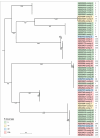
(A) Heatmap and dendrogram depicting the relationships among A. baumannii isolates based on average nucleotide identity (ANI). The color scale represents the percentage identity between whole-genome sequences, ranging from high (red) to low (white). (B) Maximum-likelihood phylogenetic tree based on concatenated amino acid sequences of conserved proteins, constructed using GTDB-Tk. Bootstrap values are shown near the nodes. The scale bar indicates 0.0002 estimated substitutions per site, and the tree is midpoint-rooted. A. baumannii KL type is indicated in the legend.
Figure 2
Figure 2
(A) Heatmap and dendrogram depicting the relationships among K. pneumoniae isolates based on average nucleotide identity (ANI). The color scale represents the percentage identity between whole-genome sequences, ranging from high (red) to low (white). (B) Maximum-likelihood phylogenetic tree based on concatenated amino acid sequences of conserved proteins, constructed using GTDB-Tk. Bootstrap values are shown near the nodes. The scale bar indicates 0.00005 estimated substitutions per site, and the tree is midpoint-rooted. K. pneumoniae KL type is indicated in the legend.
Figure 2
Figure 2
(A) Heatmap and dendrogram depicting the relationships among K. pneumoniae isolates based on average nucleotide identity (ANI). The color scale represents the percentage identity between whole-genome sequences, ranging from high (red) to low (white). (B) Maximum-likelihood phylogenetic tree based on concatenated amino acid sequences of conserved proteins, constructed using GTDB-Tk. Bootstrap values are shown near the nodes. The scale bar indicates 0.00005 estimated substitutions per site, and the tree is midpoint-rooted. K. pneumoniae KL type is indicated in the legend.
Figure 3
Figure 3
Workflow for identification and structural analysis of phage tailspike depolymerases and esterases (PTDEs).
Figure 4
Figure 4
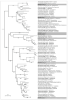
Maximum-likelihood phylogenetic tree based on amino acid sequences of MCP encoded in A. baumannii prophage regions. Bootstrap values are shown near the nodes. The scale bar indicates 0.5 estimated substitutions per site, and the tree is midpoint-rooted. A. baumannii KL type is indicated in the legend. Prophage regions are named according to the contig of the genome assembly in which they were identified.
Figure 5
Figure 5
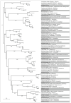
Maximum-likelihood phylogenetic tree based on amino acid sequences of MCP encoded in A. baumannii prophage regions and related phages. NCBI taxonomy is shown to the right of phage names. Bootstrap values are shown near the nodes. The scale bar indicates 0.5 estimated substitutions per site, and the tree is midpoint-rooted. Representative prophage regions are colored gray.
Figure 6
Figure 6
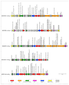
Genetic maps of prophage regions revealed in the genomes of different A. baumannii isolates. Gene annotations and predicted functions are indicated by labels and a legend. Arrows show the direction of transcription for each gene. The scale bar represents the length of the nucleotide sequence.
Figure 6
Figure 6
Genetic maps of prophage regions revealed in the genomes of different A. baumannii isolates. Gene annotations and predicted functions are indicated by labels and a legend. Arrows show the direction of transcription for each gene. The scale bar represents the length of the nucleotide sequence.
Figure 7
Figure 7
Maximum-likelihood phylogenetic tree based on amino acid sequences of MCP encoded in K. pneumoniae prophage regions and related phages. Distinct clusters from which representative prophage regions were analyzed are indicated by different colors. Bootstrap values are shown near the nodes. The scale bar indicates 0.5 estimated substitutions per site, and the tree is midpoint-rooted.
Figure 8
Figure 8
Maximum-likelihood phylogenetic tree based on amino acid sequences of MCP encoded in K. pneumoniae prophage regions and related phages. NCBI taxonomy is shown to the right of phage names. Bootstrap values are shown near the nodes. The scale bar indicates 0.5 estimated substitutions per site, and the tree is midpoint-rooted. Representative prophage regions are colored gray.
Figure 9
Figure 9
Genetic maps of prophage regions revealed in the genomes of different K. pneumoniae isolates. Gene annotations and predicted functions are indicated by labels and a legend. Arrows show the direction of transcription for each gene. The scale bar represents the length of the nucleotide sequence.
Figure 9
Figure 9
Genetic maps of prophage regions revealed in the genomes of different K. pneumoniae isolates. Gene annotations and predicted functions are indicated by labels and a legend. Arrows show the direction of transcription for each gene. The scale bar represents the length of the nucleotide sequence.
Figure 10
Figure 10
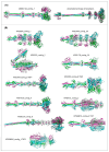
Predicted structures of putative trimeric A. baumannii and K. pneumoniae prophage-derived tailspike depolymerases and esterases, modeled using AlphaFold. (A) Structural architecture comparison of the tailspike esterase ABS82146_contig_7 and the TSP of the Acinetobacter phage Aristophanes. (B) Predicted structures of PTDE identified in K. pneumoniae prophage regions. Different monomers are distinctly colored.

Similar articles

References

    1. Ayoub Moubareck C., Hammoudi Halat D. Insights into Acinetobacter baumannii: A review of microbiological, virulence, and resistance traits in a threatening nosocomial pathogen. Antibiotics. 2020;9:119. doi: 10.3390/antibiotics9030119. - DOI - PMC - PubMed
    1. Runtuvuori-Salmela A., Kunttu H.M.T., Laanto E., Almeida G.M.F., Mäkelä K., Middelboe M., Sundberg L.-R. Prevalence of genetically similar Flavobacterium columnare phages across aquaculture environments reveals a strong potential for pathogen control. Environ. Microbiol. 2022;24:2404–2420. doi: 10.1111/1462-2920.15901. - DOI - PMC - PubMed
    1. Santajit S., Indrawattana N. Mechanisms of antimicrobial resistance in ESKAPE pathogens. BioMed Res. Int. 2016;2016:2475067. doi: 10.1155/2016/2475067. - DOI - PMC - PubMed
    1. Howard A., O’Donoghue M., Feeney A., Sleator R.D. Acinetobacter baumannii: An emerging opportunistic pathogen. Virulence. 2012;3:243–250. doi: 10.4161/viru.19700. - DOI - PMC - PubMed
    1. Sarshar M., Behzadi P., Scribano D., Palamara A.T., Ambrosi C. Acinetobacter baumannii: An ancient commensal with weapons of a pathogen. Pathogens. 2021;10:387. doi: 10.3390/pathogens10040387. - DOI - PMC - PubMed

Publication types

MeSH terms

LinkOut - more resources