. 2025 May 19;17(5):725.
doi: 10.3390/v17050725.

Discovery and Genome Characterization of Three New Rhabdoviruses Infecting Passiflora spp. in Brazil

Affiliations

Discovery and Genome Characterization of Three New Rhabdoviruses Infecting Passiflora spp. in Brazil

Andreza Henrique Vidal et al. Viruses. .

Abstract

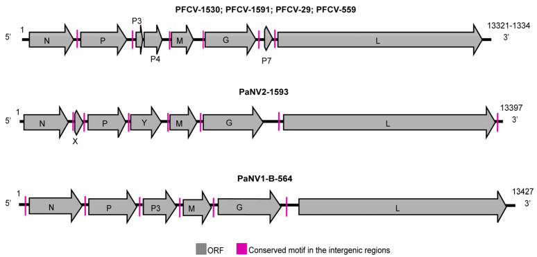
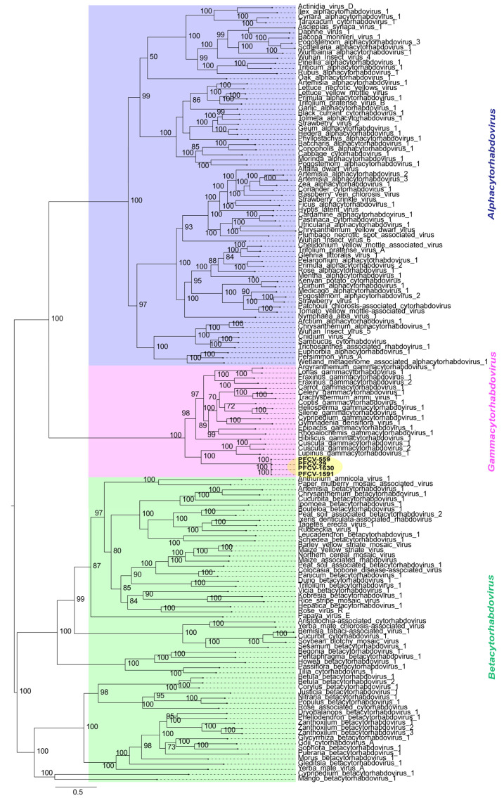
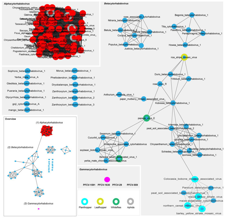
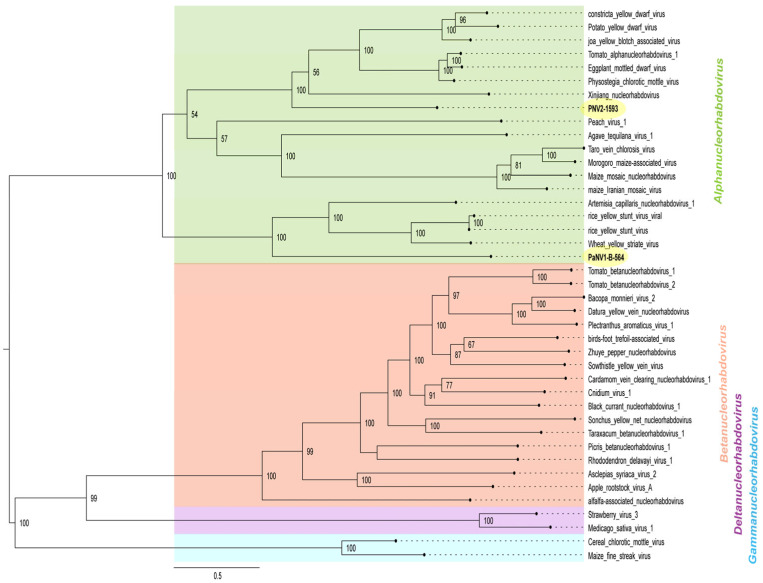
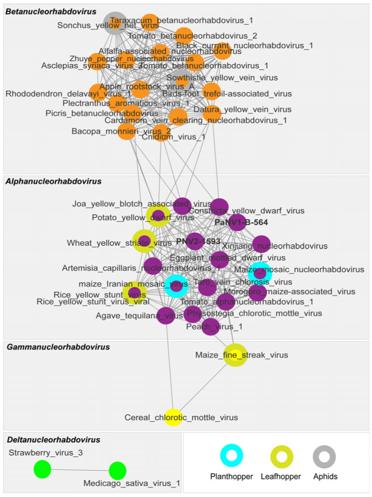
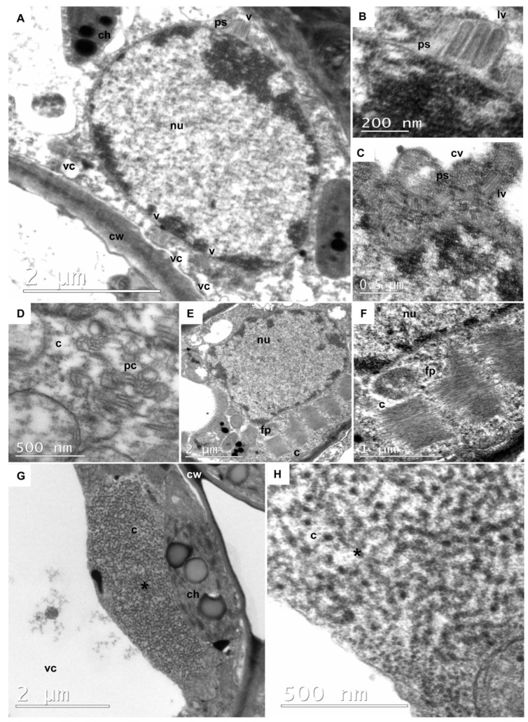
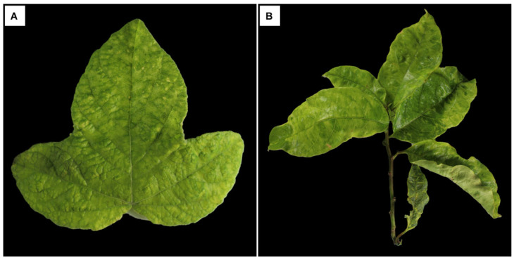
This study aimed to explore the RNA viruses affecting Passiflora species in Brazil. Our results enhance the understanding of the viruses that infect Passiflora plants by identifying and characterizing three previously unrecognized viruses: Passiflora cytorhabdovirus (PFCV), Passiflora nucleorhabdovirus 1 (PaNV1), and Passiflora nucleorhabdovirus 2 (PaNV2). These rhabdoviruses were identified through high-throughput sequencing and validated by reverse transcription-polymerase chain reaction (RT-PCR) in various Passiflora species. PFCV has a genome organization 3'-N-P-P3-P4-M-G-P7-L-5' and was classified as a novel member of the Gammacytorhabdovirus genus. A particularly noteworthy feature of PFCV is its glycoprotein, as the genomes of other gammarhabdoviruses do not contain this gene. PFCV has a high incidence across multiple locations and was identified in plants from Northeastern, Central, and Southeastern Brazil. PaNV1 with genome structure 3'-N-P-P3-M-G-L-5' and PaNV2 with genome organization 3'-N-X-P-Y-M-G-L-5' are new members of the Alphanucleorhabdovirus genus and have a more restricted occurrence. Importantly, all three viruses were found in mixed infections alongside at least one other virus. In situ observations confirmed mixed infections, with PaNV2 particles co-located in tissues with a potyvirus and a carlavirus. Phylogenetic and glycoprotein sequence similarity network analysis provided insights into their evolutionary placement and potential vector associations. These findings expand the known diversity of rhabdoviruses in Passiflora and contribute to the understanding of their evolution and epidemiology.

Keywords: alphanucleorhabdovirus; gammacytorhabdovirus; high-throughput sequencing; microscopy; plant rhabdovirus; virus particles.


Conflict of interest statement

The authors declare no conflicts of interest.

Figures

Figure 1
Figure 1
Genome organization of the new rhabdoviruses infecting Passiflora species depicting the canonical genes: nucleoprotein (N), phosphoprotein (P), matrix protein (M), glycoprotein (G), and RNA-dependent RNA polymerase—RdRP (L), and non-canonical ORFs encoding P3, P4, P7, X, Y. The rhabdovirus isolates were obtained from different passion fruit species, including PFCV-1530: P. galbana and PFCV-1591: P. eichleriana x P. gibertii from BAG-FP, PFCV-29: P. edulis from Rio de Janeiro, and PFCV-559: P. edulis from Bahia. PaNV2-1593: P. galbana from BAG-FP, and PaNV1-B-564: P. edulis from Bahia.
Figure 2
Figure 2
Midpoint-rooted maximum likelihood phylogenetic tree inferred using an amino acid alignment of RdRp (L protein) of Passiflora cytorhabdovirus (PFCV-559; PFCV-29; PFCV-1630; PFCV-1591) isolates with exemplary viruses for the species in the Alphacytorhabdovirus, Betacytorhabdovirus, and Gammacytorhabdovirus genera. The tree was generated with 1000 ultrafast bootstrap replicates and the “find and apply best model” option. Only the branch support values above 50% are shown. The viruses identified in this study are highlighted in yellow. All the accession numbers used to build the tree are listed in Supplementary Table S3.
Figure 3
Figure 3
Sequence similarity network analysis of the glycoprotein amino acid (G protein) sequences of Passiflora cytorhabdovirus isolates (PFCV-559; PFCV-29; PFCV-1630; PFCV-1591) and exemplar viruses for the species in the Alphacytorhabdovirus and Betacytorhabdovirus genera (dataset was created with an amino acid identity cut-off of 90%). Viruses with known insect vectors are highlighted. All the virus sequences accession numbers used are listed in Supplementary Table S3.
Figure 4
Figure 4
Midpoint-rooted maximum likelihood phylogenetic tree inferred using an amino acid alignment of RdRp (L protein) of Passiflora nucleorhabdovirus 1 (PaNV1-B-564) and Passiflora nucleorhabdovirus 2 (PaNV2-1593) isolates with exemplar viruses for the species in the members of Alphanucleorhabdovirus, Betanucleorhabdovirus, Deltanucleorhabdovirus, and Gammanucleorhabdovirus genera. The tree was inferred with 1000 ultrafast bootstrap replicates and the “find and apply best model” option. Only the branch support values above 50% are shown. The viruses identified in this study are highlighted in yellow. All the accession numbers used to construct the tree are listed in Supplementary Table S5.
Figure 5
Figure 5
Sequence similarity network analysis of the glycoprotein (G protein) of Passiflora nucleorhabdovirus 1 (PaNV1-B-564), Passiflora nucleorhabdovirus 2 (PaNV2-1593) isolates, and exemplar viruses for the species in the Alphanucleorhabdovirus, Betanucleorhabdovirus, Deltanucleorhabdovirus, and Gammanucleorhabdovirus genera (dataset was created with an amino acid identity cut-off of 90%). The viruses that have known insect vectors are highlighted. All the virus sequence accession numbers used in this analysis are listed in Supplementary Table S5.
Figure 6
Figure 6
Transmission electron micrographs of ultrathin sections of symptomatic rhabdovirus-infected leaves of P. gardneri (Sample 1626) (AF) and P. riparia (Sample 1624) (G,H). Sample 1626: (A) A low magnification image of a parenchyma cell showing pockets of perinuclear space (ps) containing bacilliform particles (v). (B) Detail of rhabdovirus particles sectioned longitudinally (lv) in the pockets perinuclear space (ps). (C) Detail of rhabdovirus particles in the pockets of perinuclear space (ps), cross sectioned (cv), revealing the outer membrane and the tubular inner component (nucleocapsid), as well as some of them sectioned longitudinally (lv). (D) Cross section of cylindrical inclusions, exhibiting pinwheel configuration (pc), in the cytoplasm (c) of a parenchymal cell, indicative of co-infection by potyvirus, possibly cowpea aphid-borne mosaic virus (CABMV). (E,F) Cytoplasmic inclusions of aggregates of elongated and flexuous particles (fp) in the cytoplasm (c) of the parenchymal cell. They possibly represent masses of particles of the carlavirus CPMMV. Sample 1626: (G,H) Overview of a viroplasm (*) formed by coiled filamentous material, possibly naked viral ribonucleoprotein, in the cytoplasm (c) of a parenchymal cell. Other cellular structures can be observed, including a vacuole (vc), a chloroplast (ch), a nucleus (nu), and a cell wall (cw).
Figure 7
Figure 7
Symptoms displayed by Passiflora spp. leaves. (A) P. gardneri (Sample 1626) with light mosaic and (B) P. riparia (Sample 1624) showing leaf deformation, blistering, and mosaic.

Similar articles

References

    1. Higuita M., Sanchez-Yali J., Perez A., Arias E., Gutierrez P.A. An integrated analysis of the Passifloraceae virome using public-domain data. Phytopathology. 2024 doi: 10.1094/PHYTO-08-24-0269-FI. - DOI - PubMed
    1. Bejerman N., Dietzgen R., Debat H. Novel tri-segmented rhabdoviruses: A data mining expedition unveils the cryptic diversity of cytorhabdoviruses. Viruses. 2023;15:2402. doi: 10.3390/v15122402. - DOI - PMC - PubMed
    1. Zhang S., Huang A., Zhou X., Li Z., Dietzgen R.G., Zhou C., Cao M. Natural defect of a plant rhabdovirus glycoprotein gene: A case study of virus–plant coevolution. Phytopathology. 2021;111:227–236. doi: 10.1094/PHYTO-05-20-0191-FI. - DOI - PubMed
    1. Bejerman N., Dietzgen R.G., Debat H. Illuminating the plant rhabdovirus landscape through metatranscriptomics Data. Viruses. 2021;13:1304. doi: 10.3390/v13071304. - DOI - PMC - PubMed
    1. Bejerman N., Debat H. Alpha- and Betagymnorhavirus: Two new genera of gymnosperm-infecting viruses in the family Rhabdoviridae, subfamily Betarhabdovirinae. Arch. Virol. 2024;169:193. doi: 10.1007/s00705-024-06119-y. - DOI - PubMed

Publication types

Associated data

LinkOut - more resources