Review
. 2025 Apr 25;13(5):456.
doi: 10.3390/vaccines13050456.

Host Immune Response to Bovine Viral Diarrhea Virus (BVDV): Insights and Strategies for Effective Vaccine Design

Affiliations
Review

Host Immune Response to Bovine Viral Diarrhea Virus (BVDV): Insights and Strategies for Effective Vaccine Design

Asamenew Tesfaye Melkamsew et al. Vaccines (Basel). .

Abstract

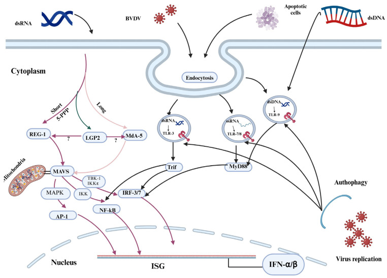
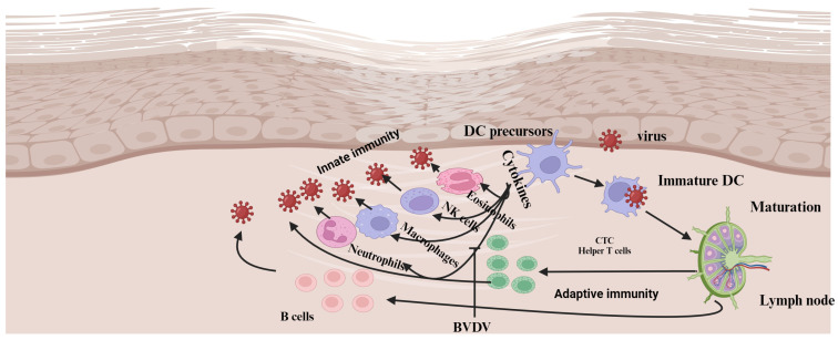
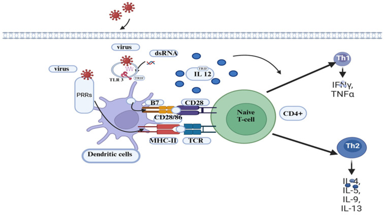
Bovine viral diarrhea (BVD) is caused by bovine viral diarrhea virus (BVDV), a member of the genus Pestivirus and in the family Flaviviridae. According to some studies, the disease incurs USD 1.5-2.5 billion per year and USD 0.50 to USD 687.80 per cow loss in beef and dairy farms, respectively. Using vaccines is among the strategies to prevent the disease. However, complete protection requires vaccines that target both the humoral and cellular immune responses of the adaptive immune system. A comprehensive literature review was made to provide insights into the interaction of BVDV with host immunity, vaccine applications, and the limitation of the currently available vaccines, as well as explore strategies used to advance the vaccines. BVDV causes immunosuppression by interfering with the innate and adaptive immune systems in a manner that is species and biotype-dependent. Interferon production, apoptosis, neutrophil activity, and antigen-processing and presenting cells are significantly affected during the viral infection. Despite maternal antibodies (MatAbs) being crucial to protect calves from early-age infection, a higher level of MatAbs are counterproductive during the immunization of calves. There are numerous inactivated or modified BVDV vaccines, most of which are made of cytopathic BVDV 1 and 2 and the BVDV 1a subgenotypes. Furthermore, subunit, marker, DNA and mRNA vaccines are made predominantly from E2, Erns, and NS3 proteins of the virus in combination with modern adjuvants, although the vaccines have not yet been licensed for use and are in the experimental stage. The existing BVDV vaccines target the humoral immune system, which never gives the full picture of protection without the involvement of the cell-mediated immune system. Several limitations were associated with conventional and next-generation vaccines that reduce BVDV vaccine efficiency. In general, providing complete protection against BVDV is very complex, which requires a multi-pronged approach to study factors affecting vaccine efficacy and strategies needed to improve vaccine efficacy and safety.

Keywords: BVD; BVDV; immunity; maternal antibody; vaccines.


Conflict of interest statement

The authors declare no conflicts of interest.

Figures

Figure 1
Figure 1
Signaling pathways for Retinoic Acid-Inducible Gene-I-like Receptors (RLRs) and Toll-like receptors (TLRs). The ssRNA of the cpBVDV biotype stimulates endosomal TLR 7/8 to induce interferon-β (IFN-β). As a result, the TRIF temporarily binds with TLR 7/8. After detaching from the receptor, the TRIF forms a speckle-like complex that relocates to interact with downstream signaling molecules such as tumor necrosis factor (TNF) receptor-associated factor 2 (TRAF2), TRAF6, and receptor-interacting protein 1 (RIP-1) [19,20]. Then, the combined effect of TRIF with TRAF3, TRAF family member-associated nuclear transcription factor-κB (NF-κB) activator (TANK)-binding kinase 1 (TBK1), inhibitor of κB (IκB) kinase-related kinase-ε (IKK-ε; also called IKK-i), and NF-κB-activating kinase (NAK)-associated protein 1 (NAP1) can be observed. Finally, the signaling pathways stimulate transcription factors, namely, IFN-regulatory factor 3/7 (IRF3/7), the TRIF-dependent NFκB, and the activator protein 1 (AP-1), thus moderating the production of type I IFNs, proinflammatory cytokines, and chemokines, respectively. AP = Activator Protein; IRF = IFN regulatory factors; IKKε = inhibitor of nuclear factor kappa-B kinase ε; ISG = IFN-stimulated genes; LGP2 = laboratory of genetics and physiology 2; MAVS = mitochondrial antiviral signaling protein; MDA5 = melanoma differentiation-associated protein 5; MAPK = mitogen-activated protein kinase; MyD88 = myeloid differentiation primary response; NF-KB = nuclear factor-kappa B; Tbk = TANK-binding kinase; Trif = TIR domain-containing adapter inducing IFN-β. Created with BioRender.com.
Figure 2
Figure 2
Model for the interaction between the innate and adaptive immune system and BVDV biotypes. Infection of mucosal or epithelial surfaces with cpBVDV activates cytokine expression, including IFNα from different cell types and dendritic cells (DCs). These cytokines induce effector cells of the innate immune response, such as eosinophils, macrophages, and natural killer cells (NK cells). Subsequently, viral replication is blocked due to the combined effect of cytokines and effector cell function. Infection with cpBVDV creates an environment that facilitates the migration of antigen-capturing DCs to the lymph nodes, where they undergo maturation. DCs probably withstand the lytic effect of cpBVDV and migrate to the local lymph node. The DCs with the antigen arrive at the lymph node and present the antigen through MHC I to antigen-specific lymphocytes. Eventually, the activated T lymphocytes (CTLs and helper T cells) migrate to the site of infection to eliminate the virus or virus-infected cells. Mature plasma cells are generated from activated B cells that reach the GC or far in the lymph node and produce virus-neutralizing antibodies. Unlike cpBVDV infection, ncpBVDV infection cannot stimulate an early cytokine response. Consequently, the viral replication is unlimited, and activation of DCs is reduced. DCs, whether they have captured the virus or not, migrate to the local lymph node. Those that encounter the virus within the lymph node produce large quantities of IFNα. The concentration of IFNα in the circulation and tissues increases, ultimately activating the DCs to inhibit viral replication. However, during this time, the ncpBVDV virus has already disseminated throughout the animal body [13]. DCs, dendritic cells; NK cells, natural killer cells; MHC I, major histocompatibility; CTLs, cytotoxic T lymphocytes; GC, germinal center; CD, cluster of differentiation; NET, neutrophil extracellular traps. Created with BioRender.com.
Figure 3
Figure 3
Antigen-presenting cell activation and interactions with T cells. On antigen-presenting cells (e.g., DCs), the viral PAMPs bind to PRRs. Matured and activated DCs migrate with the carried antigen to lymph nodes, where they encounter naïve T lymphocytes and present the processed peptides loaded onto MHC II to CD4+ cells. These CD4+ cells then differentiate into Th1 and Th2 cells in the presence of cytokines and interleukins. Th1 cells activate macrophages to phagocytize and kill the pathogen, and helper B cells produce antibodies against the pathogen. In viral infections such as BVDV, the activation of cytotoxic T cells (CTL) by CD4+ cells plays a crucial role in mounting an effective immune response. DCs with the processed peptides on MHC-I migrate to lymph nodes and bind to naïve CTLs. This activated CTL releases effector molecules such as perforin, granzymes, and granulysin to kill virus-infected cells. PRR, Pathogen Recognition Receptor; PAMP, Pathogen-Associated Molecular Patterns. TLRs, Toll-like receptors; B7/CD80/CD86 represents APCs co-receptor; CD28 denotes T cell co-receptor; and MHC II, Major Histocompatibility II. Created with BioRender.com.

Similar articles

References

    1. Szabára Á., Ózsvári L. Challenges for the Agricultural Sector in Central and Eastern Europe. Agroinform Kiadó; Budapest, Hungary: 2014. Economic impacts, control and eradication of Bovine Viral Diarrhoea virus; pp. 247–258. - DOI
    1. Khodakaram-Tafti A., Farjanikish G.H. Persistent bovine viral diarrhea virus (BVDV) infection in cattle herds. Iran. J. Vet. Res. 2017;18:154–163. doi: 10.22099/ijvr.2017.4190. - DOI - PMC - PubMed
    1. Smith D.B., Meyers G., Bukh J., Gould E.A., Monath T., Muerhoff A.S., Pletnev A., Rico-Hesse R., Stapleton J.T., Simmonds P., et al. Proposed revision to the taxonomy of the genus Pestivirus, family Flaviviridae. J. Gen. Virol. 2017;98:2106–2112. doi: 10.1099/jgv.0.000873. - DOI - PMC - PubMed
    1. Ammari M., McCarthy F.M., Nanduri B., Pinchuk L.M. Analysis of Bovine Viral Diarrhea Viruses-infected monocytes: Identification of cytopathic and non-cytopathic biotype differences. BMC Bioinform. 2010;11:9. doi: 10.1186/1471-2105-11-S6-S9. - DOI - PMC - PubMed
    1. Hilleman M.R. Strategies and mechanisms for host and pathogen survival in acute and persistent viral infections. Proc. Natl. Acad. Sci. USA. 2004;101:14560–14566. doi: 10.1073/pnas.0404758101. - DOI - PMC - PubMed

LinkOut - more resources