. 2025 May 27;11(3):00970-2024.
doi: 10.1183/23120541.00970-2024. eCollection 2025 May.

Deleterious effect of Pseudomonas aeruginosa on F508del-CFTR rescued by elexacaftor/tezacaftor/ivacaftor is clinical strain-dependent in patient-derived nasal cells

Affiliations

Deleterious effect of Pseudomonas aeruginosa on F508del-CFTR rescued by elexacaftor/tezacaftor/ivacaftor is clinical strain-dependent in patient-derived nasal cells

Caterina Allegretta et al. ERJ Open Res. .

Abstract

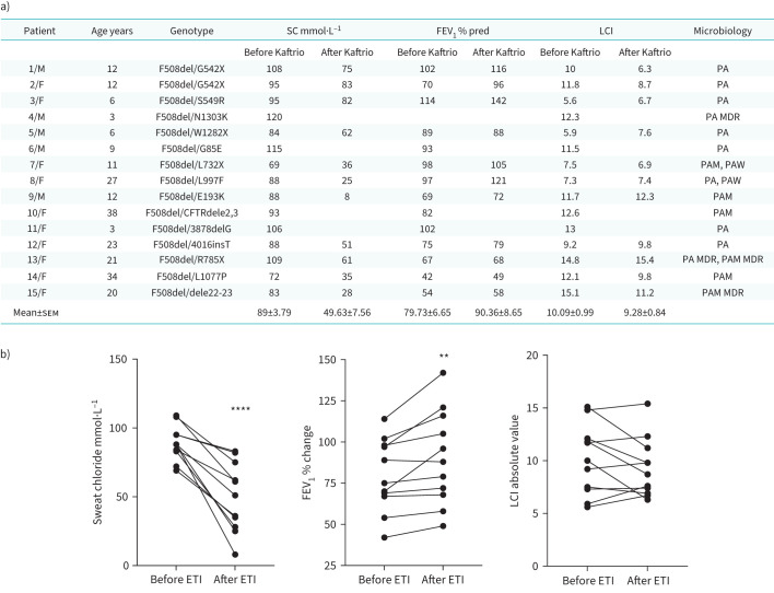
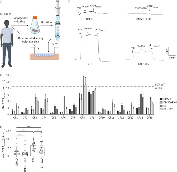
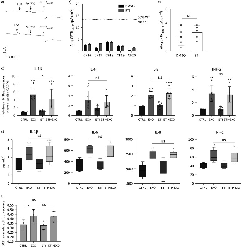
Background: The triple cystic fibrosis transmembrane conductance regulator (CFTR) modulators combination elexacaftor/tezacaftor/ivacaftor (ETI) has been approved for people with cystic fibrosis (pwCF) bearing at least one F508del allele. Despite the development of CFTR modulators having dramatically improved respiratory outcomes in pwCF, clinical studies have showed variable responses to this drug formulation. Of note, airway inflammation and bacterial colonisation persist in the upper and lower respiratory tract even in ETI-treated patients.

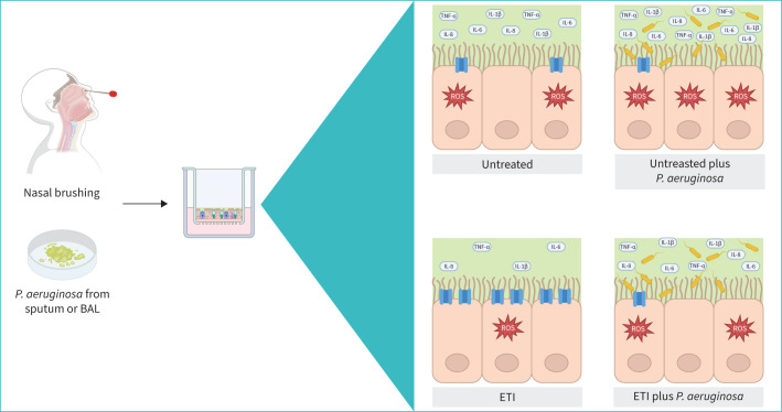
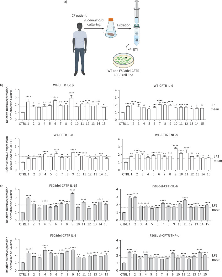
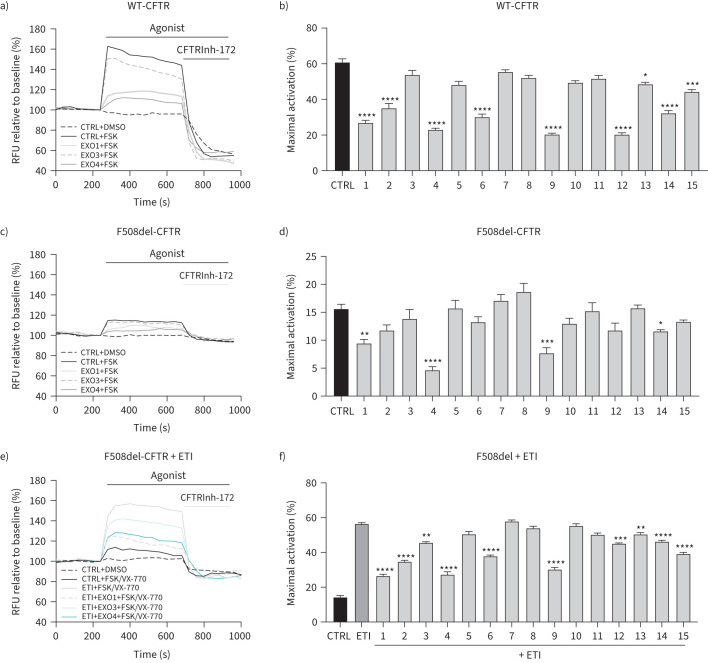
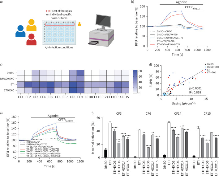
Methods: We first tested the clinical exoproducts (EXO) of Pseudomonas aeruginosa isolated from 15 CF patients in wild-type (WT) and F508del-CFTR CF bronchial epithelial (CFBE) cells. We were then prompted to evaluate the effects of EXO in ex-vivo patient-derived tissues. Therefore, we cultured primary nasal epithelial cells (HNECs) with EXO isolated from the corresponding pwCF to mimic the native status of CF airway.

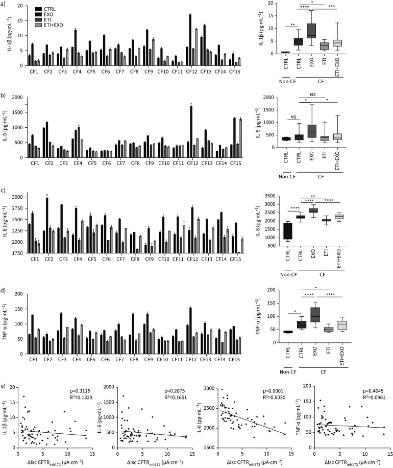
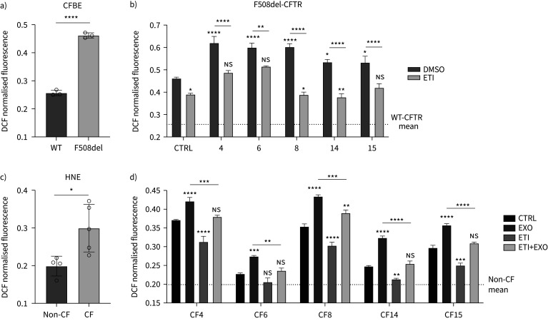
Results: We found that EXO variably decreased WT-, F508del- and ETI-dependent F508del-CFTR function and increased proinflammatory cytokines and reactive oxygen species (ROS) levels in a clinical strain-specific manner. Similarly, we observed a variable reduction of F508del-CFTR function in presence or absence of ETI and upregulation of proinflammatory cytokines and ROS levels. Interestingly, HNECs treated with EXO isolated from the corresponding donor and three different pwCF showed a variable reduction of ETI-dependent F508del-CFTR function mainly due to clinical strains with limited effect of patient background. Furthermore, we demonstrated that ETI pretreatment decreased the cytokines and ROS levels down to the levels of uninfected cells.

Conclusion: These preclinical studies suggest that in vitro screening of patient-specific response to CFTR modulators under infection/inflammation conditions could prove to be a valuable tool to enhance the prediction of clinical response.


Conflict of interest statement

Conflict of interest: The authors declare that the research was conducted in the absence of any commercial or financial relationships that could be construed as a potential conflict of interest.

Figures

None
Pseudomonas aeruginosa infection reduces ETI treatment efficacy. BAL: bronchoalveolar lavage; ETI: elexacaftor/tezacaftor/ivacaftor.
FIGURE 1
FIGURE 1
Variable inflammatory response across different clinical exoproducts of Pseudomonas aeruginosa obtained from 15 cystic fibrosis (CF) patients in CF bronchial epithelial (CFBE) cell line. a) Cartoon showing the generation of clinical exoproducts (EXO) isolated from CF patients and incubated with CFBE cell line. WT-CFTR b) and c) F508del-CFTR CFBE cells were incubated with clinical exoproducts of P. aeruginosa isolated from the sputum of 15 CF patients (1 to 15) for 24 h at 37°C. Total RNA was extracted and quantitative real-time PCR was performed in order to quantify interleukin (IL)-1β, IL-6, IL-8 and tumour necrosis factor-α (TNF-α) mRNAs. Bar graphs show the mean±sem of n=3–4. *p<0.05; **p<0.01; ***p<0.001; ****p<0.0001. ETI: elexacaftor/tezacaftor/ivacaftor; LPS: lipopolysaccharide, WT: wild-type.
FIGURE 2
FIGURE 2
Clinical exoproducts of Pseudomonas aeruginosa reduced CFTR function dependent on clinical strain in cystic fibrosis (CF) bronchial epithelial (CFBE) cell line. Representative traces of a) WT-CFTR-, c,e) F508del-CFTR-dependent chloride efflux in CFBE cells using fluorescence membrane polarisation (FMP) assay. Cells were treated for 24 h with lysogeny broth (CTRL), clinical exoproducts (EXO) of P. aeruginosa from 15 CF patients in presence or absence of 3 µM VX-661+3 µM VX-445 when reported. Bar graphs show the mean±sem of maximal activation of b) WT-CFTR, d) F508del-CFTR, f) F508del-CFTR treated with 0.1% DMSO, 3 µM VX-661+3 µM VX-445 in CFBE cells after stimulation by 1 µM FSK (for WT-CFTR) and 10 µM FSK+1 µM VX-770 (for F508del-CFTR) (n=3–4 biological replicates with 4 technical replicates each). *p<0.05; **p<0.01; ***p<0.001; ****p<0.0001. ETI: elexacaftor/tezacaftor/ivacaftor; RFU: relative fluorescence units; WT: wild-type.
FIGURE 3
FIGURE 3
Clinical data of brushed nasal cell donors. a) All 15 cystic fibrosis (CF) patients are represented by numbers, which correspond to data points throughout the text and figures. SC: sweat chloride, FEV1 % pred: % predicted forced expiratory volume in 1 s; LCI: lung clearance index; PA: Pseudomonas aeruginosa; PA MDR: P. aeruginosa multidrug resistant; PAM: P. aeruginosa mucoid isolate; PAM MDR: P. aeruginosa mucoid isolate multidrug resistant; PAW: P. aeruginosa wrinkled isolate. b) Graphs show the clinical data (sweat chloride, FEV1 and LCI) before and after 6 months of Kaftrio treatment in 11 CF patients. ETI: elexacaftor/tezacaftor/ivacaftor. **p<0.01; ****p<0.0001.
FIGURE 4
FIGURE 4
Nasal epithelial cultures from 15 cystic fibrosis (CF) patients exhibit differential phenotypic response to VX-445/VX-661/VX-770 in a clinical strain of Pseudomonas aeruginosa. a) Cartoon showing the generation of clinical exoproducts (EXO) isolated from CF patients and incubated with primary nasal epithelial cells (HNECs) from the corresponding donor. b) Representative tracings show Ussing chamber measurements of CFTR function in HNEC cultures treated for 24 h with 0.1% DMSO, 3 µM VX-661+3 µM VX-445±clinical exoproducts of P. aeruginosa (EXO) isolated from the corresponding patient. c) Bar graph shows the amplitude of the current blocked by 10 µM CFTRinh172 (ΔIscinh-172) after CFTR stimulation with 10 µM FSK+1 µM VX-770 measured in HNEC cultures from each donor. d) Bar graph shows the mean of the amplitude of the current blocked by 10 µM CFTRinh172 (ΔIscinh-172) measured in 15 CF patients. (n=2 technical replicates for each donor). ETI: elexacaftor/tezacaftor/ivacaftor. **p<0.01; ***p<0.001; ****p<0.0001.
FIGURE 5
FIGURE 5
Inflammatory response is clinical strain of Pseudomonas aeruginosa-dependent and is ameliorated by elexacaftor/tezacaftor/ivacaftor (ETI) treatment. a) Interleukin (IL)-1β, b) IL-6, c) IL-8 and d) tumour necrosis factor-α (TNF-α) secretion after stimulation with lysogeny broth (CTRL), clinical exoproducts of P. aeruginosa (EXO) isolated from the corresponding donor, 3 µM VX-661+3 µM VX-445+1 µM VX-770 (ETI)±(EXO) for 24 h in primary nasal epithelial cells from four non-cystic fibrosis (CF) donors and 15 CF patients (n=3). (e) Pearson correlation between released cytokines (pg·mL−1) and CFTR activity (ΔIscinh-172 µA·cm−2). ns: nonsignificant. *p<0.05; **p<0.01; ***p<0.001; ****p<0.0001.
FIGURE 6
FIGURE 6
Elexacaftor/tezacaftor/ivacaftor (ETI)-rescued F508del-CFTR activity is variably downregulated strain-dependent of Pseudomonas aeruginosa in primary nasal epithelial cells by high-throughput fluorescence-based assay. a) Cartoon showing nasal epithelial cells cultured in 96-transwell plate, treated with CFTR modulators in presence or absence of infection stimuli and then tested for apical CFTR activity by fluorescence membrane polarisation assay (FMP) using a plate reader. b) Representative traces of F508del-CFTR-dependent chloride efflux in HNEC cultures by FMP. Cells were treated for 24 h with 0.1% DMSO, 3 µM VX-661+3 µM VX-445±clinical exoproducts of P. aeruginosa (EXO) isolated from the corresponding patient. c) Heatmap of peak responses after 10 µM FSK+1 µM VX-770 stimulation generated from HNEC cultures. The response size is colour coded as shown in the side bar, with blue representing the highest response and white the lowest response. (n=3 technical replicates.) d) Correlation between mean donor-specific activation measured using FMP (expressed as % of maximal activation) and mean donor-specific activation measured in the Ussing chamber (ΔIscinh-172 µA·cm−2). e) Representative traces of F508del-CFTR activity in HNECs treated for 24 h with 0.1% DMSO, 3 µM VX-661+3 µM VX-445±clinical exoproducts of P. aeruginosa (EXO) isolated from patient CF3 (EXO3), CF6 (EXO6), CF14 (EXO14) and CF15 (EXO15). f) Bar graphs show the mean±sem of maximal activation after 10 µM FSK+1 µM VX-770. (n=3 technical replicates.) Statistical analysis compared to ETI control. **p<0.01; ***p<0.001; ****p<0.0001. RFU: relative fluorescence units.
FIGURE 7
FIGURE 7
Clinical exoproducts of Pseudomonas aeruginosa increased the intracellular levels of the reactive oxygen species (ROS) and were reduced by elexacaftor/tezacaftor/ivacaftor (ETI) treatment in both cystic fibrosis (CF) bronchial epithelial (CFBE) cells and primary nasal epithelial cells. Cells were treated with clinical exoproducts of P. aeruginosa isolated from the sputum of five CF patients (1 to 15) ±3 µM VX-661+3 µM VX-445+1 µM VX-770 (ETI) for 24 h at 37°C. ROS levels were measured by 10μM o5-(and 6)-chloromethyl-2′,7′-dichlorodihydrofluorescein diacetate acetyl ester (H2DCFDA)-based assay. a,b) Bar graphs show the mean±sem of ROS levels in wild-type (WT) and F508del-CFTR CFBE cells (n=3). c) Bar graph shows the mean±sem of ROS levels in primary nasal cells from four non-CF donors and five CF patients. d) Bar graph shows the mean±sem of ROS levels in nasal epithelial cells from five CF patients. Cells were treated for 24 h with 0.1% DMSO, 3 µM VX-661+3 µM VX-445±clinical exoproducts of P. aeruginosa (EXO) isolated from the corresponding patient. (n=3 technical replicates for each patient.) *p<0.05; **p<0.01; ***p<0.001; ****p<0.0001. DCF: 2′,7′-dichlorodihydrofluorescein diacetate (H2DCFDA); WT: wild-type.
FIGURE 8
FIGURE 8
ETI treatment did not decrease proinflammatory released cytokines and reactive oxygen species (ROS) levels in primary nasal epithelial cells from cystic fibrosis (CF) patients bearing elexacaftor/tezacaftor/ivacaftor (ETI)-resistant mutations. a) Representative tracings show Ussing chamber measurements of CFTR function in primary human nasal epithelial cell (HNEC) cultures from five CF patients bearing E585X/E585X (CF16), R347P/R347P (CF17), E585X/G542X (CF18), R553X/Dele2,3 (CF19), G542X/N1303 K (CF20). Cells were treated for 24 h with 0.1% DMSO, 3 µM VX-661+ 3 µM VX-445. b) Bar graph shows the amplitude of the current blocked by 10 µM CFTRinh172 (ΔIscinh-172) after CFTR stimulation with 10 µM FSK+1 µM VX-770 measured in HNEC cultures from each donor. c) Bar graphs show the mean of the amplitude of the current blocked by 10 µM CFTRinh172 (ΔIscinh-172) measured in five CF patients. d) Interleukin (IL)-1β, IL-6, IL-8 and tumour necrosis factor-α (TNF-α) mRNA levels and e) secretion after stimulation with lysogeny broth (CTRL), clinical exoproducts of Pseudomonas aeruginosa (EXO), 3 µM VX-661+3 µM VX-445+1 µM VX-770±(EXO) for 24 h in HNEC cultures from five CF patients. f) Bar graph shows the mean±sem of ROS levels in nasal epithelial cells from three CF donors. *p<0.05; **p<0.01; ***p<0.001; ****p<0.0001. DCF: 2′,7′-dichlorodihydrofluorescein diacetate.

Similar articles

References

    1. Lund-Palau H, Turnbull AR, Bush A, et al. . Pseudomonas aeruginosa infection in cystic fibrosis: pathophysiological mechanisms and therapeutic approaches. Expert Rev Respir Med 2016; 10: 685–697. - PubMed
    1. Middleton PG, Mall MA, Dřevínek P, et al. . Elexacaftor-tezacaftor-ivacaftor for cystic fibrosis with a single Phe508del allele. N Engl J Med 2019; 381: 1809–1819. doi:10.1056/NEJMoa1908639 - DOI - PMC - PubMed
    1. Keating D, Marigowda G, Burr L, et al. . VX-445-tezacaftor-ivacaftor in patients with cystic fibrosis and one or two Phe508del alleles. N Engl J Med 2018; 379: 1612–1620. doi:10.1056/NEJMoa1807120 - DOI - PMC - PubMed
    1. Schaupp L, Addante A, Völler M, et al. . Longitudinal effects of elexacaftor/tezacaftor/ivacaftor on sputum viscoelastic properties, airway infection and inflammation in patients with cystic fibrosis. Eur Respir J 2023; 62: 2202153. doi:10.1183/13993003.02153-2022 - DOI - PubMed
    1. Morgan SJ, Nichols DP, Ni W, et al. . Elexacaftor/tezacaftor/ivacaftor markedly reduces aspergillus fumigatus in cystic fibrosis. Am J Respir Crit Care Med 2024; 210: 1155–1158. doi:10.1164/rccm.202406-1128RL - DOI - PMC - PubMed

LinkOut - more resources