. 2025 May 13:16:1559446.
doi: 10.3389/fphar.2025.1559446. eCollection 2025.

Stromal vascular fraction inhibits renal fibrosis by regulating metabolism and inflammation in obstructive nephropathy

Affiliations

Stromal vascular fraction inhibits renal fibrosis by regulating metabolism and inflammation in obstructive nephropathy

Guang Yue et al. Front Pharmacol. .

Abstract

Obstructive nephropathy is one of the leading causes of kidney injury and fibrosis, which can lead to end-stage renal disease (ESRD). Stromal vascular fraction (SVF), a heterogeneous cell mixture derived from adipose tissue, has been widely used for regenerative medicine across many preclinical models and clinical applications. Recent studies have suggested that SVF can alleviate acute kidney injury in mice. However, to our knowledge, the therapeutic effects of SVF on obstructive nephropathy have not been studied before. In this study, we evaluated the therapeutic potential of SVF on obstructive nephropathy in mice with unilateral ureteral obstruction (UUO). We revealed that autologous SVF administration mitigated UUO-induced renal fibrosis. SVF treatment inhibited both the infiltration of neutrophils and CD4+ T cells, as well as the production of inflammatory cytokines. Moreover, SVF promoted metabolic reprogramming and improved mitochondrial function in the obstructed kidneys, partially through PPAR pathway activation. Mechanistically, SVF-mediated PPAR activation inhibited the epithelial-mesenchymal transition (EMT) process of tubular cells, thus alleviating renal fibrosis in UUO mice. We further confirmed that pharmacological activation of PPAR pathway significantly reduced fibrosis in UUO kidneys. Therefore, our study suggests that SVF may represent a promising therapeutic strategy for obstructive nephropathy.

Keywords: PPAR; SVF; inflammation; obstructive nephropathy; renal fibrosis.


Conflict of interest statement

The authors declare that the research was conducted in the absence of any commercial or financial relationships that could be construed as a potential conflict of interest.

Figures

FIGURE 1
FIGURE 1
Characterization of murine SVF by flow cytometry Representative flow cytometry plots of murine stromal vascular fraction (SVF). The expression of hematopoietic (CD45, CD11b, and CD11c), mesenchymal (CD29, CD90), and endothelial (CD31) markers were detected and the quantification was shown as the mean ± SEM.
FIGURE 2
FIGURE 2
SVF alleviated UUO-induced renal fibrosis and injury (A) Experimental design was shown. (B) Representative Masson staining and quantification of UUO kidneys from control and SVF-treated mice (n = 5), scale bar = 40 μm. Asterisks (*) indicate regions of interstitial collagen deposition (in blue). (C) Immunoblots and quantification of FN, Col I, and αSMA expression in UUO kidneys from control and SVF-treated mice (n = 3). (D) qPCR analysis for Col1a2 and Acta2 in UUO kidneys from control and SVF-treated mice (n = 4). (E) Serum levels of BUN and Cr in control and SVF-treated mice (n = 4). (F) qPCR analysis for Tgfb1 in UUO kidneys from control and SVF-treated mice (n = 4). The results represent mean ± SEM. *p < 0.05, **p < 0.01, NS no significant.
FIGURE 3
FIGURE 3
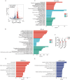
SVF contributed to metabolic reprogramming and reduced inflammation in UUO kidneys (A) Volcano plot showing the differential expressed genes in UUO kidneys between control and SVF-treated mice (fold change >1.5, p < 0.05). GO enrichment analysis of (B) the upregulated and (C) the downregulated genes in SVF-treated mice. KEGG enrichment analysis of (D) the upregulated and (E) the downregulated genes in SVF-treated mice. (F) The expression levels of Fn1, Col1a2, and Acta2 in control and SVF-treated mice as revealed by RNA-seq (n = 3). The results represent mean ± SEM. *p < 0.05.
FIGURE 4
FIGURE 4
SVF inhibited the accumulation of neutrophils and CD4+ T cells in UUO kidneys (A) Gating strategy of kidney immune cells. Representative flow cytometry plots and quantification of (B) macrophages, (C) neutrophils and monocytes, (D) B cells, (E) T, NK, and NKT cells, and (F) CD4+ T and CD8+ T cells in UUO kidneys from control and SVF-treated mice (n = 5). The results represent mean ± SEM. *p < 0.05, **p < 0.01, NS no significant.
FIGURE 5
FIGURE 5
SVF inhibited UUO-induced kidney inflammation (A,B) qPCR analysis for Il1b, Il6, Mrc1, and Retnla expression in UUO kidneys from control and SVF-treated mice (n = 4). (C) Representative plots and quantification of CD206 expression in renal macrophages from control and SVF-treated mice (n = 3). (D) Immunoblots and quantification of NF-κB p65 expression in UUO kidneys from control and SVF-treated mice (n = 3). The results represent mean ± SEM. *p < 0.05, **p < 0.01.
FIGURE 6
FIGURE 6
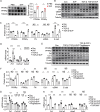
SVF promoted PPAR activation and inhibited EMT in TCMK-1 cells (A) Immunoblots and quantification of PPARα and PPARγ in UUO kidneys from control and SVF-treated mice (n = 3). (B) Immunoblots and (C) quantification of PPARα, PPARγ, FN, αSMA, and E-cad in TCMK-1 cells treated with or without SVF (n = 3). (D) qPCR analysis for Fn1, Col1a2, and Acta2 in TCMK-1 cells treated with or without SVF (n = 4). (E) Immunoblots and (F) quantification of PPARα, PPARγ, FN, Col I, αSMA, and E-cad in TCMK-1 cells treated with SVF or SVF and pioglitazone combined (n = 3). (G) qPCR analysis for Fn1, Col1a2, and Acta2 in TCMK-1 cells treated with SVF or SVF and pioglitazone combined (n = 4). (H) qPCR analysis for Fn1, Col1a2, Acta2, and Cdh1 in TCMK-1 cells treated with SVF or SVF and T0070907 combined (n = 4). The results represent mean ± SEM. *p < 0.05, **p < 0.01, ***p < 0.001, NS no significant.
FIGURE 7
FIGURE 7
Pharmacological activation of PPAR alleviated UUO-induced renal fibrosis and inflammation (A) Immunoblots and quantification of PPARα and PPARγ in UUO kidneys from control and pioglitazone-treated mice (n = 3). (B) Representative Masson staining and quantification of UUO kidneys from control and pioglitazone-treated mice (n = 5), scale bar = 40 μm. Asterisks (*) indicate regions of interstitial collagen deposition (in blue) (C) Immunoblots and quantification of FN, Col I, αSMA, E-cad, and N-cad expression in UUO kidneys from control and pioglitazone-treated mice (n = 3). (D,E) qPCR analysis for Col1a2, Il1b, and Tnf in UUO kidneys from control and pioglitazone-treated mice (n = 5). The results represent mean ± SEM. *p < 0.05, **p < 0.01.

Similar articles

References

    1. Al-Kharboosh R., Perera J. J., Bechtle A., Bu G., Quinones-Hinojosa A. (2022). Emerging point-of-care autologous cellular therapy using adipose-derived stromal vascular fraction for neurodegenerative diseases. Clin. Transl. Med. 12 (12), e1093. 10.1002/ctm2.1093 - DOI - PMC - PubMed
    1. Andia I., Maffulli N., Burgos-Alonso N. (2019). Stromal vascular fraction technologies and clinical applications. Expert Opin. Biol. Ther. 19 (12), 1289–1305. 10.1080/14712598.2019.1671970 - DOI - PubMed
    1. Atalay S., Coruh A., Deniz K. (2014). Stromal vascular fraction improves deep partial thickness burn wound healing. Burns 40 (7), 1375–1383. 10.1016/j.burns.2014.01.023 - DOI - PubMed
    1. Bateman M. E., Strong A. L., Gimble J. M., Bunnell B. A. (2018). Concise review: using fat to fight disease: a systematic review of nonhomologous adipose-derived stromal/stem cell therapies. Stem Cells 36 (9), 1311–1328. 10.1002/stem.2847 - DOI - PubMed
    1. Bensemmane L., Milliat F., Treton X., Linard C. (2023). Systemically delivered adipose stromal vascular fraction mitigates radiation-induced gastrointestinal syndrome by immunomodulating the inflammatory response through a CD11b(+) cell-dependent mechanism. Stem Cell Res. Ther. 14 (1), 325. 10.1186/s13287-023-03562-7 - DOI - PMC - PubMed

LinkOut - more resources