

Physiolysis for Metatarsal Bracketed Epiphysis
- PMID: 40432945
- PMCID: PMC12088176
- DOI: 10.55275/JPOSNA-2023-773
Physiolysis for Metatarsal Bracketed Epiphysis
Abstract
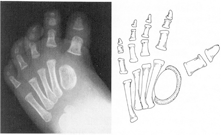
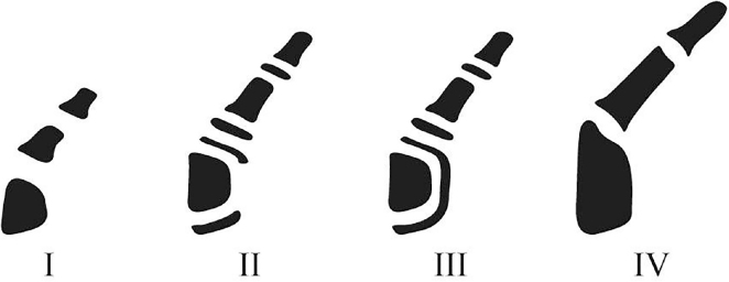
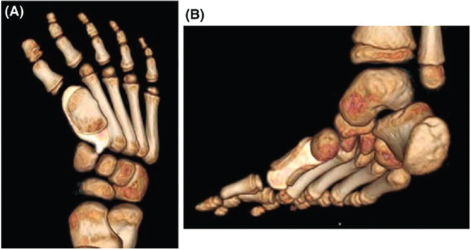
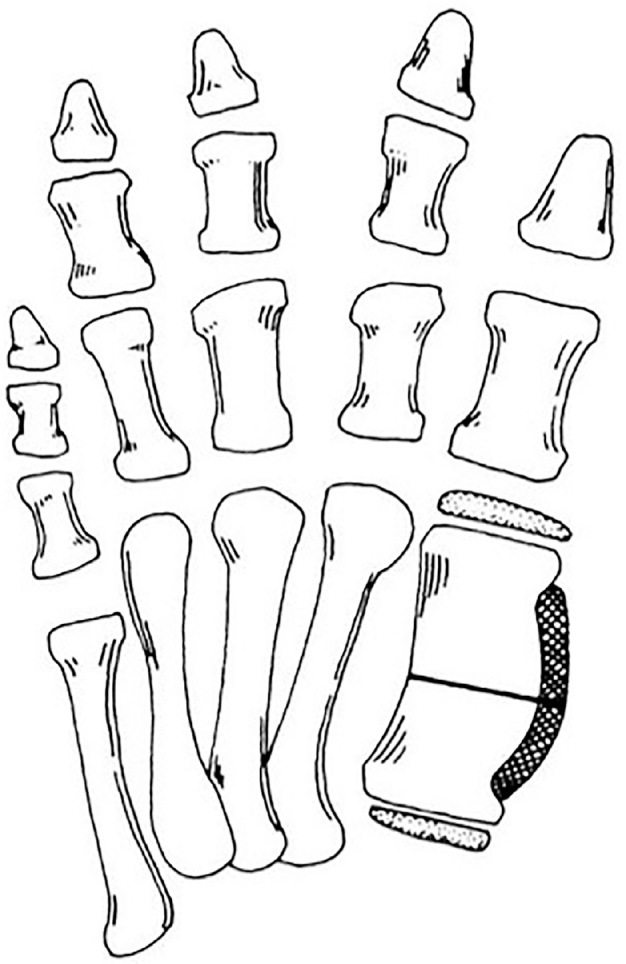
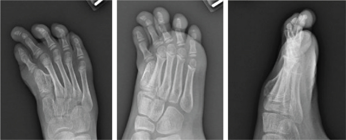
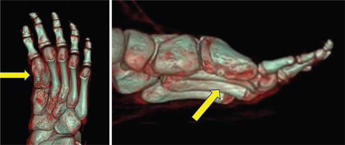
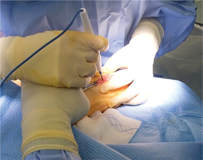
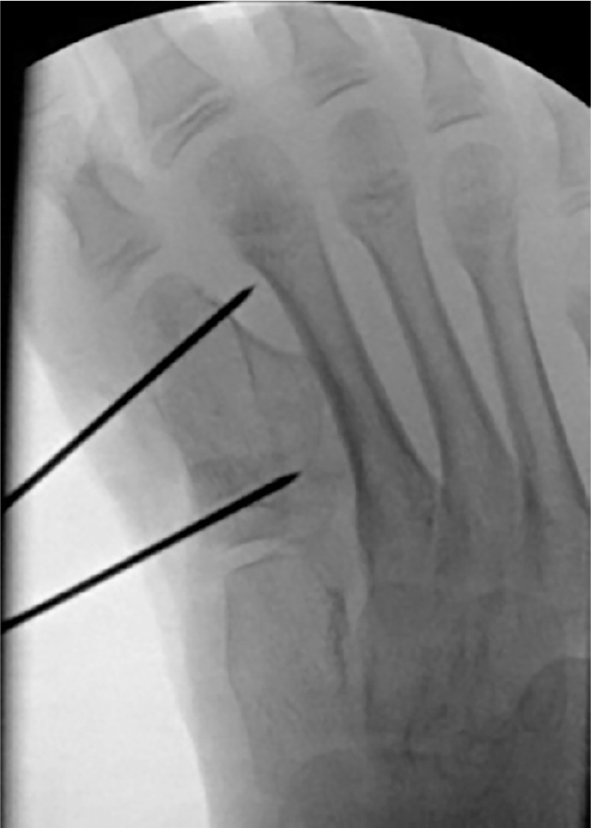
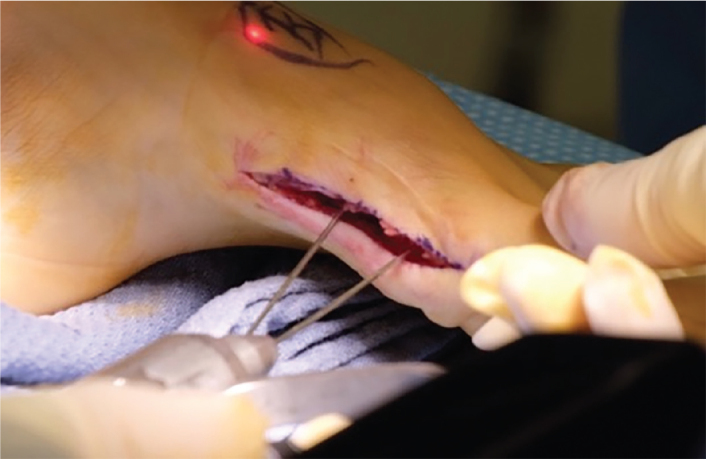
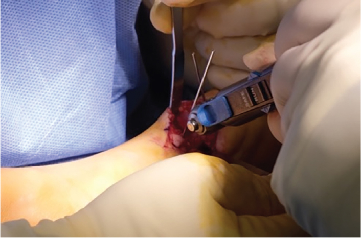
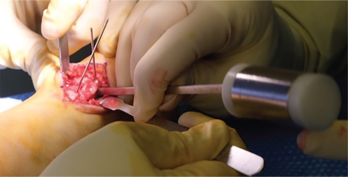
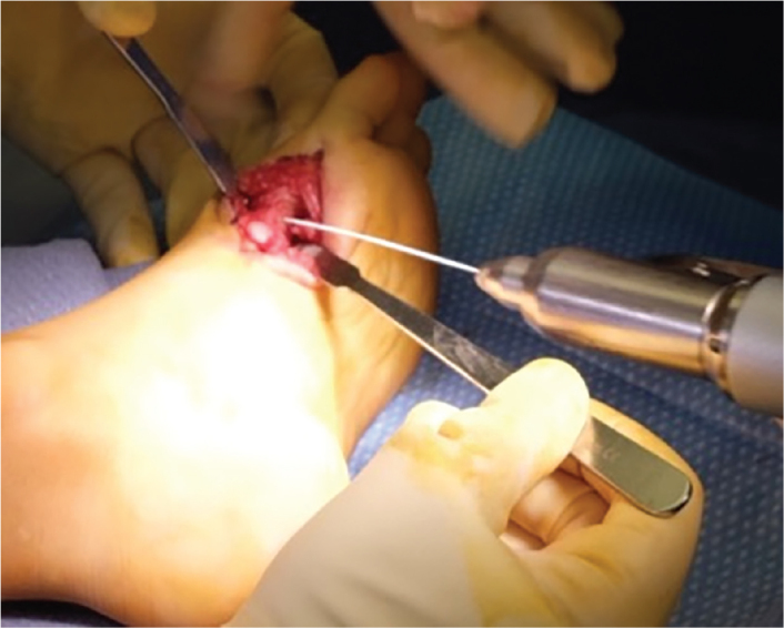
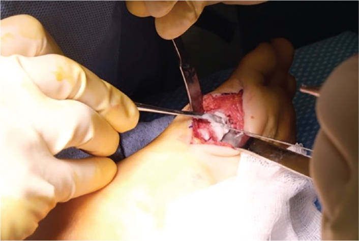
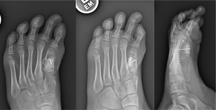
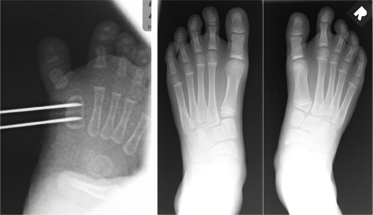
Metatarsal bracketed epiphysis is an uncommon deformity with abnormally located/continuous physeal tissue along the diaphysis leading to shortening and angular deformity of the medially convex bone. Treatment of this condition is primarily surgical and varies depending on the stage. Treatment with early physiolysis to remove the excess growth plate is a surgical technique that has been previously reported. This method gives the greatest potential for correction of longitudinal and angular deformity of the bone as the patient grows. To our knowledge, there are no visual demonstrations of surgical techniques. For ease of visual demonstration of this procedure, we document its use in an 8-year-old female with Stage 3 bracketed epiphysis. Demonstration includes central physiolysis, using polymethylmethacrylate (PMMA) to block the regrowth of the bar. Key Concepts•Treatment of stage 3 bracketed epiphysis by central physiolysis and use of PMMA to block regrowth of the bar gives the affected bone the greatest potential for angular and longitudinal correction.•Outline the area to be excised using guide wires at each end of the bone and remove the bar (cartilage and epiphysis) found in between them, down to the trabecular bone in the diaphyseal area.•Use a centrally located threaded pin to anchor bone cement (PMMA), if needed.
© 2023 JPOSNA. Published by Elsevier on behalf of the Pediatric Orthopaedic Society of North America.
Figures

Similar articles
-
Correction of thumb angulations after physiolysis of delta phalanges in a child with Rubinstein-Taybi syndrome: a case report.Case Reports Plast Surg Hand Surg. 2015 Jan 6;2(1):12-4. doi: 10.3109/23320885.2014.997236. eCollection 2015. Case Reports Plast Surg Hand Surg. 2015. PMID: 27252959 Free PMC article.
-
Cementless, Cruciate-Retaining Primary Total Knee Arthroplasty Using Conventional Instrumentation: Technical Pearls and Intraoperative Considerations.JBJS Essent Surg Tech. 2024 Sep 13;14(3):e23.00036. doi: 10.2106/JBJS.ST.23.00036. eCollection 2024 Jul-Sep. JBJS Essent Surg Tech. 2024. PMID: 39280965 Free PMC article.
-
Metatarsal epiphyseal bracket: treatment by central physiolysis.J Pediatr Orthop. 1993 Jan-Feb;13(1):5-8. doi: 10.1097/01241398-199301000-00002. J Pediatr Orthop. 1993. PMID: 8416354
-
Longitudinal epiphyseal bracket.J Child Orthop. 2013 Dec;7(6):449-54. doi: 10.1007/s11832-013-0544-1. Epub 2013 Nov 29. J Child Orthop. 2013. PMID: 24432108 Free PMC article. Review.
-
Regenerative Medicine Approaches for the Treatment of Pediatric Physeal Injuries.Tissue Eng Part B Rev. 2018 Apr;24(2):85-97. doi: 10.1089/ten.TEB.2017.0274. Epub 2017 Sep 28. Tissue Eng Part B Rev. 2018. PMID: 28830302 Free PMC article. Review.
References
-
- Mubarak S.J., O'Brien T.J., Davids J.R. Metatarsal epiphyseal bracket: treatment by central physiolysis. J Pediatr Orthop. 1993;13(1):5–8. - PubMed
-
- Shea K.G., Mubarak S.J., Alamin T. Preossified longitudinal epiphyseal bracket of the foot: treatment by partial bracket excision before ossification. J Pediatr Orthop. 2001;21(3):360–365. - PubMed
-
- Tibial Hemimelia. POSNA. Available at: https://posna.org/Physician-Education/Study-Guide/Tibial-Hemimelia#:~:te.... Accessed June 27, 2022.
LinkOut - more resources
Full Text Sources
