. 2025 May 13:12:1553215.
doi: 10.3389/fnut.2025.1553215. eCollection 2025.

The protective effect of various forms of Nigella sativa against hepatorenal dysfunction: underlying mechanisms comprise antioxidation, anti- inflammation, and anti-apoptosis

Affiliations

The protective effect of various forms of Nigella sativa against hepatorenal dysfunction: underlying mechanisms comprise antioxidation, anti- inflammation, and anti-apoptosis

Reham M Algheshairy et al. Front Nutr. .

Abstract

Introduction: The liver and kidney are vital organs that are interconnected, dealing with detoxifying and excreting xenobiotics. They are constantly exposed to oxidative stress, which can cause hepatorenal dysfunction. This study compares two forms of Nigella sativa (NS), NS oil (NSO), and NS seeds (NSS), for the first time, in their ability to mitigate hepatorenal injury induced by azathioprine (AZA), exploring potential underlying mechanisms.

Methods: Group (1): negative control; Group (2): positive control received 15 mg/kg AZA orally. Groups (3, 4, and 5) received 100 mg/kg silymarin (standard reference), 500 mg/kg NSO, and 250 mg/kg NSS, respectively, and were subjected to the same dose of AZA. A one-way analysis of variance was conducted, followed by Mann-Whitney post-hoc analysis.

Results: Administration of AZA induced hepatorenal dysfunction, evidenced by dyslipidemia, elevations in serum liver enzymes, creatinine, urea, pro-inflammatory cytokines, and cytokeratin-18. Antioxidant enzymes in liver and kidney tissues were reduced, with an elevation in caspase-3 and caspase-9. Both forms of NS significantly balanced serum pro- inflammatory cytokines (14.33 ± 2.33, 15.15 ± 1.64 vs. 24.87 ± 1.87) pg/ml, interleukin-4 (16.72 ± 1.14, 15.95 ± 1.03 vs. 10.64 ± 1.04) pg/ml, and interleukin-10 (19.89 ± 0.69, 18.38 ± 0.38 vs. 15.52 ± 1.02) pg/ml, and downregulated cytokeratin-18 (210.43 ± 21.56, 195.86 ± 19.42 vs. 296.54 ± 13.94) pg/ml for NSO and NSS vs. the positive group, respectively. NSS enhanced liver antioxidant activity (P < 0.05), normalized liver enzymes (P < 0.05, P < 0.01) for alanine aminotransferase and aspartate aminotransferase, respectively, and significantly lessened dyslipidemia (P < 0.05). Liver caspase-3 and caspase-9 improved significantly with NSS, while kidney caspase-3 and caspase-9 improved with NSO. NSO increased kidney glutathione peroxidase and catalase (P < 0.01) and corrected creatinine and urea (P < 0.05). Histopathological observations confirmed the present data.

Discussion: Conclusively, NSO and NSS mitigated hepatorenal dysfunction responses to AZA through antioxidant, anti-inflammatory, and anti-apoptosis properties that underlie their protective performance. Interestingly, NSO surpassed NSS in restoring renal oxidative damage, while NSS provided better hepatic protection than NSO, suggesting NSO for patients with kidney dysfunction and NSS for those with liver problems.

Keywords: Nigella sativa oil; Nigella sativa seed; antioxidant; apoptosis; cytokeratin-18; cytokine; hepatorenal.


Conflict of interest statement

The authors declare that the research was conducted in the absence of any commercial or financial relationships that could be construed as a potential conflict of interest.

Figures

Figure 1
Figure 1
Identify and quantify some bioactive phytochemical constituents of Nigella sativa oil detected by HPLC.
Figure 2
Figure 2
Identify and quantify some bioactive phytochemical constituents of Nigella sativa seed detected by HPLC.
Figure 3
Figure 3
Effect of Nigella sativa oil and Nigella sativa seed on lipid profile (triglyceride, total cholesterol, HDL, and LDL). Bars denote means ± SE. The marks (*) and (**) are significantly P < 0.05 and P < 0.01 comparable to the saline negative control, respectively. The letters (a, b) are significantly P < 0.05 and P < 0.01 comparable to AZA (azathioprine) positive control, respectively.
Figure 4
Figure 4
Effect of Nigella sativa oil and Nigella sativa seeds on liver function (serum ALT, alanine aminotransferase enzyme and AST, aspartate aminotransferase enzyme). Bars denote means ± SE. The mark (**) is significantly P < 0.05 comparable to the saline negative control. The letters (a, b) are significantly P < 0.05 and P < 0.01 comparable to AZA (azathioprine) positive control, respectively.
Figure 5
Figure 5
Effect of Nigella sativa oil and Nigella sativa seeds on kidney function (creatinine, urea, Na, and K). Bars denote means ± SE. The marks (*) and (**) are significantly P < 0.05 and P < 0.01 comparable to the saline negative control, respectively. The letter (a) is significantly P < 0.05 comparable to AZA (azathioprine) positive control.
Figure 6
Figure 6
Effect of Nigella sativa oil and Nigella sativa seeds on cytokines (TNF-α, Tumar necrosis factor-α; IL-4, interlukine-4; IL-10, interlukine-10) and cytokeratin 18. Bars denote means ± SE. The marks (*) and (**) are significantly P < 0.05 and P < 0.01 comparable to the saline negative control, respectively. The letters (a, b) are significantly P < 0.05 and P < 0.01 comparable to AZA (azathioprine) positive control, respectively.
Figure 7
Figure 7
Effect of Nigella sativa oil and Nigella sativa seeds on liver tissue antioxidants (GHPx, glutathione peroxidase; SOD, superoxide dismutase; CAT, catalase) and lipid peroxidation (MDA, malondialdehyde). Bars denote means ± SE. The marks (*) and (**) are significantly P < 0.05 and P < 0.01 comparable to the saline negative control, respectively. The letters (a, b) are significantly P < 0.05 and P < 0.01 comparable to AZA (azathioprine) positive control, respectively.
Figure 8
Figure 8
Effect of Nigella sativa oil and Nigella sativa seeds on kidney tissue antioxidants (GHPx, glutathione peroxidase; SOD, superoxide dismutase; CAT, catalase) and lipid peroxidation (MDA, malondialdehyde). Bars denote means ± SE. The marks (*) and (**) are significantly P < 0.05 and P < 0.01 comparable to the saline negative control, respectively. The letters (a, b) are significantly P < 0.05 and P < 0.01 comparable to AZA (azathioprine) positive control, respectively.
Figure 9
Figure 9
Effect of Nigella sativa oil and Nigella sativa seeds on liver tissue caspase-3 and caspase-9. The mark (**) is significantly different from the saline negative control at P < 0.05. Bars denote means ± SE. The mark (**) is significantly P < 0.05 comparable to the saline negative control. The letters (a, b) are significantly P < 0.05 and P < 0.01 comparable to AZA (azathioprine) positive control, respectively.
Figure 10
Figure 10
Effect of Nigella sativa oil and Nigella sativa seeds on kidney tissue caspase-3 and caspase-9. Bars denote means ± SE. The mark (*) is significantly P < 0.05 comparable to the saline negative control. The letter (a) is significantly P < 0.05 comparable to AZA (azathioprine) positive control.
Figure 11
Figure 11
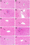
Hematoxylin and eosin (H&E; 400; scale bar, 50 m) were used to stain liver slices from each experimental group. The liver from the negative control (A, B) showed a normal photomicrograph of the hepatic lobule, including the central vein (CV) and hepatocytes (H). The liver tissue from the AZA positive control revealed activation of Kupffer cells (black arrow) (C) and collagen fiber deposition around the central vein (black arrow) (D). liver of rats administered silymarin showed no histopathological alterations (E) and few binucleations of hepatocytes (black arrow) (F). liver of rats administered NSO demonstrated activation of Kupffer cells (black arrow) (G), a small amount of localized hepatocellular necrosis, and a minor influx of inflammatory cells (red arrow) (H). The NSS-treated group showed a typical hepatic lobule, with normal hepatocytes and central vein (CV) (I). A few sections showed slight activation of the Kupffer cells (black arrow) (J).
Figure 12
Figure 12
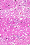
Hematoxylin and eosin (H&E; 400; scale bar, 50 m) were used to stain kidney slices from each experimental group. Kidney from negative control (A, B) illustrated normal histological architecture of renal parenchyma, including normal glomeruli (GL) and renal tubules (RT). The kidney from the AZA positive control revealed renal tubule epithelial vacuolar degeneration (black arrow) (C), vacuolar degeneration of the epithelial lining of renal tubules (black arrow), and edema (red arrow) (D). Kidney tissue from the group that received silymarin showed apparent normal renal parenchyma (E) and mild vacuolar degeneration of some renal tubule lining epithelial cells) (F). Kidney of rats administered NSO demonstrated no histopathological alterations (G, H). Most kidney sections of rats administered NSS showed slight vacuolar degeneration of some renal tubule lining epithelial cells (black arrow) (I). In contrast, some sections showed congestion of intratubular capillaries (red arrow) with glomerular tuft (blue arrow) (J).

Similar articles

References

    1. Nauroze T, Ali S, Kanwal L, Ara C, Mughal TA, Andleeb S. Ameliorative effect of Nigella sativa conjugated silver nanoparticles against chromium-induced hepatotoxicity and renal toxicity in mice. Saudi J Biol Sci. (2023) 30:103571. 10.1016/j.sjbs.2023.103571 - DOI - PMC - PubMed
    1. Barbara S. Inflammatory bowel disease and male fertility. LIJ Heal Syst. (2010) 516:734–850.
    1. Valko M, Leibfritz D, Moncol J, Cronin MTD, Mazur M, Telser J. Free radicals and antioxidants in normal physiological functions and human disease. Int J Biochem Cell Biol. (2007) 39:44–84. 10.1016/j.biocel.2006.07.001 - DOI - PubMed
    1. Amadi CN, Offor SJ, Frazzoli C, Orisakwe OE. Natural antidotes and management of metal toxicity. Environ Sci Pollut Res. (2019) 26:18032–52. 10.1007/s11356-019-05104-2 - DOI - PubMed
    1. Abdel Ghfar SS, Ali ME, Momenah MA, Al-Saeed FA, Al-Doaiss AA, Mostafa YS, et al. . Effect of Allium sativum and Nigella sativa on alleviating aluminum toxicity state in the albino rats. Front Vet Sci. (2022) 9:1042640. 10.3389/fvets.2022.1042640 - DOI - PMC - PubMed

LinkOut - more resources