

Endometrial osseous metaplasia presented as infertility cases with intra-operative obstacles: Two case reports and literature review
- PMID: 40432986
- PMCID: PMC12109679
- DOI: 10.5339/qmj.2025.24
Endometrial osseous metaplasia presented as infertility cases with intra-operative obstacles: Two case reports and literature review
Abstract
Background: Endometrial osseous metaplasia (OM) is a rare condition characterized by the transformation of endometrial tissue into bone cells. Despite its rarity, OM remains a significant contributor to infertility. Although the underlying mechanism remains debatable, an association with previous abortions and curettage has been suggested.
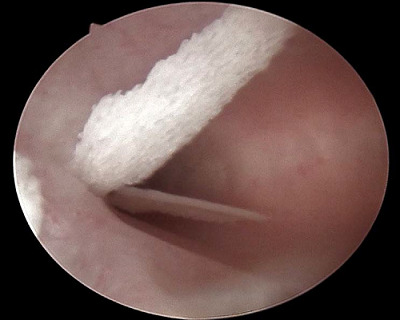
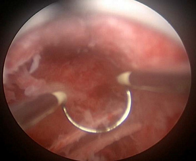
Case presentation: We present two cases of OM presented to the infertility clinic and discuss their similarities and discrepancies in presentation and risk factors. A transvaginal ultrasound raises suspicion about the diagnosis of OM with a hyperechoic mass and post-acoustic shadowing. An office hysteroscopy showed white, mesh-like bony sheets. Both cases underwent operative hysteroscopy to address surgical challenges, and the two cases were followed postoperatively for one year.
Discussion: A comprehensive literature review examined various aspects of OM, including diagnosis, therapeutic options, outcomes, prognosis, and follow-up. Our aim was to raise awareness of this intriguing condition by providing up-to-date knowledge and emphasizing the central role of hysteroscopy in diagnosis and treatment. Here, we present two cases with the same complaint, infertility. Moreover, although the same treatment method was used in both cases, only one achieved pregnancy. This highlights that OM is a possible underlying cause of infertility, in addition to considering other factors that contribute to the overall clinical picture.
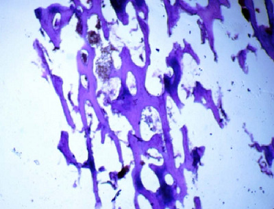
Conclusion: OM should be considered in the evaluation of infertility despite its rarity, especially with hyperechoic lesions and acoustic shadowing on ultrasound examination. Hysteroscopy is the gold standard for diagnosis and therapeutic approaches. A complete understanding of the reasons that trigger its growth is crucial. To rule out other differential diagnosis, a holistic evaluation of the patient's history, imaging, and histopathological examination is needed.
Keywords: Endometrial osseous metaplasia; abortion; curettage; diagnostic procedures; hysteroscopy; infertility.
© 2025 Helmi, Nori, licensee HBKU Press.
Conflict of interest statement
The authors have no conflicts of interest to declare.
Figures


References
-
- Cayuela E, Vilanova J, del Río M, Heredia F, Acín L, Zarco P et al. Endometrial osseous metaplasia. Hysteroscopy. 2018 Jan;:683–687. doi: 10.1007/978-3-319-57559-9_60. doi: - DOI
LinkOut - more resources
Full Text Sources