

Posterior Column Osteotomies in Adolescent Idiopathic Scoliosis
- PMID: 40433085
- PMCID: PMC12088245
- DOI: 10.55275/JPOSNA-2023-638
Posterior Column Osteotomies in Adolescent Idiopathic Scoliosis
Abstract
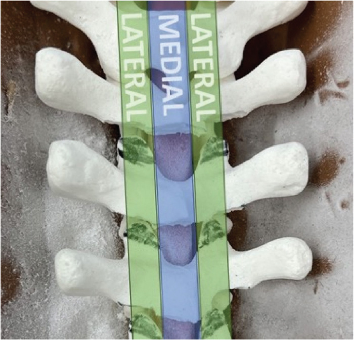
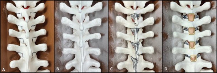
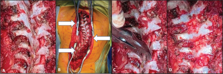
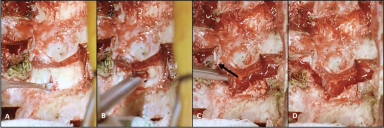
The posterior column osteotomy (PCO) is an adjunct technique for obtaining deformity correction during posterior spine fusion procedures. Full disarticulation of the posterior spinal column, including bony elements (namely the lamina and facet joints) and ligamentous complex is described as a PCO. This technique was originally described to allow for shortening of the posterior column during correction of excessive thoracic kyphosis; however, its indications have since been expanded to other spine deformities such as adolescent idiopathic scoliosis (AIS). Its expanded role in deformity surgery has been met with controversy: proponents tout increased flexibility and better spinal correction in three planes, while detractors cite lack of ostensible clinical benefit and potential for more complications. Differences in surgical technique are also prevalent. In this manuscript, we review the surgical technique of PCOs, including the traditional PCO as well as a modified posterior column release (PCR). Additionally, the controversy over when this technique should be utilized is further explored through summation of current literature on PCO outcomes. Key Concepts•The indications for posterior column osteotomies in pediatric spine deformity surgery are frequently debated, with the risk-benefit profile weighed differently among surgeons.•Proponents of PCO use in scoliosis assert improved flexibility and correction of spinal deformity in three dimensions, in particular the ability to better restore thoracic kyphosis.•Most studies acknowledge that there is an increased risk of a neuromonitoring alert when PCOs are performed, though a significant difference in postoperative deficits has not been described.•The traditional Posterior Column Osteotomy PCO (i.e., "Ponte" osteotomy) is performed by creating a gap in the posterior elements which can be closed down with deformity correction (namely kyphosis).•The modified Posterior Column Osteotomy (or Posterior Column Release [PCR]) can be performed by disarticulating the posterior tension band but leaving a smaller gap, perhaps limiting the ability for posterior compression but limiting spinal canal exposure.
© 2023 JPOSNA. Published by Elsevier on behalf of the Pediatric Orthopaedic Society of North America.
Figures
References
-
- Smith-Petersen M.N., Larson C.B., Aufranc O.E. Osteotomy of the spine for correction of flexion deformity in rheumatoid arthritis. Clin Orthop Relat Res. 1969;66:6–9. - PubMed
-
- Ponte A. Surgical treatment of Scheuermann's hyperkyphosis. Orthop Trans. 1985;9:127.
-
- Ponte A. In: Surgical Techniques for the Spine. Haher T.R., Merola A.A., editors. Thieme; New York: 2003. Posterior column shortening for Scheuermann's kyphosis: an innovative one-stage technique; pp. 107–113.
-
- Ponte A., Orlando G., Siccardi G.L. The true ponte osteotomy: by the one who developed it. Spine Deform. 2018;6(1):2–11. - PubMed
-
- Geck M.J., Macagno A., Ponte A., et al. The Ponte procedure: posterior only treatment of Scheuermann's kyphosis using segmental posterior shortening and pedicle screw instrumentation. J Spinal Disord Tech. 2007;20(8):586–593. - PubMed
Publication types
LinkOut - more resources
Full Text Sources
Miscellaneous
