. 2025 May 22:20:6515-6531.
doi: 10.2147/IJN.S510514. eCollection 2025.

Lipid-Coated Ag@MnO2 Core-Shell Nanoparticles for Co-Delivery of Survivin siRNA in Breast Tumor Therapy

Affiliations

Lipid-Coated Ag@MnO2 Core-Shell Nanoparticles for Co-Delivery of Survivin siRNA in Breast Tumor Therapy

Jing Zhang et al. Int J Nanomedicine. .

Abstract

Objective: Nanoparticles constructed with silver/manganese dioxide (Ag@MnO2) as the core, in conjunction with survivin siRNA (sis) and cyclo(RGD-DPhe-K) (Ag@MnO2-sis-c-L), were prepared for integrated tumor diagnosis and therapy.

Methods: Ag@MnO2-sis-c-L particles were prepared and characterized. The silver and manganese content were determined by inductively coupled plasma optical emission spectroscopy (ICP-OES). The stability of sis in the system was evaluated by incubation with 50% FBS before the agarose gel electrophoresis experiment. The in vitro photothermal conversion ability, cytotoxicity to 4T1 cells, and cellular uptake of preparations were evaluated. The dialysis technique was employed to determine the in vitro release profile of Ag and Mn from Ag@MnO2-sis-c-L under various pH conditions. The pharmacokinetic behavior and tissue distribution of silver in vivo were detected by ICP-OES. Animal model experiments were conducted to further evaluate the anti-tumor efficacy of Ag@MnO2-sis-c-L against breast cancer in combination with infrared irradiation.

Results: Our newly synthesized Ag@MnO2-sis-c-L nanoparticles displayed superior physicochemical properties. The combined application of these nanoparticles with photothermal therapy (PTT) exerted the strongest synergistic inhibitory effects on tumor growth. Survivin protein expression in tumor tissues were markedly suppressed following delivery of nanoparticles loaded with sis. Additionally, magnetic resonance imaging revealed the high imaging capability of hybrid nanoparticles.

Conclusion: This study supports the potential utility of Ag@MnO2-sis-c-L coupled with PTT in therapeutic and diagnostic imaging applications.

Keywords: Ag@MnO2; breast cancer; combination therapy; photothermal therapy; survivin siRNA.


Conflict of interest statement

The authors report no conflicts of interest in this work.

Figures

Figure 1
Figure 1
(A) Agarose gel electrophoresis of Ag@MnO2-sis-c-L to siRNA (Lane 1: Free siRNA; Lane 2: Ag@MnO2-c-L; Lanes 3–11: Ag@MnO2-sis-c-L N:P 10:1; 5:1; 1:1; 1:5; 1:10; 1:20; 1:40; 1:80; 1:160); (B) SEM and (C) TEM images of Ag@MnO2 (1), Ag@MnO2-sis-L (2), and Ag@MnO2-sis-c-L (3); (D) Surface element maps of Ag@MnO2 (1), Ag@MnO2-sis-L (2), and Ag@MnO2-sis-c-L (3).
Figure 2
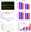
Figure 2
(A) Agarose gel electrophoresis of Free sis and Ag@MnO2-sis-c-L after incubation with simulated serum at different time-points; (B) Temperature changes of Ag@MnO2-sis-c-L under NIR radiation; (C) The photothermal conversion efficiency of Ag@MnO2-sis-c-L after repeated irradiation cycles; (D) Cytotoxicity of different preparations containing a 0.2 μg/mL of Ag in 4T1 and L929 cells; (E) Cellular uptake of Ag by 4T1 cells treated with Ag@MnO2-sis-L and Ag@MnO2-sis-c-L formulations (vs model group, *P<0.05, **P<0.01).
Figure 3
Figure 3
(A) Release of Ag and Mn from Ag@MnO2-sis-c-L at different time-points; (B) Ag concentration-time curves of the preparations in blood (n=3); (C) The tissue distribution of Ag at time points (n=3); (D) The rates of change in relative Ag contents in tissues at time-points of 0.5 h (A), 4 h (B), and 12 h (C) (**P<0.01, ***P<0.001).
Figure 4
Figure 4
(A) Tumor volumes of mice from each preparation-treated group (n=3); (B) Tumor weights of mice in each group (*P<0.05); (C) Survivin and β-actin protein expression in tumor tissues of each group (1: Ag@MnO2-sis-c-L-NIR, 2: Ag@MnO2-sis-c-L, 3: Ag@MnO2-scrsis-c-L-NIR, 4: Ag@MnO2-scrsis-c-L, 5: Ag@MnO2-c-L-NIR, 6: Ag@MnO2-c-L, 7: Free sis-NIR, 8: Free sis, 9: Model); (D) Thermography of mice from the model group (1), low-dose (2) and high-dose (3) of Ag@MnO2-sis-c-L-treatment groups (Ag equivalents of 37.5 μg/mL and 75.0 μg/mL, respectively).
Figure 5
Figure 5
(A) MRI images of Ag@MnO2-sis-c-L in mice at different time points (horizontal plane in the upper line, coronal plane in the lower line); (B) Changes in body weights of mice from each treatment group (n=3).

Similar articles

References

    1. Lei S, Zheng R, Zhang S, et al. Global patterns of breast cancer incidence and mortality: a population-based cancer registry data analysis from 2000 to 2020. Cancer Commun. 2021;41(11):1183–1194. doi:10.1002/cac2.12207 - DOI - PMC - PubMed
    1. Hou X, Chen Q, Fang Y, et al. iRGD-guided silica/gold nanoparticles for efficient tumor-targeting and enhancing antitumor efficacy against breast cancer. Int J Nanomedicine. 2024;19:8237–8251. doi:10.2147/ijn.S474135 - DOI - PMC - PubMed
    1. Bray F, Ferlay J, Soerjomataram I, Siegel RL, Torre LA, Jemal A. Global cancer statistics 2018: GLOBOCAN estimates of incidence and mortality worldwide for 36 cancers in 185 countries. CA. 2018;68(6):394–424. doi:10.3322/caac.21492 - DOI - PubMed
    1. Sung H, Ferlay J, Siegel RL, et al. Global cancer statistics 2020: GLOBOCAN estimates of incidence and mortality worldwide for 36 cancers in 185 countries. CA. 2021;71(3):209–249. doi:10.3322/caac.21660 - DOI - PubMed
    1. Slamon DJ, Leyland-Jones B, Shak S, et al. Use of chemotherapy plus a monoclonal antibody against HER2 for metastatic breast cancer that overexpresses HER2. N Engl J Med. 2001;344(11):783–792. doi:10.1056/nejm200103153441101 - DOI - PubMed

LinkOut - more resources