

The molecular architecture distinctions between compression, opposite and normal wood of Pinus radiata
- PMID: 40433164
- PMCID: PMC12106396
- DOI: 10.3389/fpls.2025.1576928
The molecular architecture distinctions between compression, opposite and normal wood of Pinus radiata
Abstract
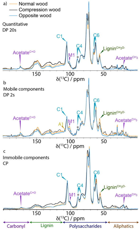
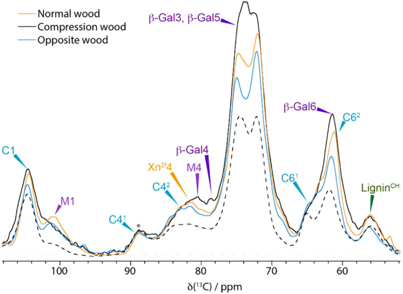
In gymnosperms compression wood is a specialised type of structural cell wall formed in response to biomechanical stresses. The differences in terms of gross structure, ultrastructure and chemistry are well-known. However, the differences between compression wood, normal wood, and opposite wood regarding the arrangements and interactions of the various polymers and water within their cell walls still needs to be established. The analysis of 13C-labelled Pinus radiata by solid-state NMR spectroscopy and other complementary techniques revealed several new aspects of compression and opposite wood molecular architecture. Compared to normal wood, compression wood has a lower water content, its overall nanoporosity is reduced, and the water and matrix polymers have a lower molecular mobility. Galactan, which is a specific marker of compression wood, is broadly distributed within the cell wall, disordered, and not aligned with cellulose, and is found to be in close proximity to xylan. Dehydroabietic acid (a resin acid) is immobilised and close to the H-lignin only in compression wood. Although the overall molecular mobility of normal wood and opposite wood are similar, opposite wood has different arabinose conformations, a large increase in the amount of chain ends, contains significantly more galactan and has additional unassigned mobile components highlighting the different molecular arrangement of cell wall polymers in opposite and normal wood.
Keywords: Pinus radiata (Monterey pine); compression wood (CW); opposite wood (OW); secondary cell wall (SCW); solid state NMR (ssNMR).
Copyright © 2025 Cresswell, Dickson, Robertson, Gallagher, Risani, Le Guen, Temple, Liszka, Donaldson, Kirby, Ralph, Hill, Dupree, Dupree and Sorieul.
Conflict of interest statement
The authors declare that the research was conducted in the absence of any commercial or financial relationships that could be construed as a potential conflict of interest.
Figures


Similar articles
-
Tracheid cell-wall structures and locations of (1 → 4)-β-D-galactans and (1 → 3)-β-D-glucans in compression woods of radiata pine (Pinus radiata D. Don).BMC Plant Biol. 2016 Sep 7;16(1):194. doi: 10.1186/s12870-016-0884-3. BMC Plant Biol. 2016. PMID: 27604684 Free PMC article.
-
Location and characterization of lignin in tracheid cell walls of radiata pine (Pinus radiata D. Don) compression woods.Plant Physiol Biochem. 2017 Sep;118:187-198. doi: 10.1016/j.plaphy.2017.06.012. Epub 2017 Jun 15. Plant Physiol Biochem. 2017. PMID: 28646704
-
Transcriptome profiling of radiata pine branches reveals new insights into reaction wood formation with implications in plant gravitropism.BMC Genomics. 2013 Nov 8;14(1):768. doi: 10.1186/1471-2164-14-768. BMC Genomics. 2013. PMID: 24209714 Free PMC article.
-
Localization of cell wall polysaccharides in normal and compression wood of radiata pine: relationships with lignification and microfibril orientation.Plant Physiol. 2012 Feb;158(2):642-53. doi: 10.1104/pp.111.184036. Epub 2011 Dec 5. Plant Physiol. 2012. PMID: 22147521 Free PMC article.
-
Molecular self-organization of wood lignin-carbohydrate matrix.Planta. 2021 Jul 16;254(2):30. doi: 10.1007/s00425-021-03675-4. Planta. 2021. PMID: 34272608 Review.
References
-
- Abe K., Yamamoto H. (2005). Mechanical interaction between cellulose microfibril and matrix substance in wood cell wall determined by X-ray diffraction. J. Wood Sci. 51, 334–338. doi: 10.1007/s10086-004-0667-6 - DOI
-
- Abe K., Yamamoto H. (2006). Change in mechanical interaction between cellulose microfibril and matrix substance in wood cell wall induced by hygrothermal treatment. J. Wood Sci. 52, 107–110. doi: 10.1007/s10086-005-0738-3 - DOI
-
- Altaner C., Hapca A. I., Knox J. P., Jarvis M. C. (2007). Detection of β-1-4-galactan in compression wood of Sitka spruce [Picea sitchensis (Bong.) Carrière] by immunofluorescence. Holzforschung 61, 311–316. doi: 10.1515/HF.2007.049 - DOI
LinkOut - more resources
Full Text Sources